Pour obtenir le grade de. Arrêté ministériel : 25 mai Emeline REBOUD
|
|
|
- Laure Malenfant
- il y a 7 ans
- Total affichages :
Transcription

1 THÈSE Pour obtenir le grade de DOCTEUR DE LA COMMUNAUTE UNIVERSITE GRENOBLE ALPES Spécialité : Virologie, Microbiologie, Immunologie Arrêté ministériel : 25 mai 2016 Présentée par Emeline REBOUD Thèse dirigée par Philippe HUBER, préparée au Laboratoire de Biologie du Cancer et de l Infection dans l'école Doctorale Chimie et Sciences du Vivant Comportement et toxicité de nouvelles souches hyper-virulentes de Pseudomonas aeruginosa Thèse soutenue publiquement le 12 octobre 2017, devant le jury composé de : Monsieur Max MAURIN Professeur, Grenoble, Président du jury Madame Suzana SALCEDO Chargée de recherche, Lyon, Rapporteur Monsieur Eric KIPNIS Professeur, Lille, Rapporteur Monsieur Thomas HENRY Directeur de Recherche, Lyon, Examinateur Monsieur Philippe HUBER Directeur de Recherche, Grenoble, Directeur de thèse
2
3 A mes parents, à ma mamy, A ma famille et amis, A Guillaume. «Il faut toujours viser la lune, car même en cas d échec, on atterrit dans les étoiles» Oscar Wilde - 1 -
4 - 2 -
5 Remerciements Je souhaite, tout d abord, remercier les membres de mon jury de thèse, le Docteur Suzana Salcedo et le Professeur Eric Kipnis d avoir accepté d être rapporteur de ma thèse ainsi que le Docteur Thomas Henry et le Professeur Max Maurin d avoir bien voulu examiner mon travail de thèse. Des remerciements additionnels aux Docteurs Thomas Henry et Anne-Marie Di Guilmi pour tous leurs conseils et ouvertures scientifiques lors de mes deux comités de suivi de thèse. Je remercie le Labex GRAL d avoir financé ma thèse pendant ces trois années ainsi que les différentes tutelles institutionnelles de notre équipe (CEA, CNRS, INSERM, Université Grenoble Alpes). Je remercie également Jérôme Garin et Jean-Jacques Feige pour m avoir accueillie au sein de l institut et du laboratoire. Un remerciement tout particulier à mon directeur de thèse, Philippe Huber, qui m a offert la possibilité de continuer en thèse et qui m a fait confiance pendant ces trois années de thèse. Merci pour sa patience, ses conseils avisés et le partage de son expérience. Un grand merci à Ina Attrée pour m avoir accueillie dans une formidable équipe aussi bien d un point de vue scientifique qu humain. Un merci à tous les membres de l équipe PB&RC (Pathogénèse Bactérienne et Réponses Cellulaires) passés et présents pour leurs conseils et tous les bons moments partagés ensembles. Un remerciement un peu spécial à ces drôles de dames (Alice, Pauline & Stéphanie) sans qui cette thèse n aurait pas été la même. Je tiens à les remercier pour tous ces fous rires, nos photos mémorables, nos soirées, nos challenges the room, les pauses thés et bien d autres, mais surtout pour leur soutien. Ce sont trois années qui s achèvent mais qui représentent aussi le début d une belle amitié. Merci à Stéphanie, mon binôme de culture cell, ma voisine de bureau, pour tous ces échanges scientifiques et «extra-professionnels», pour son aide notamment dans les manips. Je pense qu on s est bien trouvées
6 Il y a tant de personnes dans ma vie personnelle que je souhaite remercier pour avoir été d un grand soutien pendant trois ans et pour avoir tenté de comprendre l objet de mes recherches. Mes premiers remerciements vont vers ma famille, avec une attention spéciale pour mes grandsparents, cousin(e)s, mon parrain et ma marraine, pour tout le soutien qu ils m ont apporté. Un remerciement tout particulier à Sabrina et Damien pour nos soirées improvisées après le travail et leurs conseils. Merci à eux ainsi que Julie et Miguel de m avoir offert le plus beau cadeau que d être marraine de ma petite Siloé et de mon petit Théo, une pure bouffée de bonheur qui nous montre que la vie est belle et pleine de belles surprises. Une pensée pour ma mamy, qui aurait été tellement fière de voir sa petite fille soutenir sa thèse. Toi qui pensais que les analyses médicales n avaient pas de secret pour moi en tant que future docteur. Tu t es battue jusqu au bout mais la maladie a été plus forte. Mais je suis sûr que de là-haut, tu m espionnes et me guides vers les bonnes directions. Je remercie ensuite mes ami(e)s pour toutes ces «soirées décompression» et notamment mon équipe de handball qui a été très efficace dans ce domaine! Merci à Céline, mon amie de plus de 20 ans, qui a toujours été présente pour moi. Un grand merci à mes parents, pour leur patience et pour leur soutien durant ces trois années de thèse. Ils ont supporté mon caractère, ont été présents dans les bons et les mauvais moments, m ont guidé dans mes choix et surtout, ils m ont fait confiance. Pour cela et pour tout le reste, je tenais vraiment à les remercier. Un grand merci également à mon frère et ma belle-sœur notamment pour ma progression en anglais. Et enfin, le plus grand des mercis est pour mon cher et tendre, Guillaume, pour m avoir soutenu au quotidien et encouragé tout au long de ma thèse. Nous avons chacun commencé une aventure, cette thèse de mon côté, et la création d une société du sien. Et si nous avons persévéré pour réaliser nos projets, c est grâce aux encouragements et au soutien mutuel que nous nous sommes consacrés. Je sais que je pourrai compter sur lui dans l avenir comme il pourra compter sur moi
7 Avant-propos Une histoire longue de 5 ans au sein de l équipe PBRC où j ai eu l opportunité de découvrir et de participer à une grande partie des domaines de recherche de l équipe. Tout a commencé en stage de master 1 sous la supervision de François Cretin où j ai étudié le rôle du système de sécrétion de type III (SST3) dans l internalisation de Pseudomonas aeruginosa par les macrophages. Le SST3 est le facteur de virulence majeur de ce pathogène. Après un stage de master 1 côté immunologie, j ai découvert le monde de la régulation de gènes bactériens lors de mon stage de master 2, sous la supervision de Sylvie Elsen. C est lors de ce stage que l histoire avec la toxine ExlA a débuté. ExlA est une toxine formant des pores ou «pore-forming toxin» découverte par l équipe. Elle a été mise en évidence à partir d un isolat clinique prélevé sur un patient du CHU de Grenoble. Lors de mon stage de master 2, j ai étudié la régulation de l expression du gène exla ainsi que des gènes impliqués dans la résistance aux antibiotiques de la bactérie. Grâce à un financement Labex Gral (Laboratory of excellence Grenoble Alliance for Integrated Structural Cell Biology) obtenu par Philippe Huber, j ai découvert le monde de l interaction «hôte-pathogène» lors de ma thèse. Et mon histoire avec ExlA a continué. Pendant ma première année de thèse j ai cherché à savoir si ExlA était produite par d autres souches de Pseudomonas aeruginosa afin de comprendre la prévalence de cette toxine. Nous avons pu montrer que les souches possédant le gène exla appartenaient à une nouvelle famille de souches de type PA7-like de Pseudomonas aeruginosa. Les deux années suivantes ont été consacrées à l effet de la toxine ExlA sur les cellules eucaryotes afin de comprendre son rôle dans la pathogénicité. J ai étudié le rôle d ExlA dans la dégradation des protéines jonctionnelles (la E et la VE-cadhérine) qui permettent l intégrité tissulaire. J ai également participé à d autres projets au sein de l équipe notamment dans l étude de la compréhension du mécanisme de formation du pore dans les cellules eucaryotes induit par ExlA. Enfin, j ai collaboré à une étude in vivo chez la souris afin de caractériser les effets d ExlA lors d infections pulmonaires
8 Dans ce mémoire, j aborde le contexte scientifique de mon étude à travers cinq chapitres d introduction. Les deux premiers chapitres se focalisent sur les caractéristiques du pathogène Pseudomonas aeruginosa et des facteurs de virulence importants pour la toxicité de la bactérie. Dans le chapitre III, je détaille les barrières tissulaires de l organisme, les jonctions intercellulaires et le franchissement de ces barrières par les pathogènes bactériens. Je continue dans le chapitre IV avec une présentation générale des pore-forming toxins. Enfin je conclue mon introduction avec un chapitre V dédié à la présentation de la nouvelle famille de souches PA7-like de Pseudomonas aeruginosa. Le travail réalisé au cours de ma thèse est présenté dans la partie «Résultats & Discussions» à travers la présentation de quatre articles scientifiques. Dans cette partie, le chapitre VI décrit l étude phénotypique des souches PA7-like collectées à travers le monde. Dans les chapitres VII et VIII, je présente brièvement les deux études auxquelles j ai participé, décrivant le mécanisme de formation du pore d ExlA et le rôle d ExlA dans l infection pulmonaire. Et enfin, je termine avec le chapitre IX où je présente l effet d ExlA sur la rupture des protéines jonctionnelles impliquées dans l intégrité de la barrière tissulaire
9 Table des Matières Remerciements Avant-propos Liste des Figures Liste des Tableaux Abréviations Introduction I. Pseudomonas aeruginosa, un pathogène opportuniste Généralités Caractéristiques biologiques Mode de vie Les infections à P. aeruginosa Les principaux sites et risques d infection Les infections aiguës i. Les pneumonies ii. Les autres infections aiguës Les infections chroniques i. Infection de patients atteints de mucoviscidose ii. Les autres infections chroniques La résistance aux antibiotiques II. Les facteurs de virulence de Pseudomonas aeruginosa Les systèmes de sécrétion et leurs effecteurs Le système de sécrétion de type 3 (SST3) i. Composition ii. Les effecteurs Le système de sécrétion de type 5 (SST5) i. Les systèmes autotransporteur (AT) ii. Les systèmes TPS
10 3. Les autres systèmes de sécrétion i. Le système de sécrétion de type 1 (SST1) ii. Le système de sécrétion de type 2 (SST2) iii. Le système de sécrétion de type 6 (SST6) Les facteurs de virulence de surface Le flagelle Les pili de type IV Les lipopolysaccharides (LPS) Autres facteurs de virulence Production de chromophores Rhamnolipides Production de cyanure d hydrogène III. Les barrières tissulaires & l infection La barrière épithéliale Caractéristiques Les jonctions intercellulaires i. Les jonctions serrées ii. Les jonctions d ancrages iii. Les jonctions communicantes La barrière endothéliale Caractéristiques Les jonctions intercellulaires i. Les jonctions serrées ii. Les jonctions adhérentes iii. Les jonctions communicantes de type gap Les jonctions adhérentes des cellules épithéliales et endothéliales La E et VE-cadhérines, des protéines des jonctions adhérentes i. Structure ii. Fonction iii. Régulation de l activité biologique des cadhérines
11 Franchissement des barrières tissulaires Les différentes voies i. Transcellulaire ii. Paracellulaire iii. Mécanisme du «cheval de troie» Franchissement de la barrière pulmonaire i. Structure ii. Franchissement de la barrière pulmonaire par les pathogènes iii. Franchissement de la barrière pulmonaire par P. aeruginosa IV. Les pore-forming toxins Généralité Les pore-forming toxins Les pore-forming toxins bactériennes Les α-pfts La famille des Colicines La famille des cytolysines A (ClyA) Les β-pfts La famille des Hémolysines La famille des aérolysines La famille des cholesterol-dependent cytolysins (CDC) Toxicité des PFTs Disfonctionnement des barrières tissulaires in vivo Interaction avec le système immunitaire i. Toxicité envers les cellules immunitaires ii. Survie des bactéries phagocytées La mort cellulaire induite par les PFTs Réponses cellulaires Réponse au stress Réparation de la membrane plasmique
12 V. Une nouvelle famille de souches de Pseudomonas aeruginosa PA7, une souche «marginale» Origine Absence de certains facteurs de virulence De nouveaux facteurs de virulence i. Un nouveau SST ii. PA7 et la réponse immunitaire innée iii. Résistance aux antibiotiques Les points communs entre PA7 et PAO CLJ1, une souche hypervirulente Un nouveau facteur de virulence : ExlBA Système de sécrétion à deux partenaires ExlBA i. Le système TPS ExlBA ii. La régulation d ExlBA ExlA, une pore-forming toxin i. La structure d ExlA ii. Une pore-forming toxin iii. Les homologues d ExlA Résultats & Discussions VI. Etude phénotypique de souches PA7/CLJ1-like Résumé Publication Participation au projet : Article Résultats complémentaires & discussion Evolution de l arbre phylogénétique Prévalence des souches exla-positive Caractérisation de la souche IHMA
13 VII. ExlA et les pili de type IV coopèrent pour induire la mort cellulaire Résumé ExlBA est un nouveau TPS de P. aeruginosa ExlA est une pore-forming toxin dont l activité est favorisée par les pili Publication Participation au projet : Article VIII. ExlA provoque des dommages pulmonaires qui facilitent la dissémination bactérienne Résumé Effet drastique d ExlA sur les alvéoles pulmonaires Dissémination CLJ1 échappe au système immunitaire Publication Participation au projet : Article IX. ExlA induit le clivage des E- et VE-cadhérines des jonctions adhérentes Résumé Publication Participation au projet : Article Résultats complémentaires & discussion Les flux calciques sont médiés par le pore d ExlA La Streptolysine O induit le clivage des cadhérines Les jonctions intercellulaires : une cible pour les pathogènes i. Effets des PFTs ii. La E-cadhérine, une cible pour d autres toxines que les PFT Les flux calciques et rupture des jonctions intercellulaires
14 Conclusions PA7-like : une nouvelle famille de souches de P. aeruginosa ExlA : une pore-forming toxin aux multiples facettes ExlA une PFT de la famille des two-partner secretion systems Récepteur cellulaire et formation du pore d ExlA Effets sur le système immunitaire Annexes Annexe 1 : Revue sur les souches PA7-like Annexe 2 : Lettre scientifique de BIG N Annexe 3 : CEA Sciences Annexe 4 : Poster n Annexe 5 : Communication orale n Annexe 6 : Communication orale n Annexe 7 : Poster n Bibliographies
15 Liste des Figures Figure 1 : Dynamique d un biofilm Figure 2 : Prévalence de différents pathogènes lors d infections pulmonaires chez des patients atteints de mucoviscidose Figure 3 : Les systèmes de sécrétion de Pseudomonas aeruginosa Figure 4 : Organisation des domaines protéiques des différents systèmes de sécrétion de type Figure 5 : Structure de la protéine FhaC, un TpsB de Bortella pertussis Figure 6 : Les facteurs de virulence de surface de P. aeruginosa Figure 7 : Les jonctions intercellulaires des cellules épithéliales Figure 8 : Structure de la E/VE-cadhérine Figure 9 : Organisation des jonctions adhérentes Figure 10 : Franchissement de la barrière gastro-intestinale par Listeria, Salmonella et Shigella Figure 11 : Anatomie des voies aériennes Figure 12 : Composition des parois d une alvéole pulmonaire Figure 13 : Transmigration de P. aeruginosa à travers la barrière pulmonaire Figure 14 : Mécanisme de formation du pore par les pore-forming toxins de S. aureus Figure 15 : Divergence phylogénétique des souches PA7-like Figure 16 : Effet de la protéine PumA de PA7 sur la réponse immunitaire innée Figure 17 : (A) Organisation génétique et (B) protéomique du TPS ExlBA Figure 18 : Mécanisme de régulation des gènes exlba Figure 19 : Structure protéique d ExlA Figure 20 : Arbre phylogénétique des souches PA7-like
16 Figure 21 : Arbre phylogénétique basé sur l étude des SNP de 97 souches de P. aeruginosa Figure 22 : Modèle de coopération entre les pili et ExlA pour induire une cytotoxicité Figure 23 : Les stocks intracellulaires de calcium n interviennent pas dans le clivage de la E- cadhérine dépendante d ExlA Figure 24 : Clivage de la VE-cadhérine par la Streptolysine O Figure 25 : Mécanisme d action d ExlA sur les cellules épithéliales et endothéliales et sur les macrophages Liste des Tableaux Tableau 1 : Les sites d infection de P. aeruginosa, facteurs de risque et pathologies associées Tableau 2 : Liste des agents pathogènes prioritaires pour la recherche-développement de nouveaux antibiotiques Tableau 3 : Exemples de pore-forming toxins exprimées par différents organismes Tableau 4 : Les gènes codant pour les facteurs de virulence dans PA Tableau 5 : Les pore-forming toxins homologues à ExlA Tableau 6 : Comparaison phénotypique des souches CLJ1 et IHMA Tableau 7 : Les flux calciques induits par les PFTs ciblent les jonctions intercellulaires
17 Abréviations ADAM10 : A Disintegrin And Metalloprotéase-10 ADPRT : ADP ribosyltransferase AMPc : AMP cyclique AT : Autotransporteur BPCO : Bronchopneumopathie chronique obstructive CFTR : cystic fibrosis transmembrane conductance regulator E. coli : Escherichia coli E-cadhérine : Epithélial-cadhérin ExlA : Exolysine A ExoS, T, Y et U : Exotosines S, T, Y et U GAP : GTPase activating protein IL : Interleukine LPS : Lipopolysaccharide MMP : métalloprotéinase matricielle P. aeruginosa : Pseudomonas aeruginosa PFT : Pore-forming toxin Pili : Pili de type IV S. aureus : Staphylococcus aureus S. marcescens : serratia marcescens SST1 à 6 : Système de Sécrétion de type 1 à 6 TLR : Toll-like receptor TPS : Two partner secretion system VE-cadhérine : Vascular Endothélial-cadhérin
18 - 16 -
19 Introduction
20 - 18 -
21 I. Pseudomonas aeruginosa, un pathogène opportuniste
22 - 20 -
23 Introduction Chapitre I Généralités 1. Caractéristiques biologiques Pseudomonas aeruginosa (P. aeruginosa) est une bactérie à gram négatif de 1 à 5 µm de long. Découverte en 1862 sur des plaies infectées, P. aeruginosa a été isolée pour la première fois en 1882 par le Docteur Gessard (Lyczak et al., 2000). Elle est également appelée «bacille pyocyanique» du fait de la production de pyocyanine, un pigment qui donne aux plaies infectées une couleur bleu-vert caractéristique de P. aeruginosa. C est une bactérie mésophile qui se développe dans une plage de température allant de 4 C à 45 C. La température optimale de croissance se situe entre 30 C et 37 C. Elle tolère également des variations du ph dans son environnement. Bactérie aérobie, elle dégrade un grand nombre de substrats carbonés, mais sa capacité d adaptation lui permet de se développer en condition anaérobie en utilisant comme accepteur final d électron le nitrate. Elle peut également convertir l arginine en ornithine, générant de l ATP (Vasil, 1986). Son adaptation à l environnement (ph, température, substrat) lui permet d être une bactérie ubiquitaire capable de coloniser différents habitats (sols, eaux douces) ou différents hôtes (végétaux, animaux, nématodes, insectes) (Green et al., 1974; Lyczak et al., 2000; Filloux and Vallet, 2003). L utilisation du nitrate en condition anaérobie permet à P. aeruginosa de se développer dans les sols et dans un environnement inhospitalier. Sa capacité à produire des protéases pour dégrader l arginine lui permet de survivre lorsque peu de molécules d oxygène sont disponibles (Vasil, 1986). P. aeruginosa possède plusieurs appendices capables de mouvements et permettant différents types de motilité : un flagelle polaire pour le déplacement en milieu liquide ou sur des milieux semi-solides («swarming»), et des pili de type IV qui favorisent l adhérence et le déplacement sur milieux solides («twitching»). Le premier génome de P. aeruginosa à avoir été séquencé est celui de la souche PAO1 en Il est constitué de 6,3 millions de paires de bases (pb) et renferme 5570 gènes prédits (Stover et al., 2000). Parmi les bactéries à gram négatif, c est l un des plus grands génomes. De nos jours, un grand nombre de génomes de P. aeruginosa ont été séquencés et sont accessibles en
24 Introduction Chapitre I ligne sur une base de données : Pseudomonas Genome Database ( Leur génome varie de 5,5 à 7 Mpb. Le génome de P. aeruginosa se divise en deux parties (Kung et al., 2010) : - Un «génome cœur», conservé par toutes les souches de P. aeruginosa, il représente 90% du génome. - Un génome accessoire, spécifique à chaque souche de P. aeruginosa, qui détermine souvent la virulence de la bactérie. Il est réparti en différentes régions insérées dans le «génome cœur», appelées «îlots de pathogénicité». Ces îlots proviennent principalement d éléments d ADN mobiles qui sont insérés dans le génome par transfert horizontal. Dans les différents projets réalisés au cours de ma thèse, la souche PAO1 est considérée comme une souche de référence représentative des souches classiques de P. aeruginosa. 2. Mode de vie P. aeruginosa peut avoir deux modes de vie, planctonique ou sessile/biofilm, suivant l environnement dans lequel elle se trouve. Lors d un mode de vie sous forme planctonique, les bactéries se déplacent librement de façon isolée les unes des autres, contrairement au mode de vie sessile/biofilm, où les bactéries vivent en communauté au contact d une surface et sont protégées par une matrice d exopolysaccharides. Les bactéries sont capables de passer d un mode de vie à un autre en réponse à des signaux environnementaux entrainant des changements d expression génique qui inhibent ou augmentent l expression de certains gènes. Suite à un stimulus, les bactéries sont capables de communiquer entre elles grâce à la production de phéromones diffusibles. Ces phéromones jouent sur la régulation des gènes de virulence, d adaptation et de persistance. Ce mécanisme est appelé quorum sensing (Moradali et al., 2017). Le passage d un mode de vie planctonique à la formation d un biofilm se fait par un recrutement des bactéries mobiles vers le site d attachement. Le rassemblement de bactéries entraine la formation de micro-colonies au sein desquelles les bactéries vont croître et adopter une structure en forme de champignon pour former un biofilm (Figure 1) (Filloux and Vallet, 2003; Filloux and Ventre, 2006)
 ou lors de pathologies pulmonaires chroniques.")
25 Introduction Chapitre I Figure 1 : Dynamique d un biofilm. (D après (Filloux and Vallet, 2003)) Les infections à P. aeruginosa P. aeruginosa est retrouvée dans plusieurs types d infections, contractées au cours de séjours hospitaliers (infections aiguës) ou lors de pathologies pulmonaires chroniques. Environ 5% des patients qui séjournent à l hôpital contractent une infection nosocomiale. P. aeruginosa est responsable de 8,4% des infections nosocomiales contractées dans les hôpitaux français et affecte plus de deux millions de patients dans le monde chaque année. Il est le 3ème agent responsable de maladies nosocomiales après Escherichia coli (26%) et Staphylococcus aureus (16%). Il existe deux types d infections, les infections aiguës associées à un mode de vie planctonique des bactéries et les infections chroniques pour lesquelles les bactéries sont inclues dans un biofilm et qui touchent principalement les poumons. Dans ce cas, les bactéries colonisent les bronches, établissent un biofilm et provoquent une inflammation chronique qui est elle-même nocive pour l hôte. Au cours de ce processus, P. aeruginosa évolue génétiquement et phénotypiquement en développant une multirésistance aux antibiotiques et en inhibant l expression de facteurs impliqués dans la motilité (flagelle, pili) et la toxicité (SST et LPS), limitant en même temps la réponse immune (Breidenstein et al., 2011; Cullen and McClean, 2015; Varga et al., 2015)
26 Introduction Chapitre I 1. Les principaux sites et risques d infection P. aeruginosa est capable d infecter différents organes, comme les poumons, le sang, le système urinaire ou encore la cornée. Les principales causes d infection sont (Tableau 1) : - Les dispositifs médicaux (cathéter, chambre implantatoire, sonde, intubateur, lentille de contact). En effet, P. aeruginosa est capable de se déplacer sur des surfaces acellulaires grâce à ses facteurs de motilité tels que le flagelle et les pili. De plus, il a été démontré que P. aeruginosa pouvait former des biofilms à la surface de ces dispositifs, entrainant un danger important pour le patient (Mulcahy et al., 2014). - L état de santé du patient (plaies chirurgicales et traumatiques, grands brûlés, dysfonctionnements du système immunitaire) qui peut faciliter l implantation de la bactérie dans l organisme et sa multiplication. Site d infection Facteurs de risque Maladie associée Poumon Ventilation mécanique / Pneumonie Bronchopneumopathie chronique obstructive (BPCO) / mucoviscidose Peau, tissus mous Brûlures, plaies Infection cutanée Système urinaire Sonde urinaire Infection urinaire Sang Cathéter / chambre implantatoire Bactériémie Tympan Endommagement des tissus / rétention d eau dans le canal auditif Cornée Lentille de contact (solution de nettoyage, contact prolongé) Otite Kératite (infection de la cornée) Tableau 1 : Les sites d infection de P. aeruginosa, facteurs de risque et pathologies associées. 2. Les infections aiguës L infection aiguë est une infection soudaine et brève qui évolue rapidement. Contrairement aux infections chroniques, les pathogènes comme P. aeruginosa, expriment de nombreux facteurs de virulence qui les rendent agressifs entrainant une destruction tissulaire. De plus, les bactéries expriment les facteurs de motilité qui leurs permettent de disséminer dans l organisme et de coloniser d autres organes
27 Introduction Chapitre I Les infections aiguës sont majoritairement associées à des infections nosocomiales où l état du patient joue un rôle important dans l établissement de l infection. Une infection est considérée comme nosocomiale lorsqu elle est contractée après 48 h passées dans un établissement de santé. i. Les pneumonies Les infections pulmonaires nosocomiales sont potentiellement très dangereuses. P. aeruginosa est le pathogène à gram négatif le plus représenté et le plus dangereux dans ce type d infection. Les pneumonies les plus fréquentes sont celles acquises sous ventilation mécanique où le taux de mortalité peut atteindre 30 % dans certains établissements. La pathogénicité de P. aeruginosa est souvent associée à un endommagement de l épithélium dû à la pose d un système d intubation. Cette brèche dans l épithélium favorise la dissémination de P.aeruginosa dans l organisme conduisant généralement à un choc septique et au dysfonctionnement de la plupart des organes. Lors d une suspicion de pneumonies nosocomiales, une antibiothérapie contre Pseudomonas est rapidement mise en place (Sadikot et al., 2005; Williams et al., 2010). ii. Les autres infections aiguës Ce pathogène n affecte pas seulement les voies aériennes. En effet, il est l un des pathogène fréquemment rencontré dans les infections urinaires avec Escherichia coli et Staphylococcus aureus. Les infections urinaires peuvent être des infections aiguës ou chroniques. P. aeruginosa peut infecter différents organes de patients immunodéprimés (grands-brulés, cancereux traités par chimiothérapie, leucémiques, sidéens). La capacité de P. aeruginosa à se développer dans différents milieux, surtout humides, lui permet de coloniser les yeux. En effet, un contact prolongé de la cornée avec des lentilles de contact peut entrainer un traumatisme de la cornée et favoriser ainsi les infections à P. aeruginosa si les lentilles sont contaminées. P. aeruginosa peut provoquer également des otites, notamment chez les nageurs. Un autre type d infection à P. aeruginosa souvent fatal pour le patient est l infection sanguine ou bactériémie. Ces infections peuvent avoir lieu soit par accès direct des bactéries via un dispositif médical tel qu un cathéter soit par franchissement des barrières tissulaires à partir d un premier site d infection. Les sondes urinaires et les cathéters sanguins sont à l origine de
28 Introduction Chapitre I la plupart des bactériémies à P. aeruginosa (Juan et al., 2017). La persistance d une bactériémie peut conduite à une septicémie (infection diffusée dans tout l organisme transportée par le sang) ou encore à un choc septique (défaillance circulatoire aiguë). 3. Les infections chroniques Les personnes touchées par les infections chroniques sont principalement les patients atteints de mucoviscidose ainsi que les patients souffrant d infections pulmonaires telles que les bronchiectasies répétées ou les bronchopneumopathies chroniques obstructives (BPCO). i. Infection de patients atteints de mucoviscidose La mucoviscidose ou fibrose kystique est une maladie génétique rare qui touche principalement les voies respiratoires et digestives. Elle est provoquée par la mutation du gène CFTR (cystic fibrosis transmembrane conductance regulator) présent sur le chromosome 7. Ce gène code pour des canaux chloriques dépendants de l AMP cyclique (AMPc) qui permettent le transport du chlore à travers la membrane des cellules épithéliales. La mutation du gène CFTR peut entraîner deux situations : une absence ou une anomalie de la protéine, qui dans les deux cas entraine une absence du canal. Ce dysfonctionnement provoque une augmentation de la viscosité du mucus empêchant son élimination par le système de clairance muco-ciliaire. Les voies aériennes partiellement obstruées deviennent alors un lieu favorable pour l installation de bactéries opportunistes, entraînant des infections respiratoires (Gellatly and Hancock, 2013). P. aeruginosa est la première cause de décès par infection chez les patients souffrant de mucoviscidose. En effet 80% des patients atteints de mucoviscidose développent une infection à P. aeruginosa à l âge adulte. A la naissance, plusieurs pathogènes colonisent les poumons de ces patients comme Staphylococcus aureus ou encore Haemophilus influenza. Le nombre de bactéries pulmonaires augmente avec l âge et leur proportion varie. P. aeruginosa devient la bactérie la plus représentée chez l adulte. De la naissance à l adolescence des phases d infections intermittentes et récurrentes à P. aeruginosa ont lieu, puis elles évoluent en infections chroniques ; cette évolution est corrélée avec la diminution des fonctions pulmonaires des patients. (Figure 2) (Folkesson et al., 2012)

29 Introduction Chapitre I Figure 2 : Prévalence de différents pathogènes lors d infections pulmonaires chez des patients atteints de mucoviscidose (d après (Folkesson et al., 2012). Dans ce contexte chronique, P. aeruginosa se développe sous forme de biofilm au sein du mucus. Dans cet environnement, la bactérie se protège du système immunitaire et de toutes les attaques antimicrobiennes. Malgré la présence des macrophages pulmonaires et le recrutement de neutrophiles, P. aeruginosa persiste, et cette situation entraine la destruction progressive des tissus pulmonaires par exacerbation de l inflammation locale. Le traitement ultime pour la guérison du patient est la transplantation pulmonaire (Hoiby et al., 2010). ii. Les autres infections chroniques Les infections chroniques à P. aeruginosa sont également associées à d autres infections pulmonaires comme les bronchiectasies ou les BPCO. La bronchiectasie est associée à une dilation et un épaississement permanents des voies respiratoires ainsi qu à un mauvais fonctionnement des cils vibratiles. Des poches se créent dans les voies respiratoires entrainant une accumulation de mucus et une mauvaise clairance muco-ciliaire ce qui favorise les infections chroniques. Comme chez les patients atteints de mucoviscidose, P. aeruginosa s adapte à cet environnement muqueux et se développe sous forme de biofilm, la protégeant du système immunitaire et des antibiothérapies (Woo et al., 2016; Hilliam et al., 2017)
30 Introduction Chapitre I Les BPCO sont également un terrain favorable à l infection chronique par P. aeruginosa. En effet il s agit d une inflammation chronique qui conduit à un rétrécissement des voies respiratoires et une diminution du flux d air (Gellatly and Hancock, 2013). P. aeruginosa peut également engendrer d autres infections chroniques non pulmonaires. Les infections urinaires chroniques sont souvent associées à la pose de sondes (Kirmusaoglu et al., 2017). Chez les patients diabétiques, le risque de développer une infection chronique sur une blessure est très élevé du fait de problèmes de circulation et d un mauvais fonctionnement nerveux (Mulcahy et al., 2014). P. aeruginosa est également responsable d otites chroniques qui peuvent avoir des conséquences importantes dans les premières années de l enfant (Mittal et al., 2014). La résistance aux antibiotiques La mise en place d antibiothérapies et l utilisation de désinfectants pour lutter contre les infections en milieu hospitalier ont entrainé une adaptation et une résistance de certaines bactéries pathogènes. En février 2017, l organisation mondiale de la santé (OMS) a classé la bactérie P. aeruginosa dans le groupe des pathogènes les plus critiques du point de vue de l antibiorésistance, et pour lesquels la recherche et le développement de nouveaux antibiotiques est prioritaire. Ce groupe est composé de bactéries résistantes à un grand nombre d antibiotiques à large spectre dont les carbapénèmes, très utilisés pour lutter contre les bactéries multirésistantes en milieu hospitalier (Tableau 2)
31 Introduction Chapitre I Classement Pathogène Résistance aux antibiotiques Priorité 1 : CRITIQUE 1 Acinetobacter baumannii Carbapénèmes 2 Pseudomonas aeruginosa Carbapénèmes 3 Enterobacteriaceae Carbapénèmes Priorité 2 : ELEVÉE 1 Enterococcus faecium Vancomycine 2 Staphylococcus aureus Méthicylline / Vancomycine 3 Helicobacter pylori Clarithromycine 4 Campylobacter spp. Fluoroquinolones 5 Salmonellae Fluoroquinolones 6 Neisseria gonorrhoeae Céphalosporines / Fluoroquinolones Priorité 3 : MOYENNE 1 Streptococcus pneumoniae Insensible à la penicilline 2 Haemophilus influenzae Ampicilline 3 Shigella spp. Fluoroquinolones Tableau 2 : Liste des agents pathogènes prioritaires pour la recherche-développement de nouveaux antibiotiques (D après l OMS-2017) P. aeruginosa utilise plusieurs mécanismes de défense contre les antibiotiques : - Lors d infections chroniques, les bactéries regroupées en biofilm se protègent grâce à une matrice d exopolysaccharides composée principalement d alginates (Kipnis et al., 2006). - P. aeruginosa exprime ou non à sa surface des pompes d efflux ou des porines qui jouent un rôle dans la résistance aux antibiotiques. Prenons l exemple de la porine OprD qui permet l entrée des carbapénèmes (famille des β-lactames) dans la bactérie. Une régulation négative du gène oprd ou une inhibition de la porine OprD entraine une antibiorésistance de la bactérie. A l inverse l expression de pompes d efflux à la membrane de la bactérie favorise le transport d antibiotiques à l extérieur de la bactérie, ce qui lui confère une résistance à certains antibiotiques. Des gènes codant pour 10 pompes d efflux différentes ont été identifiés parmi les souches de P. aeruginosa qui permettent l export d un certain nombre d antibiotiques comme les fluoroquinolones, les β-lactames et les aminoglycosides. (Lister et al., 2009)
32 Introduction Chapitre I L utilisation abusive d antibiotiques a entrainé une adaptation de P. aeruginosa par le biais de mutations sur son génome. Mais le plus préoccupant à l heure actuelle est l adaptation de ce pathogène chez des patients au cours d un traitement par antibiothérapie. Cette capacité à s adapter et à persister dans son environnement tissulaire est un véritable défi pour la recherche en infectiologie. Le développement de nouveaux antibiotiques visant à détruire les bactéries est pour le moment sans succès. Le recul sur les expériences précédentes suggère que des antibiotiques de ce type ne peuvent qu entrainer de nouvelles résistances de la bactérie à ces traitements. En revanche les recherches de molécules ciblant des facteurs de virulence ou des mécanismes de régulation, tel que le quorum sensing qui est impliqué dans la régulation de nombreux facteurs de virulence, sont des projets prometteurs (Kipnis et al., 2006; Le Berre et al., 2006)
33 II. Les facteurs de virulence de Pseudomonas aeruginosa
34 - 32 -
35 Introduction Chapitre II P. aeruginosa possède un arsenal de facteurs de virulence qui lui confère sa toxicité envers des cellules hôtes diverses, telles que les cellules animales, les cellules végétales mais également les bactéries. Ces facteurs permettent l adhésion, la pathogénicité, l adaptation et la survie des bactéries dans leur environnement. Les facteurs de virulence de la bactérie sont finement régulés en fonction du contexte d infection (aiguë ou chronique). En effet, lors d infections aiguës, les facteurs de motilité et la sécrétion de toxines via des systèmes de sécrétion jouent un rôle essentiel dans l inflammation, la dissémination et la survie de la bactérie. En revanche, lors d infections chroniques, ces facteurs sont sous-régulés et les facteurs impliqués dans la formation de biofilm sont surexprimés permettant ainsi à P. aeruginosa de s installer durablement chez l hôte. (Ben Haj Khalifa et al., 2011; Gellatly and Hancock, 2013) Les systèmes de sécrétion et leurs effecteurs Une des propriétés essentielles des bactéries est le transport de molécules, protéines ou ADN du cytoplasme bactérien vers d autres compartiments tels qu une cellule hôte, l environnement extérieur ou un autre pathogène. Pour cela, les bactéries à gram négatif ont mis en place des machineries d export protéique appelées systèmes de sécrétion (SST). Il existe 5 systèmes (SST1 à SST6 ou TSS1 à TSS6 en anglais), aucun SST4 n a été mis en évidence chez P. aeruginosa (Figure 3). Pour les bactéries à gram négatif, possédant donc deux membranes, il existe des systèmes de sécrétion traversant les deux membranes (SST1, SST3 et SST6) ou d autres systèmes (SST2, SST5) nécessitant la présence de transporteurs de la membrane interne, comme Sec et Tat, pour la translocation de leurs effecteurs dans le périplasme
 est considéré comme étant le facteur de virulence principal de P. aeruginosa et d autres bactéries pathogènes à gram négatif.")
36 Introduction Chapitre II Figure 3 : Les systèmes de sécrétion de Pseudomonas aeruginosa (d après (Filloux, 2011)). 1. Le système de sécrétion de type 3 (SST3) i. Composition Le système de sécrétion de type 3 (SST3) est considéré comme étant le facteur de virulence principal de P. aeruginosa et d autres bactéries pathogènes à gram négatif. Ce système est exprimé lors d infections aiguës et est associé à une augmentation de la mortalité des patients infectés à P. aeruginosa (Hauser et al., 2002; Le Berre et al., 2011; El-Solh et al., 2012). Il permet l injection d exotoxines directement dans le cytoplasme des cellules eucaryotes à travers une nano-aiguille (Figure 3). La machinerie du SST3 est composée d un corps basal inséré dans les membranes internes et externes de la bactérie, d une nano-aiguille présente dans le milieu extracellulaire et d un translocon qui s insère dans la membrane plasmique des cellules eucaryotes
37 Introduction Chapitre II - Le corps basal est composé entre autres, de l ATPase PscN qui permet la dissociation du complexe exotoxine-chaperonne ; les travaux concernant le rôle de PscN sont menés par un doctorant de l équipe. - L aiguille est formée par la polymérisation de la protéine PscF. Elle mesure nm de long et 7 nm de large (Pastor et al., 2005). - Le translocon est constitué des protéines PopB, PopD et PcrV. Ce complexe protéique forme un pore dans la membrane plasmique qui permet la sécrétion des toxines directement dans le cytoplasme des cellules eucaryotes ciblées (Hauser, 2009; Filloux, 2011). ii. Les effecteurs Le SST3 permet l injection des exotoxines ExoS, ExoT, ExoY et ExoU. Ces toxines sont exprimées de façon variable d un isolat clinique à un autre. Les gènes exos et exou sont généralement exclusifs (Shaver and Hauser, 2004) et leur distribution n est pas équivalente : 80% des souches cliniques ont le gène exos contre 20% pour exou (Sawa et al., 2014). ExoU est une phospholipase qui a pour cible les phospholipides, les lysophospholipides et les lipides neutres. Elle induit une rupture rapide de la membrane plasmique conduisant à une mort par nécrose des cellules eucaryotes ciblées. La toxicité d ExoU a été montrée sur différents types cellulaires : des cellules épithéliales, endothéliales, des macrophages et des neutrophiles (Hauser, 2009; Gendrin et al., 2012). ExoS et ExoT sont des protéines bifonctionnelles qui possèdent une activité GAP (GTPase activating protein) et une activité ADPRT (ADP ribosyltransferase). Le domaine GAP des deux toxines cible les petites GTPases Rho, Rac et CDC42 (cell division cycle 42) impliquées dans la régulation et le maintien du cytosquelette d actine et dans la phagocytose des bactéries (Hauser, 2009; Bleves et al., 2010; Filloux, 2011). Les activités ADPRT d ExoS et ExoT ont des substrats différents. Les cibles du domaine ADPRT d ExoS sont les protéines ERM (erzin, radixin, moesin) et certaines GTPases (Ras, Rac, CDC42, Rab5 et 9) (Kazmierczak and Engel, 2002; Deng and Barbieri, 2008). Ce domaine est impliqué dans la rupture du cytosquelette d actine (conduisant à un arrondissement des
38 Introduction Chapitre II cellules), la mort cellulaire, l inhibition de la synthèse d ADN, de l endocytose et du transport de vésicules (Hauser, 2009). En revanche, les cibles du domaine ADPRT d ExoT sont les protéines de la famille des CRK (CT10 regulator of kinase) et certaines phosphoglycerate kinases. ExoT est capable de convertir les CRK en cytotoxine conduisant à la disparition des contacts focaux (Wood et al., 2015). ExoS et ExoT affectent également la voie de la Lim kinase-cofilin impliquée dans la stabilité des filaments d actine des cellules endothéliales (Huber et al., 2014). ExoY est une nucleotidyl cyclase jouant un rôle mineur dans l infection par le SST3 (Raoux- Barbot et al., 2016). Elle est responsable d une élévation de la concentration en nucléotides cycliques dans la cellule hôte, modifiant l expression de certains gènes (Beckert et al., 2014). Cependant, son importance dans la pathogénicité est controversée. Certains groupes montrent l absence d effet d ExoY sur les cellules épithéliales et endothéliales (Ganter et al., 2009; Huber et al., 2014), tandis que d autres montrent, dans des temps tardifs d infection, un effet sur le cytosquelette d actine et sur l assemblage des microtubules. (Hauser, 2009; Balczon et al., 2013; Belyy et al., 2016). Une étude in vivo, basée sur la mortalité, la persistance des bactéries dans les poumons et la dissémination a montré qu ExoU était l effecteur du SST3 le plus toxique, suivi par ExoS ; ExoT ne jouant qu un rôle mineur dans l infection. De plus la toxicité d ExoS est essentiellement dépendante de son activité ADPRT (Shaver and Hauser, 2004). Il a été montré dans un modèle murin d infection pulmonaire que l injection de la toxine ExoS dans les pneumocytes de type I facilitait le franchissement du tissu épithélial par les bactéries (Rangel et al., 2015)
 Le système de sécrétion de type 5 (SST5) permet la sécrétion d une protéine, généralement impliquée dans la virulence ou dans l adhésion de la bactérie.")
39 Introduction Chapitre II 2. Le système de sécrétion de type 5 (SST5) Le système de sécrétion de type 5 (SST5) permet la sécrétion d une protéine, généralement impliquée dans la virulence ou dans l adhésion de la bactérie. L export de la protéine à travers le SST5 se fait en 2 étapes. Dans un premier temps, la protéine est transportée à travers la membrane interne via la machinerie Sec. Puis, la formation d un canal dans la membrane externe par un transporteur composé de feuillets β permet le transport des protéines vers le milieu extracellulaire. Il existe 2 types de SST5, les autotransporteurs (AT : SST5a, SST5c, SST5d) et les systèmes de sécrétion à 2 partenaires (TPS/SST5b) (Figure 4). Figure 4 : Organisation des domaines protéiques des différents systèmes de sécrétion de type 5 (D après (Fan et al., 2016)). Toutes les systèmes sont composés d un peptide signal (rouge), d un domaine passenger (bleu), d un linker (violet) et d un domaine en feuillet β (vert). Seuls les SST5b et d sont composés de domaines POTRA (orange). i. Les systèmes autotransporteur (AT) Le système AT permet une sécrétion autonome des protéines. En effet, les protéines sécrétées sont composées d un peptide signal, d un domaine «passenger» et d un domaine à feuillets β. Le peptide signal est localisé à l extrémité N-terminal, il permet l export vers le périplasme à travers la machinerie Sec. Une fois dans le périplasme, ce peptide signal est clivé. Le domaine
40 Introduction Chapitre II transmembranaire est situé du côté C-terminal ; il est composé de 12 feuillets β qui permettent l ancrage de la protéine dans la membrane externe et la translocation du domaine «passenger» à la surface de la bactérie. Le domaine «passenger» est ensuite clivé et libéré dans le milieu extracellulaire (Figure 3) (Bleves et al., 2010; Filloux, 2011; Costa et al., 2015; Fan et al., 2016; Green and Mecsas, 2016). L autotransporteur SST5a le plus connu de P. aeurginosa est la protéine EstA. Cependant, son domaine «passenger» n est pas clivé et reste donc en surface de la bactérie. EstA possède une activité estérase qui est impliquée dans la formation des rhamnolipides, la mobilité bactérienne et la formation du biofilm (Wilhelm et al., 2007). Le SST5c est formé d un homo-trimère composé d adhésines qui sont impliquées dans l adhérence bactérienne. Ce système est très peu étudié. Le SST5d est très proche du SST5a, il diffère par la présence d un domaine périplasmique additionnel appelé POTRA dont la fonction est inconnue. Un exemple de SST5d retrouvé chez P. aeruginosa est la protéine PlpD (Patatin-like protein) (Salacha et al., 2010). Le «passenger» de la protéine est un domaine patatine qui est libéré dans le milieu extracellulaire. Ce domaine est doté d une activité phospholipase envers les phosphatidylinositoles ainsi que d autres lipides analogues (Salacha et al., 2010; da Mata Madeira et al., 2016). Le SST5e est appelé autotransporteur inversé, en effet le domaine «passenger» est localisé du côté C-terminal tandis que le domaine responsable de la formation du pore se trouve côté N- terminal (Fan et al., 2016). Il n y a pas d exemple connu chez P. aeruginosa. ii. Les systèmes TPS Les systèmes TPS sont composés de deux protéines (Figure 3) (Bernstein, 2010; van Ulsen et al., 2014; Fan et al., 2016) : - Une protéine sécrétée, appelée TpsA - Un transporteur ancré dans la membrane externe, appelé TpsB Les gènes codant pour le TpsA et le TpsB sont généralement regroupés génétiquement dans un même opéron. (Jacob-Dubuisson et al., 2013). Le TpsB est une protéine de la famille des Omp85 (outer membrane protein) d environ 60 kda (Gentle et al., 2005). Elle est composée d un peptide signal reconnu par la machinerie Sec permettant le transport du cytoplasme vers le périplasme ainsi que le repliement de la protéine
 appelés POTRA 1 et POTRA 2 ; localisés dans le périplasme.")
41 Introduction Chapitre II Elle possède également un domaine β-barrel comprenant une dizaine de feuillets β permettant l ancrage de la protéine dans la membrane externe. Et enfin les TpsB possèdent deux domaines POTRA (Periplamic Polypeptide Transport-Associated) appelés POTRA 1 et POTRA 2 ; localisés dans le périplasme. Ces domaines jouent un rôle essentiel dans le transport de la protéine TpsA (Jacob-Dubuisson et al., 2004; Simmerman et al., 2014; Guérin et al., 2017). Le TpsB le mieux décrit est la protéine FhaC de Bordetella pertussis (Figure 5) (Clantin et al., 2007). Figure 5 : Structure de la protéine FhaC, un TpsB de Bortella pertussis (D après (Clantin et al., 2007)) Les TpsA sont des grosses protéines de 100 à 650 kda. Elles sont composées d un peptide signal reconnu par la machinerie Sec qui est ensuite clivé une fois la protéine repliée dans le périplasme. Sa partie linker appelée également domaine TPS est essentielle puisqu elle interagit avec les domaines POTRA du TpsB permettant la sécrétion du TpsA ainsi qu un second repliement de la protéine. La partie «passenger» de la protéine est ensuite sécrétée dans le milieu extracellulaire (Jacob-Dubuisson et al., 2004; Guérin et al., 2017). Les TpsA peuvent être divisées en différents groupes, basés sur leur fonction (Guérin et al., 2017) : - Les cytolysines/hémolysines, - Les adhésines, - Les CDI ou «contact dependent growth inhibition» impliquées dans la compétition bactérienne, - Les protéases, - Les protéines fixant le fer, - Les fonctions inconnues
42 Introduction Chapitre II Les premiers TPS à avoir été caractérisés sont les systèmes ShlBA de Serratia marcescens, FhaBC de Bordetella pertussis et HpmAB de Proteus mirabilis. Depuis, de nombreux systèmes ont été décrits (Guérin et al., 2017). P. aeruginosa possède trois types de TPS. - Le TPS PdtBA dont la fonction moléculaire est inconnue, mais qui est impliqué dans la virulence envers les nématodes (Faure et al., 2014). - Plusieurs systèmes CDI impliqués dans l adhésion, la formation de biofilm et dans la compétition bactérienne qui jouent un rôle important dans l adaptation de P. aeruginosa (Mercy et al., 2016). - Et enfin le TPS ExlBA sur lequel j ai travaillé et qui sera décrit dans le chapitre V (Elsen et al., 2014; Basso et al., 2017b). 3. Les autres systèmes de sécrétion i. Le système de sécrétion de type 1 (SST1) Le SST1 est le système de sécrétion le plus simple. Il est retrouvé dans un grand nombre de bactéries à Gram négatif. Il permet le transport de protéines en une seule étape du cytoplasme vers le milieu extracellulaire. Il est composé d une protéine de la membrane externe, d un transporteur ABC (cassette de fixation de l ATP) inséré dans la membrane interne et d une protéine adaptatrice qui permet la connexion des deux protéines (Figure 3) (Bleves et al., 2010; Green and Mecsas, 2016). Deux SST1 ont été bien décrits chez P. aeruginosa. Le système Apr, qui est le plus connu, est composé des protéines AprD (transporteur ABC), AprE (protéine adaptatrice) et AprF (protéine de la membrane externe). Ce système permet la sécrétion de la protéase alkaline AprA et de la protéine AprX dont la fonction est inconnue (Guzzo et al., 1991; Duong et al., 2001). AprA est impliquée dans la lyse des globules rouges médiée par le complément. Elle bloque la phagocytose et la lyse des bactéries par les neutrophiles en dégradant les protéines du complément C1q, C2 et C3 (Laarman et al., 2012). La protéase permet également la dégradation de la flagelline qui est impliquée dans l activation du TLR5 (Toll like receptor 5) ce qui empêche la reconnaissance des bactéries par le système immunitaire (Bardoel et al., 2012). AprA est une protéase essentielle pour la survie de la bactérie dans l organisme
43 Introduction Chapitre II Le deuxième SST1 de P. aeruginosa est le système HasDEF qui permet la sécrétion de la protéine HasAp. Cette dernière a la capacité de se fixer à l hème de l hémoglobine. L hème est ensuite internalisé par la bactérie (Letoffe et al., 1998). D autres SST1 sont présents chez P. aeruginosa, mais aucun substrat spécifique n a été mis en évidence. ii. Le système de sécrétion de type 2 (SST2) Le SST2 permet le transport de protéines repliées du périplasme vers le milieu extracellulaire. Le transport des protéines du cytoplasme vers le périplasme s opère via les machineries Tat ou Sec (Figure 3). Le SST2 est formé de 11 à 16 protéines différentes permettant la formation d un complexe dans la membrane externe, d une plateforme insérée dans la membrane interne, d un système d hydrolyse de l ATP et d un pseudopilus. L hydrolyse de l ATP permet au pseudopilus d expulser les protéines à travers le complexe présent dans la membrane externe de la bactérie (Bleves et al., 2010; Costa et al., 2015). Il existe trois SST2 chez P. aeruginosa. Les souches classiques possèdent deux SST2 (Xcp et Hxc). Un troisième SST2 (Txc) a été mis en évidence dans la souche PA7 et sera décrit dans le chapitre V. Le système Hxc est actif uniquement en absence de phosphate et sécrète un seul substrat, LapA qui est une phosphatase alcaline (Ball et al., 2002). A l opposé, le système Xcp permet la sécrétion d un grand nombre de substrats, dont les plus importants lors d une infection aiguë sont les élastases LasA et LasB, l exotoxine A (ToxA), la protéase IV (PrpL) ainsi que des lipases et des phospholipases (Bleves et al., 2010). Ce système est le mieux caractérisé. ToxA (appelée également ETA) fait partie des composés les plus toxiques de P. aeruginosa. Cette toxine est composée d un domaine B qui possède une activité ADPRT et un domaine A qui permet l interaction avec la cellule hôte. Après internalisation par la cellule hôte, ToxA interagit avec le facteur d élongation EF-2 entrainant un arrêt de la synthèse protéique et la mort des cellules (Bleves et al., 2010; Ben Haj Khalifa et al., 2011). ToxA est exprimée et active principalement au cours de l infection aiguë. En effet, il a été montré que lors d infections chroniques chez des patients souffrant de mucoviscidose, ToxA perdait son activité ADPRT, et ne pouvait donc pas être responsable des lésions observées (Gallant et al., 2000)
44 Introduction Chapitre II LasB est une protéase qui joue un rôle majeur lors de l infection ; elle intervient dans la destruction des tissus et dans la dissémination des bactéries en dégradant des protéines de la matrice extracellulaire comme l élastine, la fibronectine, le collagène mais également les protéines des jonctions serrées (les claudines et les occludines) et la VE-cadhérine, protéine des jonctions adhérentes des cellules endothéliales (Beaufort et al., 2013; Golovkine et al., 2014; Nomura et al., 2014). Une étude portant sur 56 isolats cliniques de P. aeruginosa a montré dans un modèle murin de pneumonie aiguë que les toxines du SST3 et la production de l élastase LasB par le SST2 étaient les deux facteurs majeurs dans la virulence de P. aeruginosa lors d infections aiguës (Le Berre et al., 2011). iii. Le système de sécrétion de type 6 (SST6) Le système de sécrétion de type 6 (SST6) est très conservé dans de nombreuses bactéries à gram négatif. La particularité de ce système par rapport aux autres systèmes de sécrétion, est qu il permet l injection de toxines à la fois dans le cytoplasme de cellules eucaryotes mais également procaryotes ce qui lui confère un rôle à la fois dans la pathogénicité et dans la compétition bactérienne (Figure 3). Il existe trois SST6 chez P. aeruginosa, le SST6-1 n a pour cible que les bactéries, contrairement aux SST6-2 et SST6-3 qui peuvent cibler les cellules eucaryotes et procaryotes. Le SST6-1 joue un rôle essentiel lors d infections chroniques. Ce système permet la colonisation du poumon par P. aeruginosa au détriment d autres bactéries (Mougous et al., 2006). Les SST6-2 et SST6-3 sont impliqués dans les infections aiguës. Ils favorisent l internalisation de la bactérie, en ciblant le réseau de microtubules des cellules épithéliales (Sana et al., 2016)
 (Figure 6). Figure 6 : Les facteurs de virulence de surface de P.")
45 Introduction Chapitre II Les facteurs de virulence de surface Pour infecter son hôte et disséminer dans l organisme, P. aeruginosa a besoin de se déplacer et d adhérer à des surfaces. Pour cela, elle dispose de facteurs de virulence de surface : le flagelle, les pili de type IV et les lipopolysaccharides (LPS) (Figure 6). Figure 6 : Les facteurs de virulence de surface de P. aeruginosa (D après (Kipnis et al., 2006)). 1. Le flagelle Le flagelle est un appendice extracellulaire qui joue un rôle primordial dans l infection. En effet, il est impliqué dans la nage des bactéries mais également dans l adhésion des bactéries à un support solide. P. aeruginosa est une bactérie monotriche, c est-à-dire qu elle possède un seul flagelle polaire (Figure 5). Le flagelle est divisé en 3 parties : - Le corps basal, ancré dans la membrane bactérienne, il fournit l énergie nécessaire à la rotation du flagelle. - Le filament rigide, composé de polymère de flagelline FliC, il agit comme une hélice. - Le coude, qui permet le lien entre le corps basal et le filament
46 Introduction Chapitre II Le flagelle permet à la bactérie de se déplacer suivant 2 types de motilité : la nage (swimming) et «l essaimage» (swarming). La nage permet à la bactérie de se déplacer dans un milieu liquide. Suivant le sens de rotation du flagelle, la bactérie est capable d aller en avant ou en arrière à une vitesse de l ordre de dizaines de micromètres par seconde, ce qui lui permet de couvrir une large zone en peu de temps (Conrad et al., 2011). Cependant sa vitesse est ralentie au contact de cellules (Golovkine et al., 2016b). Le swarming est un mode de déplacement collectif sur une surface semi-solide. Il est dépendant du quorum sensing, des pili de type IV et de la production de biosurfactants (rhamnolipides) (Wang et al., 2014). Ces deux modes de motilité peuvent être observés in vitro en laboratoire sur des géloses contenant 0,3% d agar pour la nage et 0,5% d agar pour le swarming. La nage peut également être observée par microscopie. Le flagelle intervient dans des mécanismes autres que la motilité. En effet, il joue un rôle essentiel dans l initiation de l adhésion des bactéries à une surface cellulaire ou acellulaire. Pour l adhésion aux cellules, elle s effectue principalement sur les protéoglycanes à héparane sulfate présents du côté basolatéral des cellules épithéliales (Bucior et al., 2012; Bruzaud et al., 2015), ce qui ne favorise pas le contact de la bactérie à la surface apicale. Le flagelle a un rôle important dans la réaction de l hôte à l infection puisqu il participe à la réponse inflammatoire NFκB-dépendante en activant les TLR5 et 2 et les canaux calciques qui induisent une production d interleukine-8 (IL-8) (Adamo et al., 2004; Kipnis et al., 2006), conduisant à une réponse immunitaire innée. En outre, il active la phagocytose par les cellules de l immunité innée et donc l élimination de la bactérie. Mais le flagelle est également primordial pour l établissement de la virulence de la bactérie lors d une infection aigüe. En effet, il a été montré dans un modèle murin de pneumonie aiguë qu un mutant sans flagelle n était pas capable de disséminer dans l organisme du fait d une absence de motilité (Feldman et al., 1998). En revanche, lors d infections chroniques, il a été montré que P. aeruginosa était capable de s adapter en ne produisant plus de flagelle, afin de limiter la réponse de l hôte (Mahenthiralingam et al., 1994)
47 Introduction Chapitre II Par conséquent, le flagelle semble jouer deux rôles apparemment contradictoires : il permet la motilité et l adhésion des bactéries aux cellules hôtes, mais il permet également une activation du système immunitaire et l élimination de la bactérie. Le flagelle est primordial dans un premier temps pour l établissement de la virulence, il est ensuite éliminé par la bactérie afin de persister dans l organisme et d échapper à la réponse immunitaire. 2. Les pili de type IV Les pili de type IV sont des appendices filamenteux de surface de P. aeruginosa (Figure 6). Ils sont divisés en 4 sous-complexes : - Un complexe dans la membrane externe composé d une sécrétine PilQ. - Un complexe dans la membrane interne, composé de 3 ATPases (PilB, PilT et PilU) impliquées dans la polymérisation et dépolymérisation du pilus. - Un complexe d alignement qui assure le lien entre les complexes des membranes externes et internes. - Un pilus composé de sous-unités de la piline majeure PilA et de pilines mineures ainsi que l adhésine PilY1 à l extrémité du pilus. Les pili sont impliqués dans deux types de motilité qui reposent sur la polymérisation et la dépolymérisation en continue de la piline majeure PilA. Comme décrit précédemment, les pili ainsi que le flagelle coopèrent pour permettre un déplacement en communauté, appelé swarming. Les pili permettent surtout le déplacement des bactéries sur une surface solide par reptation, appelé également «twitching». Après adhésion de PilY1 à une surface, la dépolymérisation du pili au sein du complexe interne entraine un raccourcissement progressif du pili, et génère ainsi une force de traction qui met en mouvement la bactérie. L intervention successive de plusieurs pili entraine un mouvement de reptation saccadé. Le twitching s effectue en réponse à des signaux participant au guidage des bactéries (ADN extracellulaire, exopolysaccharides) (Burrows, 2012). Le pilus, comme le flagelle, permet l attachement simple des bactéries à une surface cellulaire ou acellulaire. - PilY1 possède un domaine RGD (Arginine-Glycine-Acide Aspartique) qui permet la reconnaissance et la fixation aux intégrines des cellules eucaryotes (Johnson et al., 2011)
48 Introduction Chapitre II - Le pilus interagit avec les protéines asialo-gm1 et asialo-gm2 (glycosphingolipides) présentes à la surface des cellules épithéliales favorisant l adhérence de la bactérie (Sheth et al., 1994; Comolli et al., 1999a). Les pili sont directement impliqués dans la virulence : en absence de pili, les bactéries n ont pas de toxicité médiée par le SST3. En effet, la toxicité du SST3 est dépendante des pili qui permettent un rapprochement entre la cellule hôte et la bactérie pour faciliter l'injection de toxines du SST3 (Heiniger et al., 2010; Hayashi et al., 2015; Golovkine et al., 2016a). En outre, l attachement du pilus à une surface entraine une augmentation de la production d AMPc, activant la transcription par le régulateur Vfr (Virulence factor regulator) d'une large gamme de gènes, dont les facteurs de virulence (Siryaporn et al., 2014; Persat et al., 2015). L absence des ATPases PilT et PilU (impliquées dans la rétraction du pilus) et l absence de la piline majeur PilA diminuent la toxicité et l attachement des bactéries sur des cellules épithéliales pulmonaires et limitent la virulence sur un modèle murin de pneumonie aiguë (Hahn, 1997; Comolli et al., 1999b; Comolli et al., 1999a). Ces résultats suggèrent un rôle important dans la pathogénicité bactérienne in vivo. Cependant, des résultats plus récents semblent indiquer que les mutants dépourvus de pili se développent mieux dans un modèle murin d infection intestinale (Skurnik et al., 2013). En effet, les pili font parties des composants immunogènes de P. aeruginosa. 3. Les lipopolysaccharides (LPS) La membrane externe des bactéries à gram négatif est essentiellement recouverte de lipopolysaccharides (LPS) (Figure 6). Le LPS est composé de 3 parties : - Une chaine lipidique ou Lipide A, - Un cœur polysaccharidique, - Un antigène O, constitué d une région polysaccharidique variable d une souche à une autre. Les LPS jouent un rôle dans l adhésion des bactéries avec les cellules épithéliales via les récepteurs CFTR (Cystic fibrosis transmembrane conductance regulator) et asialogm1, et à ce titre ils sont classés dans les facteurs de virulence de la bactérie. Ils entrainent également la
49 Introduction Chapitre II production de cytokine (IL-8) par activation de la réponse inflammatoire via son interaction avec le TLR4 (Kipnis et al., 2006). L antigène O permet l identification antigénique du sérotype des bactéries. Les sérotypes permettent de classifier les bactéries au sein d une espèce. Il existe 20 variants chez P. aeurigonsa mais en pratique seulement 16 antisérums sont utilisés pour le sérotypage. Ils sont nommés de O:1 à O:16. Il existe des souches non sérotypables, non agglutinables ou polyagglutinables. Les sérotypes O:6 et O:11 sont les plus fréquemment isolés en bactériologie médicale. Les bactéries de sérotype O:12 sont associées à une forte résistance aux antibiotiques (Legakis et al., 1982; Lam et al., 2011; Lu et al., 2014; Thrane et al., 2015). Autres facteurs de virulence 1. Production de chromophores P. aeruginosa produit plusieurs chromophores dont la pyoverdine et la pyocyanine, impliqués dans la virulence. La pyocyanine est un pigment bleu-vert qui joue un rôle important dans la virulence de la bactérie. Elle induit une mort par apoptose des macrophages et elle possède des propriétés oxydoréductrices entrainant la nécrose des tissus respiratoires. Cependant, comme pour beaucoup de ces facteurs, elle favorise la production d IL-8 et ainsi déclenche la réponse inflammatoire. La pyoverdine est un chélateur du fer, un métal nécessaire au métabolisme de la bactérie. Ce sidérophore est impliqué dans la régulation transcriptionnelle de certains facteurs de virulence. En effet, la liaison de la pyoverdine au fer entraine un changement de conformation de son récepteur déclenchant la libération des facteurs sigma qui initient la transcription des gènes de synthèse de la pyoverdine, du gène PrpL et indirectement, du gène de l'exotoxine A (toxa) (Kipnis et al., 2006; Minandri et al., 2016)
50 Introduction Chapitre II 2. Rhamnolipides Les rhamnolipides sont des glycolipides sécrétés par la bactérie qui ont plusieurs actions au cours de l infection : - Ils favorisent la virulence en dégradant les phospholipides du surfactant pulmonaire, facilitant l accès des phospholipases aux cellules pulmonaires (Ben Haj Khalifa et al., 2011). - Les rhamnolipides affectent la structure de l'épithélium des voies respiratoires humaines aux premiers stades de l'infection par altération des jonctions serrées ce qui favorise l invasion paracellulaire de P. aeruginosa (Zulianello et al., 2006). - Ils permettent également l invasion pulmonaire en perturbant les mouvements ciliaires de l épithelium bronchial (Read et al., 1992). - Ils protègent la bactérie du système immunitaire en inhibant la phagocytose (McClure and Schiller, 1996). - Ils jouent également un rôle dans le maintien de l architecture du biofilm (Davey et al., 2003). - Ils sont impliqués dans la nage des bactéries de type «swarming». 3. Production de cyanure d hydrogène Le cyanure d hydrogène (HCN) est un composé volatile relativement spécifique des souches de P. aeruginosa. Il est produit en grande quantité par les souches non-mucoïdes par rapport aux souches mucoïdes. Des études sont en cours afin de détecter la production d HCN en tant que biomarqueur d une infection à P. aeruginosa notamment pour les personnes atteintes de mucoviscidose (Gilchrist et al., 2011; Smith et al., 2013)
51 III. Les barrières tissulaires & l infection
52 - 50 -
53 Introduction Chapitre III Un tissu est un groupe de cellules semblables qui remplissent ensemble, une fonction spécialisée. Le corps est composé de plus de 25 types de tissus, classés en cinq groupes : tissu épithélial, tissu endothélial, tissu mésenchymateux (composé de matrices extracellulaires abondantes), tissu musculaire et tissu nerveux. Mon projet de thèse a consisté à étudier l effet d ExlA sur les cellules de la barrière pulmonaire qui est composée principalement d un épithélium et d un endothélium juxtaposés. La barrière épithéliale Les barrières tissulaires sont essentielles pour la compartimentation et la fonction des organes. Les épithélia (épiderme, muqueuses) forment la première ligne de protection du corps. 1. Caractéristiques La barrière épithéliale ou épithélium est constituée d un groupe de cellules associées les unes aux autres en mono ou multi-couches. Elle forme une barrière cellulaire continue à l extérieur de l organisme (épiderme) ou en bordures de cavités muqueuses, glandulaires ou séreuses à l intérieur de l organisme. L épithélium est séparé du tissu mésenchymateux par une fine membrane appelée lame basale (Marchiando et al., 2010). Dans les muqueuses, l épithélium est composé de cellules adhérentes polarisées. Le côté apical, se trouvant face à la lumière de la cavité, contient la majorité des protéines essentielles au fonctionnement de l organe. Le côté baso-latéral permet deux interactions : - la partie latérale maintient les cellules entre elles grâce aux jonctions intercellulaires - la partie basale interagit avec la lame basale. La lame basale est une structure d'ancrage des cellules épithéliales ; elle intervient comme filtre pour leur nutrition et elle est indispensable à leur survie et à leur cicatrisation. Elle est composée de protéoglycanes, de collagènes de type IV, de fibronectine, de laminines et d entactines. L épithélium tapisse la majeure partie des organes du corps humain. Il assure différentes fonctions suivant les organes. Il peut avoir un rôle de protection (épithélium de revêtement) et
 ou constitués de couches peu épaisses (cornée). 2.")
54 Introduction Chapitre III de sécrétion de molécules (épithélium glandulaire). Suivant la fonction et l organe, le type d épithélium est différent. Les épithélia qui peuvent être traversés par P. aeruginosa sont de type pavimenteux simples (alvéoles pulmonaires, système urinaire) ou constitués de couches peu épaisses (cornée). 2. Les jonctions intercellulaires Les jonctions intercellulaires sont impliquées dans l intégrité tissulaire. Elles maintiennent les cellules les unes aux autres et sont reliées au cytosquelette d actine, formant ainsi un tissu cellulaire robuste. Elles jouent un rôle crucial dans la prolifération cellulaire, la polarisation et la différentiation, grâce à l expression de composants impliqués dans la régulation des voies de signalisation cellulaire. Il existe trois types de jonctions : serrées, d ancrages (jonctions adhérentes et desmosomes) et communicantes (jonctions de type gap) (Figure 7) (Farquhar and Palade, 1963; Cereijido et al., 2004). Figure 7 : Les jonctions intercellulaires des cellules épithéliales. Elles sont composées de jonctions serrées, d ancrages (adhérentes et de desmosomes) et communicantes (de type gap)
55 Introduction Chapitre III i. Les jonctions serrées Les jonctions serrées sont localisées du côté apical des jonctions intercellulaires (Figure 7). Elles permettent un rapprochement très étroit des cellules, ce qui conduit à une limitation forte et régulée des échanges moléculaires entre les compartiments apicaux et sub-cellulaires. Elles contribuent au maintien de la polarité de la cellule en empêchant la diffusion de lipides et de protéines membranaires entre le domaine membranaire baso-latéral et le domaine apical (Aijaz et al., 2006). Elles sont composées de protéines transmembranaires (Occludine, Claudine, JAM et CRB3), de protéines adaptatrices (ZO-1, ZO-2, ZO-3) ainsi que des protéines régulatrices (Shin et al., 2006; Campbell et al., 2017). Cet ensemble de protéines permet une interaction forte entre les protéines transmembranaires et le cytosquelette d actine des cellules. ii. Les jonctions d ancrages Les jonctions d ancrages intercellulaires sont localisées sous les jonctions serrées dans le domaine baso-latéral (Figure 7). Elles sont composées de jonctions adhérentes et de desmosomes. Les jonctions adhérentes forment, comme les jonctions serrées, des cordons continus («zonula») en périphérie des cellules, et se trouvent juste au-dessus des desmosomes. Elles sont composées de la juxtaposition de molécules de E-cadhérine, qui sont des protéines transmembranaires de la famille des cadhérines. Les cadhérines se lient avec d autres cadhérines de même type formant une liaison homophile intercellulaire. Les cadhérines interagissent, par leur partie cytoplasmique à des caténines, permettant l association au cytosquelette d actine, et à d autres protéines impliquées dans la signalisation cellulaire. Les jonctions adhérentes sont les premières à se former lors du contact cellule-cellule et leur formation conditionne la mise en place des autres structures jonctionnelles et leur maintien. Les desmosomes sont des structures en forme de disque d environ 0,1 à 0,5 µm de diamètre et 100 nm d épaisseur («macula») qui relient physiquement et fermement les cellules épithéliales. Les cellules sont solidement connectées par la liaison d autres types de cadhérines (desmogleines et desmocollines). Ces molécules sont en relation avec les filaments intermédiaires du cytosquelette, comme les cytokératines (Kowalczyk and Green, 2013; Nekrasova and Green, 2013; Hatzfeld et al., 2017)
56 Introduction Chapitre III iii. Les jonctions communicantes Les jonctions communicantes de type gap sont composées de deux canaux ioniques reliés en séries (Figure 7). Ces jonctions servent de chemins de connexion entre cytoplasmes, à travers lesquels les ions et d'autres petites molécules se déplacent librement vers la cellule adjacente. Les jonctions sont formées de protéines appelées connexines qui s assemblent sous forme d hexamères formant un connexon. L alignement des connexons de deux cellules adjacentes forme un canal laissant passer des ions calciums, de l ATP et des petites molécules (Goodenough and Paul, 2009). La barrière endothéliale 1. Caractéristiques La barrière endothéliale ou endothélium est un épithélium pavimenteux constitué d une monocouche de cellules endothéliales qui tapissent la paroi interne des vaisseaux sanguins (artères, veines et capillaires) et des vaisseaux lymphatiques. L endothélium est plus perméable que l épithélium, notamment au niveau des capillaires et des vénules post-capillaires, ce qui lui permet d assurer différentes fonctions biologiques : il contient et canalise le sang à l intérieur des vaisseaux sanguins, il contrôle la coagulation sanguine et la vasomotricité des vaisseaux, il permet les échanges de substances nutritives et de gaz (O2, CO2) entre le sang et les tissus au niveau des capillaires sanguins, par des voies paracellulaires (aux jonctions) ou transcellulaires (à travers la cellule). Lors d une infection ou d une inflammation, les cellules pro-inflammatoires (leucocytes, monocytes) sont capables de transmigrer à travers l endothélium par la voie paracellulaire principalement ou transcellulaire afin de s échapper des vaisseaux et rejoindre les tissus (pour le système sanguin) ou des tissus vers la lymphe (pour le système lymphatique). La transmigration des leucocytes à travers les jonctions est un phénomène qui s accompagne de l ouverture et de la fermeture des jonctions (Allport et al., 2000; Shaw et al., 2001; Dimasi et al., 2013; Kroon et al., 2014). Le phénotype des cellules endothéliales varie d un organe à un autre mais également au sein d un même organe. Dans les artères, les cellules sont arrondies ou cuboïdes, tandis que dans les
57 Introduction Chapitre III capillaires ou les veines, elles sont pavimenteuses et extrêmement plates (épaisseur inférieure à 0.3 µm) (Aird, 2007; Wallez and Huber, 2008; Aird, 2012). 2. Les jonctions intercellulaires Comme l épithélium, l endothélium est doté d un système d adhérence cellule-cellule par le biais de jonctions intercellulaires. L espace intercellulaire varie de 10 à 20 nm. Comme pour les cellules épithéliales, les cellules endothéliales possèdent des jonctions serrées, des jonctions adhérentes et des jonctions communicantes mais elles diffèrent sur le plan organisationnel et biochimique (Wallez and Huber, 2008). En outre, les cellules endothéliales ne possèdent pas de desmosomes. Il est à noter que dans l endothélium les jonctions serrées et adhérentes sont souvent entremêlées. i. Les jonctions serrées La composition des jonctions serrées varie le long du segment vasculaire jouant un rôle dans la perméabilité et le passage des leucocytes. Les cellules endothéliales présentes aux artères possèdent des jonctions serrées plus développées afin de résister au flux sanguin, contrairement aux capillaires. Comme pour les cellules épithéliales, les jonctions serrées sont composées des protéines transmembranaires : occludine, claudine, JAM et nectine. Cependant, les cellules endothéliales expriment fortement la protéine claudine-5 qui leur est propre. Les occludines sont quant à elles exprimées seulement à la barrière hémato-encéphalique. Les cellules endothéliales expriment également les protéines adaptatrices (ZO-1, ZO-2, ZO-3 ) ainsi que les protéines régulatrices présentes dans les cellules épithéliales. ii. Les jonctions adhérentes Elles sont formées d une cadhérine spécifique, la VE-cadhérine (Wallez and Huber, 2008), qui sera décrite plus en détail dans la partie suivante
.")
58 Introduction Chapitre III iii. Les jonctions communicantes de type gap Les jonctions communicantes de type gap des cellules endothéliales sont similaires aux cellules épithéliales. Elles diffèrent par l expression de connexines différentes. Elles expriment les connexines 37, 40 ou 43 suivant la position dans l arbre vasculaire et le type de vaisseaux (Haefliger et al., 2004). Les jonctions adhérentes des cellules épithéliales et endothéliales 1. La E et VE-cadhérines, des protéines des jonctions adhérentes i. Structure La E-cadhérine (Epithélial-cadhérin ou CDH1) et la VE-cadhérine (Vascular Endothélialcadhérin ou CDH5) sont des glycoprotéines d environ 125 kda. Les E- et VE-cadhérines sont composées d un domaine intracellulaire du côté C-terminal, d un domaine transmembranaire (TM) et de 5 régions extracellulaires répétées (EC1 à EC5) comprenant des sites de liaison au calcium (Figure 8). Cette association avec le calcium permet le repliement tri-dimensionnel de la protéine et est requise pour son activité adhésive. (Wallez and Huber, 2008; Hoy et al., 2010; Gul et al., 2017). Figure 8 : Structure de la E/VE-cadhérine (D après (Hoy et al., 2010). Les E- et VE-cadhérines sont composées d un domaine intracellulaire, d un domaine transmembranaire et de 5 régions extracellulaires répétées comprenant des sites de liaison au calcium (étoiles vertes)

59 Introduction Chapitre III ii. Fonction Deux cadhérines de cellules adjacentes interagissent par leur domaine extracellulaire EC1 (les plus distaux). Les E-cadhérines se lient par une interaction homophile en trans au niveau d un résidu tryptophane du domaine EC1. Les VE-cadhérines interagissent de la même façon, mais en impliquant deux résidus tryptophanes (Harrison et al., 2011; Biswas and Zaidel-Bar, 2017; Gul et al., 2017). Le calcium joue un rôle essentiel pour les cadhérines, en leur permettant d adopter une conformation biologiquement active. En effet, il participe au repliement de la partie extracellulaire, il joue également un rôle important dans la régulation de la liaison des cadhérines. In vitro, à faible concentration en calcium (<50µM), le domaine extracellulaire adopte une conformation repliée. Une concentration supérieure à 1 mm est nécessaire à la formation d une interaction entre deux protéines de cadhérines (Pertz et al., 1999). Le domaine intracellulaire est associé à différentes protéines de la famille des caténines (α, β et p120). Le domaine cytoplasmique interagit directement avec la β-catenine et la p120-caténine. L α-caténine, et d autres protéines comme l EPLIN et la vinculine, participent à l établissement de la liaison entre la β-caténine et les filaments d actine (Figure 9) (Meng and Takeichi, 2009; Chervin-Petinot et al., 2012; Cadwell et al., 2016; Coopman and Djiane, 2016). Figure 9 : Organisation des jonctions adhérentes. (D après (Chervin-Petinot et al., 2012)). Les jonctions adhérentes sont reliées au cytosquelette d actine par un ensemble de protéines
60 Introduction Chapitre III iii. Régulation de l activité biologique des cadhérines Les cadhérines sont finement régulées afin de maintenir la cohésion tissulaire ou à l inverse favoriser la migration cellulaire. Il existe différents mécanismes de régulation des cadhérines. - Régulation par phosphorylation La VE-cadhérine est le siège de phosphorylations sur tyrosine dans son domaine cytoplasmique. La VE-cadhérine a été identifiée comme un substrat de la tyrosine kinase Src en réponse au VEGF (Vascular endothelial growth factor). Cette phosphorylation Y685 permet la rupture des jonctions adhérentes et la migration des cellules endothéliales (Wallez et al., 2007). D autres sites de phosphorylations sont impliqués dans la régulation des cadhérines. Les sites Y658 et Y731 modifient les interactions de la VE-cadhérine avec les caténines, β-caténine et p120, entrainant une dissociation des jonctions (Potter et al., 2005; Allingham et al., 2007). La phosphorylation sur la sérine 665 de la VE-cadhérine a également été documentée. Cette phosphorylation est également activée par le VEGF et permet le recrutement de la protéine β- arrestine qui favorise l internalisation de la VE-cadhérine (Gavard and Gutkind, 2006). A l inverse la phosphorylation d autres sérines Ser-840, Ser-853, et Ser-849 dans la partie cytoplasmique de la cadhérine par la CK II (caséine kinase II) et la GSK-3β (glycogène synthase kinase-3β) augmente l'affinité de la cadhérine pour la β-caténine et ceci maintient l intégrité tissulaire. (Lickert et al., 2000). - Régulation par clivage protéolytique Les cadhérines sont particulièrement sensibles aux clivages enzymatiques. Il a été montré que l exposition de la VE-cadhérine à des protéases libérées par les neutrophiles, comme l élastase et la cathepsine G, induisait le clivage du domaine extracellulaire dans des cellules en culture. (Carden et al., 1998; Hermant et al., 2003). Les cadhérines sont également la cible des métalloprotéinases matricielles (MMP). Cette famille d'enzymes à domaine lié au zinc possède une activité protéolytique contre une large gamme de protéines extracellulaires. Leur expression est généralement liée au remodelage tissulaire associé à des processus biologiques
61 Introduction Chapitre III Il a été montré que la MMP-7 (matrilysine) et la MMP-3 (stromélysine-1) étaient capable de cliver la E-cadhérine entre le domaine EC5 et le domaine transmembranaire (Noe et al., 2001; McGuire et al., 2003). La MMP-7 est exprimée de manière prédominante dans les voies aériennes lésées et est nécessaire pour la réparation des muqueuses des voies respiratoires. Elle est également exprimée de manière prédominante par l'épithélium alvéolaire chez les patients atteints de mucoviscidose (Cosgrove et al., 2002). La métalloprotéase ADAM10 (A Disintegrin And Metalloprotease-10) induit également la dégradation des cadhérines au même site de clivage que les MMP-3 et 7 coupant la protéine en deux fragments : le fragment N-terminal ou NTF de 90 kda et le fragment C-terminal et transmembranaire ou CTF1 de 30 kda (Figure 8). Suite au clivage protéolytique médié par ADAM10 et les MMP-3 et 7, CTF-1 est clivé dans la membrane par le complexe Preseniline-1 (PS1) / γ-secretase, générant un fragment sans domaine transmembranaire (CTF-2), libéré dans le cytosol et rapidement dégradé par le protéasome. (Marambaud et al., 2002; Uemura et al., 2006; Ferber et al., 2008). La famille «ADAM» est un ensemble de protéines transmembranaires à domaine zinc impliquées dans le clivage des domaines extracellulaires des protéines membranaires régulant ainsi l adhésion et la migration cellulaire. ADAM10 et ADAM17 sont les deux protéines majeures de la famille ADAM. ADAM17 permet l activation du pro-tnf-α et le clivage d un grand nombre de protéines membranaires, excepté les cadhérines, qui sont dégradées par ADAM10 (White, 2003; Edwards et al., 2008; Saftig and Reiss, 2011; Dreymueller et al., 2012). La surproduction de la protéine ADAM10 est impliquée dans l inflammation, le cancer et la maladie d Alzheimer. ADAM10 fait l objet de nombreuses études dans ces domaines de recherche (Endres and Fahrenholz, 2010). Des études récentes ont montré qu ADAM10 pouvait également jouer un rôle dans la pathogénicité des bactéries et notamment comme récepteur de l α-hémolysine de Staphylococcus aureus (Wilke and Wardenburg, 2010)
62 Introduction Chapitre III Franchissement des barrières tissulaires Les voies respiratoires et le tube digestif représentent une surface d environ 300 m² constituant les sites majeurs d entrée des bactéries dans l organisme. Les bactéries opportunistes peuvent transmigrer à travers un tissu dans trois contextes : - Lorsque la barrière tissulaire est endommagée par une agression physique (dispositif médical), chimique ou biologique (autre pathogène). - Lors de maladies génétiques ou immunitaires - Lors de traitements immuno-suppresseurs. En dépit des mécanismes de défenses mis en place par l hôte, les pathogènes opportunistes développent de nombreuses stratégies pour proliférer, transmigrer à travers le tissu et disséminer dans l organisme. 1. Les différentes voies Afin de transmigrer à travers l épithélium et l endothélium, les bactéries peuvent utiliser 3 voies. La voie transcellulaire, la voie paracellulaire ou le mécanisme du «cheval de troie». i. Transcellulaire De nombreuses bactéries utilisent la voie transcellulaire pour franchir les barrières de l organisme en utilisant différentes stratégies. Prenons l exemple de la barrière gastro-intestinale. Elle est composée en grande partie d un épithélium cylindrique simple où l on retrouve des cellules M, des cellules caliciformes, des cellules dendritiques et des agrégats de follicules appelés «plaque de Peyer» contenant des cellules dendritiques, des lymphocytes B et T. Les cellules M permettent le transport par endocytose des antigènes de la lumière de l intestin vers la plaque de Peyer afin de les présenter aux cellules immunitaires. Plusieurs pathogènes tels que Salmonella typhimurium ou Shigella flexneri profitent de la fonction des cellules M pour être internalisées et traverser l épithélium intestinal pour ensuite coloniser l hôte. Salmonella typhimurium cible également les fonctions des cellules dendritiques insérées dans l épithélium (Figure 10) (Ribet and Cossart, 2015)

63 Introduction Chapitre III Figure 10 : Franchissement de la barrière gastro-intestinale par Listeria, Salmonella et Shigella (D après (Ribet and Cossart, 2015)). Salmonella typhimurium utilise un mécanisme appelé «trigger» afin d être internalisée par la cellule. Ce mécanisme est activé après l injection de toxines par un de ses deux SST3 (SST3-1) dans le cytoplasme de la cellule hôte. Les toxines injectées activent des effecteurs cellulaires de la famille des Rho GTPase (Cdc42) entrainant un réarrangement du cytosquelette d actine et la formation de protrusions dans la membrane plasmique conduisant à l entrée de la bactérie dans la cellule hôte. Grâce à son second SST3 (SST3-2), la bactérie peut ensuite proliférer ou traverser la cellule pour accéder à la lamina propria (tissu conjonctif sous l épithélium intestinal) (Schlumberger and Hardt, 2006; Muller et al., 2012). Les bactéries non mobiles comme Shigella flexneri sont capturées par des filopodes présents à la surface apicale afin d être internalisées par les cellules épithéliales (Romero et al., 2011; Carayol and Nhieu, 2013). Pour franchir la barrière endothéliale, certains pathogènes induisent la formation de tunnels transcellulaires. Le tunnel résulte de la dissolution des filaments d actine par l inactivation de la GTPase Rho par une toxine bactérienne. C est le cas de l exoenzyme EDIN de S. aureus (Munro et al., 2010). D autres pathogènes comme Bacillus pertussis et Bacillus anthracis sont également capables d induire la formation de tunnels transcellulaires (Maddugoda et al., 2011)
64 Introduction Chapitre III ii. Paracellulaire La voie paracellulaire consiste en un franchissement des barrières tissulaires par les pathogènes, en perturbant les jonctions intercellulaires. Les bactéries peuvent ensuite passer entre les cellules. Elles ciblent principalement les jonctions serrées des cellules épithéliales. Des protéases sécrétées par Vibrio cholerea et Clostridium perfringes clivent des protéines des jonctions serrées, comme les occludines et les claudines. D autres pathogènes comme Salmonella, Shigella, Listeria et E. coli, ciblent de manière indirecte les jonctions serrées : l injection de toxines dans le cytoplasme des cellules hôtes perturbe le cytosquelette d actine entrainant la dissociation des jonctions serrées. (Guttman and Finlay, 2009; Zihni et al., 2014). Les jonctions serrées peuvent également être dégradées par les bactéries via l expression de molécules du quorum sensing. Le quorum sensing coordonne la production de facteurs de virulence et de biofilms. Il peut également causer la perte de la fonction de la barrière épithéliale via la modification de la signalisation calcique intracellulaire entrainant une altération des protéines des jonctions serrées. Il peut également activer les metalloprotéinases MMP-2 et MMP-3 impliquées dans le clivage des protéines membranaires des jonctions serrées (Vikstrom et al., 2010; Eum et al., 2014). Les jonctions adhérentes sont également la cible des pathogènes. Les mécanismes seront décrits dans le chapitre IX. iii. Mécanisme du «cheval de troie» Le mécanisme du «cheval de troie» est utilisé par les parasites et les bactéries intracellulaires dont Mycobactérium tuberculosis, Legionella pneumophila, Listeria monocytogenes, Salmonella typhimurium et typhi, Shigella flexneri et Yersinia pestis. Cette tactique consiste à utiliser comme véhicule, les cellules phagocytaires mobiles de l organisme pour franchir les différentes barrières tissulaires. Les principales cellules phagocytaires sont les cellules du système immunitaire : macrophages, monocytes et cellules dendritiques. Ces cellules ont pour but de dégrader les bactéries internalisées. En effet les bactéries sont piégées dans des vacuoles appelées phagosomes. Ces phagosomes sont maturés en s acidifiant. Puis, ils fusionnent avec d autres vacuoles appelées lysosomes formant ainsi un phagolysosome contenant des enzymes de dégradation telles que
65 Introduction Chapitre III des lipases et des protéases. En règle générale, les bactéries ne résistent pas aux ph acides ni aux différentes enzymes présentes dans le phagolysosome. Cependant les bactéries intracellulaires ont développé des mécanismes de résistance en empêchant la fusion entre le phagosome et le lysosome, en bloquant l acidification de la vacuole ou encore en s échappant de la vacuole pour survivre dans le cytosol. En résistant à la lyse intracellulaire, les bactéries sont ainsi véhiculées par les cellules du système immunitaire au sein de l organisme (Kumar and Valdivia, 2009). Les cellules dendritiques sont principalement utilisées par les pathogènes pour franchir la barrière intestinale (Niedergang et al., 2004). D autres pathogènes comme Mycobactérium tuberculosis, Listeria monocytogenes, Salmonella typhimurium utilisent les cellules dendritiques comme stratégies pour franchir la barrière hémato-encéphalique (Doran et al., 2013). 2. Franchissement de la barrière pulmonaire i. Structure Lors d une infection pulmonaire, les pathogènes arrivent aux poumons par inhalation ou par un système de ventilation mécanique. Par inhalation, les bactéries franchissent le nez et la trachée avant d arriver dans les voies aériennes. Celles-ci se ramifient en bronches puis en bronchioles, de diamètre de plus en plus petit, pour finalement arriver dans les alvéoles pulmonaires où ont lieu des échanges gazeux avec les vaisseaux sanguins (Figure 11). Figure 11 : Anatomie des voies aériennes. Les voies aériennes débutent à la trachée, elles se ramifient ensuite en bronches, en bronchioles puis en alvéoles où ont lieu les échanges gazeux avec les vaisseaux sanguins
.")
66 Introduction Chapitre III La barrière pulmonaire est le tissu séparant l alvéole des vaisseaux sanguins. Il est composé principalement d un épithélium et d un endothélium et de cellules accessoires (fibroblastes et macrophages). Plusieurs protéines matricielles sont présentes, notamment des collagènes et de l élastine. L épithélium est localisé à la surface des parois alvéolaires, il comprend deux types de cellules : les cellules alvéolaires pavimenteuses de type I (pneumocytes de type I) et les cellules alvéolaires cuboïdes de type II (pneumocytes de type II). Ces dernières sont des cellules sécrétrices de surfactant dont le rôle est d éviter l affaissement des alvéoles et de protéger les cellules des agressions chimiques ou microbiologiques. On retrouve également les cellules endothéliales des capillaires qui sont étroitement associées aux cellules alvéolaires de type I, et sont séparées par une simple membrane basale. Des macrophages libres qui proviennent de la moelle osseuse sont présents dans la lumière des alvéoles (Figure 12). Figure 12 : Composition des parois d une alvéole pulmonaire. ii. Franchissement de la barrière pulmonaire par les pathogènes Le tissu alvéolaire est la principale barrière franchie par les pathogènes responsables d infections pulmonaires graves. La rupture de cette barrière permet à la bactérie de disséminer dans l organisme. Pour cela, la bactérie doit franchir l épithélium dans un premier temps puis l endothélium dans un deuxième temps afin de rejoindre la circulation sanguine et disséminer dans d autres organes
67 Introduction Chapitre III S. pneumoniae utilise l'inflammation comme principal facteur de perturbation de la barrière pulmonaire. En effet, la bactérie profite de l'infiltration des cellules immunitaires (neutrophiles et cellules dendritiques), qui entraine une ouverture des jonctions, pour disséminer à travers la barrière pulmonaire (Rosendahl et al., 2013; Penaloza et al., 2015). iii. Franchissement de la barrière pulmonaire par P. aeruginosa Pour transmigrer à travers l épithélium puis l endothélium pulmonaire, P. aeruginosa utilise plusieurs stratégies. Bien que P. aeruginosa ait une localisation essentiellement extracellulaire, son internalisation dans des cellules non-phagocytaires, comme les cellules épithéliales, a été rapportée (Chi et al., 1991; Hauser et al., 1998; Darling et al., 2004; Lepanto et al., 2013; Mittal et al., 2014). Différents facteurs de virulence sont impliqués dans le mécanisme d internalisation de P. aeruginosa : - Il a été montré que P. aeruginosa était capable d induire la formation d un domaine membranaire de type baso-latéral dans le domaine apical des cellules épithéliales afin de favoriser son internalisation (Engel and Eran, 2011; Lepanto et al., 2011). - L internalisation de P. aeruginosa dans les cellules épithéliales est facilitée par l'interaction d'un SST6 avec le réseau de microtubules (Sana et al., 2015). - La diffusion de la pyocyanine à travers la membrane de la cellule épithéliale active la production de dérivés réactifs de l'oxygène (ROS) déstabilisant les filaments d actine et favorisant ainsi l internalisation de P. aeruginosa (Liu et al., 2017). Au final, même si des mécanismes d internalisation épithéliale ont été décrits, on ne sait pas si cette internalisation se poursuit par une transmigration bactérienne. A l inverse, le SST3 inhibe l internalisation de P. aeruginosa par les cellules épithéliales; cette inhibition est provoquée par le domaine GAP des toxines ExoS et ExoT (Hauser et al., 1998; Garrity-Ryan et al., 2000). En outre, le domaine ADPRT de la toxine ExoS semble essentiel à la survie de la bactérie internalisée par les cellules épithéliales. En effet, ExoS entraîne la formation de vacuoles dans la cellule permettant à la bactérie d échapper aux vésicules acides responsables de la lyse bactérienne (Angus et al., 2010; Heimer et al., 2013)
68 Introduction Chapitre III Une étude précédente, réalisée au laboratoire, a montré que la transmigration à travers une monocouche épithéliale était un phénomène rare qui se produisait uniquement aux jonctions intercellulaires, et seulement aux sites de division cellulaire ou d expulsion de cellules sénescentes (Golovkine et al., 2016a). En effet lors de ces deux évènements, les jonctions adhérentes sont transitoirement permissives, favorisant ainsi le passage paracellulaire de cohortes de bactéries ayant de ce fait accès au compartiment subcellulaire. Les phénomènes de réparation tissulaire semblent donc être favorables à la transmigration bactérienne. Pour le franchissement de l endothélium, plusieurs mécanismes ont été décrits : - La protéase LasB, sécrétée par le SST2, clive la VE-cadhérine in vitro et in vivo, entrainant une ouverture des jonctions inter-endothéliales (Golovkine et al., 2014). Il est à noter que LasB n a pas d action sur la E-cadhérine. - Les toxines ExoS et ExoT facilitent également le franchissement du tissu endothélial en perturbant le cytosquelette d actine ; la toxine ExoY permettrait quant à elle le démantèlement des microtubules, mais ces résultats sont controversés (Huber et al., 2014; Stevens et al., 2014). La bactérie se retrouve alors dans le système sanguin et peut disséminer dans l organisme (Figure 13)
) 1) Entrée bactérienne dans les voies aériennes, et recrutement des neutrophiles dans l'espace alvéolaire. P.")
69 Introduction Chapitre III Figure 13 : Transmigration de P. aeruginosa à travers la barrière pulmonaire. (D après (Berube et al., 2016)) 1) Entrée bactérienne dans les voies aériennes, et recrutement des neutrophiles dans l'espace alvéolaire. P. aeruginosa utilise le SST3 pour injecter ExoS dans les neutrophiles, ce qui bloque la phagocytose de la bactérie et permet une persistance bactérienne. 2) P. aeruginosa injecte ExoS dans les pneumocytes de type I conduisant à la mort cellulaire et la perturbation de la barrière épithéliale. 3) P. aeruginosa sécréte LasB par le SST2, une protéase qui clive la VE-cadhérine et conduit à la perturbation de la jonction adhérente des cellules endothéliales vasculaires. 4) P. aeruginosa a accès au flux sanguin et dissémine dans l organisme. Les mécanismes de franchissement des épithéliums et des endothéliums ont été décrits pour des souches de P. aeruginosa exprimant le SST3 (telle que PAO1). Dans mon projet de thèse, je me suis intéressée aux mécanismes de franchissement d un épithélium et d un endothélium utilisés par une famille de souches de P. aeruginosa qui ne possède pas le SST3, mais sécrète un nouveau facteur de virulence, appelé exolysine ou ExlA
70 Introduction Chapitre III
71 IV. Les pore-forming toxins
72 - 70 -
73 Introduction Chapitre IV Généralité 1. Les pore-forming toxins Les toxines formant des pores ou «pore-forming toxins» (PFT) sont exprimées par différents organismes tels que les bactéries, les animaux, les cnidaires (anémones, verre de terre d eau), les plantes, les champignons et les parasites (Tableau 3) (Gonzalez et al., 2008; Bischofberger et al., 2012). Un exemple de PFT retrouvée chez l homme est la protéine du complément C9 qui permet la perforation des bactéries ou des parasites. Les PFTs sont capables de former des pores ou des canaux dans la membrane plasmique de cellules eucaryotes, de mammifères ou de bactéries conduisant à la rupture de la membrane et à la mort de la cellule. Type d organisme PFT Cibles Référence Métazoans Animaux (Humain, souris ) Anémone (A. Equina) Verre de terre d eau (C. viridissima) Perforine Cellules eucaryotes infectées ou malignes (Law et al., 2010) C9 (complément) Bactéries / parasites (Kondos et al., 2010) Bax, Bac Mitochondries (Westphal et al., 2011) Equinatoxine II Cellules eucaryotes (Kristan et al., 2009) Hydralysine Lysenine Cellules eucaryotes Cellules eucaryotes (Sher et al., 2005) (Yamaji-Hasegawa et al., 2003) Plante E. contortiliquum Enterolobine Cellules eucaryotes (Sousa et al., 1994) Champignon P. ostreatus Pleurotolysine Cellules eucaryotes (Tomita et al., 2004) Protiste Parasite (T. gondi) PLP1 Vacuoles parasitophores (Kafsack et al., 2009) Bactérie Bactéries à gram négatif et positif α-hémolysine Aérolysine Listeriolysine O Cellules de mammifères (Bischofberger et al., 2012) Tableau 3 : Exemples de pore-forming toxins exprimées par différents organismes (D après (Bischofberger et al., 2012)
74 Introduction Chapitre IV 2. Les pore-forming toxins bactériennes Les PFTs sont des protéines exprimées par de nombreuses bactéries à gram négatif ou positif. Elles sont responsables de la pathogénicité et représentent 25 à 30% des protéines bactériennes cytotoxiques (Los et al., 2013). Etant retrouvées dans un grand nombre de pathogènes, les PFTs sont une cible importante pour la recherche médicale. Les PFTs sont sécrétées sous forme de protéines monomériques solubles. Un certain nombre de mécanismes de formation du pore ont été décrits. En règle générale, après reconnaissance d un récepteur spécifique qui permet son rapprochement de la membrane plasmique, les protéines monomériques s oligomérisent pour former un pré-pore. Le récepteur spécifique peut être de nature protéique, lipidique ou un sucre. La protéine multimérique subit alors un changement de conformation exhibant une structure hydrophobe qui s insère spontanément dans la couche lipidique de la cellule hôte. La stœchiométrie de l assemblage multimérique (c est-à-dire le nombre de monomères assemblés) varie d une PFT à une autre. En effet, Cry1Aa de Bacillus thuringirnsis s assemble en tétramère, l aérolysine de Aeromonas spp. est un heptamère tandis que les PFTs s associant aux cholestérols ont une structure à plus de 30 monomères (Iacovache et al., 2010; Groulx et al., 2011; Bischofberger et al., 2012; Hotze and Tweten, 2012). Les petits pores formés par les PFTs ont un diamètre d environ 1 à 2 nm. C est le cas de l αhémolysine de S. aureus, de l α-hémolysine d E. coli ou de l aérolysine de Aeromonas spp. En revanche, les PFTs s associant au cholestérol forment des pores de plus grandes tailles avec un diamètre de l ordre de nm (Alouf, 2003). Les PFTs diffèrent par la taille de la protéine, la taille du pore, le récepteur membranaire, le mécanisme de formation du pore, le mécanisme de sécrétion de la toxine par la bactérie, l architecture structurale de la protéine et la fonction de la protéine. Elles sont regroupées suivant deux classes basées sur la structure secondaire de la partie de la protéine responsable de la formation du pore. On retrouve les classes α et β correspondant à la présence d hélices α ou de feuillets β, respectivement
75 Introduction Chapitre IV Les α-pfts Les toxines de la classes des α-pfts ont des structures très diverses malgré une structure commune en hélices α. 1. La famille des Colicines Les colicines E1, Ia, A et N sont des PFTs sécrétées par la bactérie E. coli. Elles ciblent et forment un pore dans la membrane des bactéries et sont donc impliquées dans la compétition bactérienne. La formation de pores par les colicines nécessite l'insertion de l'épingle hélicoïdale hydrophobe dans la membrane interne des bactéries. Cette insertion conduit à la dépolarisation de la membrane, à une déplétion en ATP et, finalement, à la mort cellulaire. La structure exacte et la stœchiométrie des colicines sont inconnues, en raison de leur incapacité à former des complexes oligomériques stables susceptibles d'être analysés structurellement. Cependant, il est admis que, lors de l'interaction avec la membrane cible, les hélices amphipathiques s'éloignent de l'épingle hélicoïdale hydrophobe, ce qui permet son insertion dans la membrane. Ce réarrangement conduit à la formation d'une structure en forme de «parapluie» qui semble être insuffisante pour la formation du pore. Il semblerait que d autres mécanismes soient impliqués. La complémentarité électrostatique des colicines et des membranes bactériennes chargées négativement favoriserait l'insertion de certaines hélices amphipathiques dans la membrane. (Tilley and Saibil, 2006; Dal Peraro and van der Goot, 2016). 2. La famille des cytolysines A (ClyA) La famille des cytolysines A regroupe la toxine ClyA ou Haemolysine E (HlyE) produite par E. coli, S. enteritica et S. flexneri ainsi que les toxines Nhe (Non-haemolytic tripartite entérotoxine) et Hbl (Haemolysin BL) de Bacillus cereus (Madegowda et al., 2008; Hunt et al., 2010). Les cytolysines A ciblent les cellules eucaryotes et ont pour récepteur membranaire le cholestérol. Elles diffèrent par la longueur de l hélice α positionnée en N-terminale. Cette variation de longueur pourrait avoir des conséquences majeures sur le processus de formation
76 Introduction Chapitre IV des pores. En effet, Nhe possède une hélice α beaucoup plus courte, il semblerait qu elle utilise un mécanisme différent, inconnue à l heure actuelle (Ganash et al., 2013). En solution, ClyA est une protéine allongée, entièrement composée d α-hélices, à l'exception d un feuillet β hydrophobe qui s insère dans la bicouche lipidique. Cet événement déclenche un réarrangement massif des hélices α et une oligomérisation circulaire de la toxine formant ainsi une structure en anneaux (Wallace et al., 2000; Mueller et al., 2009; Vaidyanathan et al., 2014). Un autre processus de formation du pore semble être observé pour ClyA. Des pré-pores solubles de ClyA seraient encapsulés dans les vésicules de la membrane externe bactérienne (OMV) de E. coli. Les OMV contenant des pré-pores seraient protégés de la formation des pores dus à l absence de cholestérol dans les membranes bactériennes. Cependant le mécanisme de libération et de formation du pore dans la membrane cible est inconnu (Fahie et al., 2013). Les β-pfts 1. La famille des Hémolysines S. aureus est un pathogène opportuniste qui possède un arsenal de facteur de virulence composé principalement de PFTs de la famille des hémolysines (Vandenesch et al., 2012). La plus étudiée de nos jours, est l α-hémolysine (Hla ou α-toxine). Sous forme de monomère, la protéine est composée d un domaine à trois feuillets β. L oligomérisation de la protéine entraine la formation d un noyau héptamérique de feuillets β à 14 tronçons. Ce noyau hydrophobe s'insère spontanément dans la bicouche lipidique (Dal Peraro and van der Goot, 2016). Son recrutement à la membrane plasmique est médié par l interaction avec son récepteur protéique spécifique ADAM10 (Figure 14A) (Wilke and Wardenburg, 2010; von Hoven et al., 2016). De plus, Hla entraine une dégradation des E- et VE-cadhérines par l activation d ADAM10 (Inoshima et al., 2011; Kwak et al., 2012). Les autres β-pfts de S. aureus sont des systèmes à deux composants. On retrouve les γ- hémolysines AB (HlgAB) et CB (HlgCB), les leucocidines AB (LukAB) et ED (LukED) ainsi que la leucocidine Panton-Valentine (PVL) (Vandenesch et al., 2012). Ces systèmes sont

 (Seilie and Bubeck Wardenburg, 2017; Spaan et al., 2017).")
77 Introduction Chapitre IV composés de deux sous-unités F (Fast) et S (Slow) qui s assemblent en hétéro-oligomères. Le composant S interagit dans un premier temps avec un récepteur spécifique présent à la surface de la cellule eucaryote. Cette interaction permet un changement de conformation qui favorise la dimérisation des composants S et F. Puis le dimère s oligomérise en octamère afin de former un pré-pore qui s insère dans la membrane (Figure 14B) (Seilie and Bubeck Wardenburg, 2017; Spaan et al., 2017). Figure 14 : Mécanisme de formation du pore par les pore-forming toxins de S. aureus. (D après (Seilie and Bubeck Wardenburg, 2017) (A) Modèle représentant la formation du pore par Hla de S. aureus. (B) Modèle de formation du pore par les leucocidines à deux composants de S. aureus. Toutes ces PFTs sécrétées par S. aureus possèdent des récepteurs spécifiques qui leur permettent de cibler différents types cellulaires. Hla cible les cellules endothéliales et épithéliales ainsi que les plaquettes du fait de l expression de la protéine ADAM10 à leur surface (Powers et al., 2012; Powers et al., 2015). En revanche les leucocidines ciblent principalement les cellules du système immunitaire (monocytes, neutrophiles ) (Figure 15) (Vandenesch et al., 2012; Seilie and Bubeck Wardenburg, 2017). Cette diversité de récepteurs permet à S. aureus de rompre des barrières épithéliales des cellules eucaryotes mais également d échapper au système immunitaire et faciliter ainsi la dissémination bactérienne
 (A) Les récepteurs spécifiques ainsi que (B) les cellules cibles humaines des PFTs de S. aureus.")
78 Introduction Chapitre IV Figure 15 : Les cibles des pore-forming toxins de S. aureus. (D après (Seilie and Bubeck Wardenburg, 2017) (A) Les récepteurs spécifiques ainsi que (B) les cellules cibles humaines des PFTs de S. aureus. La famille des hémolysines regroupe de nombreuses autres β-pfts comme la toxine (VCC) de Vibrio cholerae, l haemolysine (VVH) de Vibrio vulnificus, ShlA de Serratia marcescens ou encore la δ-toxine de Clostridium perfringens (Dal Peraro and van der Goot, 2016). Dans la famille des hémolysines, les PFTs à un composant s oligomérisent en heptamère, tandis que les toxines à deux composants forment des octamères. 2. La famille des aérolysines Les aérolysines forment une grande famille de protéines, dont l'aérolysine sécrétée par les espèces Aeromonas, l ε-toxine et l entérotoxine de Clostridium perfringens et bien d autres. L'aérolysine d Aeromonas est synthétisée sous la forme d'une pro-toxine avec une extension carboxy-terminale qui empêche son oligomérisation prématurée. Après clivage du peptide C- terminal, les monomères s'unissent d'abord dans une structure pré-pore heptamerique. Puis le contact avec la membrane déclenche un changement de conformation formant un tonneau en feuillets-β transmembranaire. Les pores des aérolysines ont donc une structure extracellulaire large, plate, en forme de disque (Dal Peraro and van der Goot, 2016)
79 Introduction Chapitre IV 3. La famille des cholesterol-dependent cytolysins (CDC) Les cholesterol-dependent cytolysins sont des PFTs ayant pour récepteurs le cholestérol. Elles sont principalement exprimées par les bactéries à gram positif tel que Bacillus, Clostridium et Streptococcus. Les PFTs les plus étudiées sont entre autres la streptolysine O et S (SLO et SLS) de Streptococcus pyogenes et la pneumolysine (PLY) de Streptococcus pneumoniae. Les monomères de CDC sont des protéines allongées, riches en feuillets β. L oligomérisation de ces monomères entraine la formation d un tonneau β géant composé de brins β, selon la stoechiométrie des pores. L'alignement correct des brins β est essentiel pour l'assemblage des pores, en particulier pour un tel complexe. Le diamètre des pores formés est de l ordre de 30 nm. Toxicité des PFTs Les pores formés par les PFTs sont responsables de flux d ions (Ca 2+, K+) entre le milieu intra et extracellulaire du fait de la perméabilisation membranaire et des différences de concentration en ions entre ces deux milieux. Ces changements de concentration jouent un rôle important dans la signalisation cellulaire et la mort cellulaire. Les PFTs sont également liées à la rupture des barrières tissulaires et interagissent avec le système immunitaire afin de faciliter la prolifération et la dissémination bactérienne. 1. Disfonctionnement des barrières tissulaires in vivo In vivo, les PFTs induisent une rupture de l intégrité de l épithélium et de l endothélium, soit par un endommagement direct des tissus, soit par l effet inflammatoire induit par les PFTs. Cependant, ces deux mécanismes sont souvent dépendants l un de l autre. Les PFTs sont capables d induire la rupture de plusieurs tissus de l organisme : - La PFT Hla de S. aureus provoque une rupture de la barrière alvéolo-capillaire. En effet, comme mentionné précédemment, Hla induit la dégradation de la E-cadhérine suite à l activation de la protéine eucaryote ADAM10 (Inoshima et al., 2011). - La toxine PLY sécrétée par Streptococcus pneumoniae entraine une destruction du tissu pulmonaire, médiée par l induction de l apoptose et le recrutement massif de neutrophiles
80 Introduction Chapitre IV au site d infection. La protéine purifiée induit également la perméabilité des alvéoles pulmonaires chez la souris, ainsi qu une hypertension pulmonaire sévère et un disfonctionnement de la barrière pulmonaire (Garcia-Suarez Mdel et al., 2007). - La leucocidine PVL de S. aureus induit de façon indirecte des effets délétères sur les poumons. PVL entraine une augmentation d IL-8 et de la chemiokine MCP-1 (monocyte chemotactic protein 1) par les cellules immunitaires qui induit une infiltration massive des monocytes au site d infection. Cette infiltration est responsable de la nécrose du tissu pulmonaire, de l hémorragie alvéolaire, conduisant à la rupture de la barrière pulmonaire (Diep et al., 2010). 2. Interaction avec le système immunitaire En plus de rompre les barrières de l organisme, ce qui facilite l accès des bactéries à d autres organes à travers la circulation sanguine, les PFTs sont capables de contourner le système immunitaire. Pour cela, les PFTs ont soit une activité cytotoxique envers les cellules du système immunitaire, soit elles sont impliquées dans la survie de la bactérie phagocytée. i. Toxicité envers les cellules immunitaires Un certain nombre de bactéries possède une cytotoxicité envers les cellules immunitaires du fait de la spécificité tissulaire des récepteurs impliqués dans la formation du pore. C est le cas notamment des leucocidines sécrétées par S. aureus (PVL, HlgAB, HlgCD, LukAB, LukED) qui ont pour cible les monocytes, les cellules dendritiques et les granulocytes principalement (Seilie and Bubeck Wardenburg, 2017). En plus de ses capacités à infecter les cellules endothéliales et épithéliales, Hla de S. aureus induit la mort cellulaire des monocytes et des lymphocytes T et B dans le sang humain, facilitant ainsi l infection systémique d autres organes (Nygaard et al., 2012). In vivo et in vitro, les toxines SLO et SLS produites par Streptococcus pyogenes entrainent la mort des macrophages. Là encore, cette cytotoxicité permet à la bactérie de disséminer plus facilement dans l organisme.(goldmann et al., 2009)
81 Introduction Chapitre IV ii. Survie des bactéries phagocytées Certains pathogènes survivent à l internalisation par les cellules phagocytaires du système immunitaire. Il a été montré que certaines PFTs étaient impliquées dans le mécanisme de survie. C est le cas notamment des PFTs de S. aureus qui est majoritairement extracellulaire. Hla bloque l acidification du lysosome (Cohen et al., 2016). Le mécanisme n a pas encore été élucidé, mais une des possibilités serait que les flux calciques engendrés par la formation du pore perturberaient la maturation du phagolysosome (Seilie and Bubeck Wardenburg, 2017). Ce phénomène a également été démontré pour la bactérie intracellulaire Listeria monocytogenes qui sécrète la lystériolysine O (LLO). LLO facilite la réplication de la bactérie dans le phagolysosome des macrophages (Birmingham et al., 2008). 3. La mort cellulaire induite par les PFTs A haute concentration en PFTs, la mort cellulaire suit la voie de la nécrose, provoquée par une dérégulation de l activité mitochondriale due à une diminution en ATP et en potassium cytoplasmique. A faible concentration en PFTs, la mort cellulaire est provoquée par induction de mécanismes de nécropotose, pyropotose ou apoptose (Bischofberger et al., 2012). L apoptose est une mort cellulaire programmée indépendante de l inflammation, contrairement à la pyroptose qui est une mort cellulaire dans un contexte pro-inflammatoire, résultant de l activation des caspase-1 ou caspase-11 de la cellule hôte (Storek and Monack, 2015). La nécroptose est, quant à elle, une mort cellulaire programmée indépendante des caspases qui déclenche une inflammation par la libération de DAMPs (Damage Associated Molecular Patterns) (Jorgensen et al., 2017). Cependant, différentes voies de mort cellulaires peuvent être mises en jeu par les PFTs, soit simultanément, soit en fonction de la concentration en PFT. Et surtout, les PFTs n induisent pas les mêmes voies de mort suivant les types cellulaires. Par exemple, il a été montré que VacA de helicobacter pylori provoquait une mort par apoptose des cellules épithéliales. VacA cible la membrane interne des mitochondries, qu'elle perméabilise, dissipant ainsi le potentiel de la membrane, interférant avec le métabolisme énergétique et déclenchant la mort par apoptose (Rassow, 2011). A l inverse, il a été montré
82 Introduction Chapitre IV que VacA induisait la mort par nécroptose des cellules épithéliales intestinales (Radin et al., 2011). De la même façon, Hla induit une mort par apoptose de divers types de cellules, tels que les cellules épithéliales, les cellules endothéliales, les kératinocytes, les ostéoblastes ainsi que les lymphocytes et les macrophages. En revanche il a été montré qu Hla pouvait induire une mort par pyroptose des monocytes (Essmann et al., 2003; Craven et al., 2009). Réponses cellulaires 1. Réponse au stress En réponse à un stress de la cellule provoqué par un stimulus, une activation de voies de défense est mise en place. Il a été montré qu un grand nombre de PFTs entrainait une activation par phosphorylation des voies p38-mitogen activated protein kinase (p38-mapk) et c-jun N- terminal kinase (JNK). Ces voies sont des modules de transduction de signaux impliqués dans la défense des cellules hôtes. Lorsque les PFTs sont présentes à de faibles concentrations, l'activation de la voie p38-mapk peut déclencher différentes stratégies de survie (Porta et al., 2011), dont quelques exemples sont donnés ci-dessous. La toxine Hla de S. aureus entraine une dépolarisation de la membrane plasmique qui provoque une augmentation des taux de phosphorylation de p38-mapk. En effet, les changements dans l'équilibre des ions (notamment en K+), le stress osmotique et la modification du potentiel de la membrane plasmique sont des activateurs importants de la voie p38-mapk (Eiffler et al., 2016). Plusieurs membres de la famille des CDCs induisent également la phosphorylation des p38- MAPK et de JNK (Porta et al., 2011). Il a également été montré qu Hla était capable d activer la phosphorylation du facteur eucaryote d initiation de la traduction 2α (eif2α), suite à la perturbation de l'intégrité de la membrane. Il semblerait que la phosphorylation d eif2α inhibe de la formation des pores dans la membrane par Hla (von Hoven et al., 2015)
83 Introduction Chapitre IV 2. Réparation de la membrane plasmique La concentration en PFTs joue un rôle essentiel dans la toxicité de la protéine. Nous avons pu voir que cette concentration était impliquée dans les mécanismes de défense de la cellule cible par activation des MAP kinases, mais également dans le type de mort cellulaire engendré. La concentration en PFTs intervient aussi dans un mécanisme de réparation de la membrane plasmique. Une faible concentration ou un court temps d exposition des PFTs permet à la cellule de réparer sa membrane endommagée par exclusion de la portion contenant le pore. Ce phénomène est principalement observé pour les PFTs de la famille des CDCs. Il existe deux mécanismes de réparation de la membrane : la voie de l endocytose et celle de l ectocytose. Ces deux mécanismes sont dépendants des flux calciques provoqués par la formation du pore. La réparation par endocytose est un mécanisme rapide qui consiste à internaliser le domaine membranaire contenant le pore en mettant en jeu les lysosomes. En effet, le pore formé permet l'afflux de Ca 2+ dans le cytosol, ce qui facilite la fusion des lysosomes avec la membrane plasmique et la libération de sphingomyélinase acide (ASM) dans l'espace extracellulaire. L'ASM convertit la sphingomyéline de la membrane en céramide, ce qui permet l'invagination et l'endocytose de la toxine dans la cellule (Tam et al., 2010; Cassidy and O'Riordan, 2013). Dans la réparation par ectocytose, la formation d évagination de la membrane contenant les pores et l augmentation du Ca 2+ cytosolique activent l'annexine A1 qui s'accumule à la membrane endommagée. Cette accumulation forme un repliement de la membrane contenant le pore qui est ensuite éliminé par exocytose (Cassidy and O'Riordan, 2013). Récemment, il a été démontré que les PFTs des CDCs pouvaient également être éliminées suite à la formation de microvésicules extracellulaires, des structures plus grandes que les exosomes produites par activation de l annexine 1. Ce mécanisme, qui n est pas dépendant des flux calciques, induit la réparation rapide de la membrane (Romero et al., 2017). L ensemble de ces études montre qu une réparation de la membrane des cellules ciblées par certaines PFTs est possible. Cette réparation est rapide et majoritairement dépendante du calcium. En revanche, la réparation de la membrane suite à la formation de petits pores par d autres types de PFT, notamment β-pfts, est lente voire nulle, et indépendante du calcium
84 Introduction Chapitre IV dans les cas où elle a lieu. Il a été montré que la largeur et la nature du canal formé par les β- PFTs était un facteur déterminant de la sensibilité à la réparation dépendante du calcium. En général, les PFTs formant des petits pores provoquent des perturbations insidieuses qui permettent à la toxine d échapper à la réparation de la membrane. C est notamment le cas de la cytolysine VCC de Vibrio cholera, qui à travers la formation d un pore de 1,2 nm de diamètre, induit des flux calciques qui n activent pas de mécanismes de réparation de la membrane (von Hoven et al., 2017)
85 V. Une nouvelle famille de souches de Pseudomonas aeruginosa
86 - 84 -
87 Introduction Chapitre V PA7, une souche «marginale» 1. Origine En 2010, Paul H. Roy et son équipe publient le génome de la souche PA7 de P. aeruginosa. Cette souche a été isolée à Buenos Aires en Argentine, sur un patient atteint de brûlure. Plusieurs souches similaires ont été isolées depuis, par plusieurs laboratoires dont le nôtre (voir la description dans le chapitre VI). Une première étude phylogénétique publiée par Kos et al. de 405 souches de P. aeruginosa montre une grande divergence de génome entre les souches de type PA7 et les souches classiques de type PAO1 (exos+) ou PA14 (exou+). En effet PA7 et PAO1 partagent seulement 93,5 % d identité (contre 98,7 à 99,4% entre des souches classiques) (Figure 15) (Roy et al., 2010; Kos et al., 2015). Le génome de PA7 possède 6,5 Mpb et code pour 6286 gènes. L étude des gènes de ménage d un ensemble de souches de P. aeruginosa a confirmé que les souches de type PA7 formaient un nouveau clade au sein de cette espèce. Figure 15 : Divergence phylogénétique des souches PA7-like (D après (Kos et al., 2015)). Arbre phylogénétique basé sur l étude du polymorphisme d'un seul nucléotide (SNP) du génome cœur de 405 souches de P. aeruginosa
88 Introduction Chapitre V 2. Absence de certains facteurs de virulence L étude du génome de PA7 a montré un certain nombre de différences avec le génome de PAO1. La plus grande particularité est l absence des 36 gènes codant pour la machinerie du SST3 et les gènes codant pour les exotoxines exos, exou, exot et exoy. Comme nous le verrons plus loin, la souche PA7 est effectivement peu toxique. Les gènes codant pour ToxA, pour un SST2 hypothétique, pour la phospolipase D sont également absents du génome de PA7. Et enfin, les gènes impliqués dans la biosynthèse des rhamnolipides n ont pas été retrouvés chez PA7 (Tableau 4). Gènes absents dans PA7 Gènes supplémentaires dans PA7 Gènes de virulence communs entre PAO1 et PA7 SST3 + exotoxines Exolysin A ou ExlA Synthèse et régulation des alginates SST2 hypothétique SST2-Txc SST6-1 (Hcp) ExotoxineA PumA Type IV pili PhospholipaseD Biosynthèse des rhamnolipides Flagelle Pyocheline AprA Protease IV Elastases Phospholipase Tableau 4 : Les gènes codant pour les facteurs de virulence dans PA7 (D après (Roy et al., 2010))
89 Introduction Chapitre V 3. De nouveaux facteurs de virulence Hormis l absence de systèmes de virulence importants pour la pathogénicité de P. aeruginosa, la souche PA7 possède quelques particularités qui n ont pas été mises en évidence dans les souches classiques de P. aeruginosa. Il a été retrouvé dans le génome de PA7, les gènes codant pour (Tableau 4) : - Un nouveau SST2. - La protéine PumA à domaine TIR. - Des porines et des pompes d efflux impliquées dans la résistance aux antibiotiques. - L Exolysine A ou ExlA. i. Un nouveau SST2 L étude du génome de PA7 a permis de mettre en évidence la présence d un nouveau SST2. Ce système, unique à PA7, est appelé Txc pour «Third Xcp homolog». Il est très proche du système Xcp présent dans de nombreuses souches de P. aeruginosa et décrit précédemment. Ce système est codé par la région RGP69 présent dans le génome accessoire. Il est composé de : - 11 gènes formant le SST2 (txcp à txcz), - 2 gènes codant pour des substrats (cbpe et PSPA7_1418) - 1 gène codant pour une protéine senseur ttss - 1 gène régulateur ttsr localisé à côté de la région RGP69. Ce SST2 permet la sécrétion de la protéine appelée chitin-binding protéin E (CbpE) du fait de sa liaison aux chitines d insecte. CbpE possède un signal peptide en position N-terminale qui permet la reconnaissance par la machinerie Sec et le transport à travers la membrane interne. Elle est ensuite sécrétée dans le milieu extracellulaire via le SST2-Txc. La protéine CbpE appartient à la famille des protéines se fixant aux N-acetyl-glucosamine et partage 28% d identité avec la protéine CbpD (chitin-binding protein D) sécrétée par le SST2- Xcp. Ce nouveau SST2 est régulé par un système à deux composants comprenant les gènes ttss et ttsr (Cadoret et al., 2014; Varga et al., 2015)

90 Introduction Chapitre V ii. PA7 et la réponse immunitaire innée Lors d une infection bactérienne, la réponse immunitaire innée est déclenchée par la reconnaissance de différents stimuli bactériens tels que le LPS ou le flagelle qui activent les TLR4 ou 5. La reconnaissance des TLRs entraine une signalisation en cascade via son domaine TIR (Toll/interleukin-1 receptor) entrainant la production de cytokines pro-inflammatoires. Lorsque le TLR4 est activé, le domaine TIR (TIRAP) interagit avec la protéine MyD88 (myéloid differenciation primary response) qui après oligomérisation permet le recrutement de kinases induisant la translocation de NF-κB dans le noyau entrainant l expression de gènes proinflammatoires (Horng et al., 2001). Afin d échapper à la réponse inflammatoire innée, certains pathogènes sont capables de sécréter des protéines à domaine TIR perturbant l interaction TIR dépendante (Newman et al., 2006; Cirl et al., 2008; Salcedo et al., 2008; Salcedo et al., 2013). Il a été mis en évidence chez PA7, la protéine PumA contenant un domaine TIR. Cette protéine joue un rôle essentiel dans la virulence de PA7. En effet le domaine TIR de PumA est capable d interagir avec le domaine TIRAP (domaine TIR) et MyD88 du TLR4 ainsi qu avec la protéine UBAP1 (Ubiquitinassociated protein 1) un composant de la protéine ESCRT-I (endosomal-sorting complex required for transport I) impliqué dans la signalisation dépendante du TNFR1 (Tumor necrosis factor receptor 1). Ces deux voies de signalisation sont impliquées dans le transport de NF-κB vers le noyau. La sécrétion de la protéine PumA entraine une inhibition de NF-κB et bloque la production de cytokines pro-inflammatoires. In fine, cela permet à la bactérie d échapper à la réponse immunitaire innée et de se développer dans l organisme (Figure 16) (Imbert et al., 2017). Figure 16 : Effet de la protéine PumA de PA7 sur la réponse immunitaire innée. (D après (Imbert et al., 2017))
91 Introduction Chapitre V iii. Résistance aux antibiotiques PA7 est une souche de sérotype O12, un sérotype souvent associé à une multi-résistance aux antibiotiques. En effet, PA7 est résistante aux pénicillines, céphalosporines, monobactame aztreoname, fluoroquinolones, aminoglycosides, chloramphénicol et β-lactames mais est sensible aux carbapénèmes. (Roy et al., 2010; Thrane et al., 2015). La résistance de PA7 aux fluoroquinolones est associée à des mutations sur les gènes gyra et parc mais également à l expression d un nouveau régulateur MexS, impliqué dans la surexpression de la pompe d efflux MexEF-OprN, présent uniquement dans le génome de la souche PA7. PA7 possède également les gènes codant pour les pompes MexAB-OprM et MexCD-OprJ (Morita et al., 2015). La résistance de PA7 aux fluoroquinolones, aux aminoglycosides, aux macrolides, aux tétracyclines, et à certaines céphalosporines est associée à l expression de la pompe d efflux MexXY-OprA présente dans la membrane externe. MexXY-OprA est absente de nombreuses souches de P. aeruginosa. La présence du locus mexxy-opra est corrélée à un sérotype O12 (Morita et al., 2012). La mutation des pompes d efflux MexEF-OprN, mexab-oprm et MexXY-OprA entraine une sensibilité de PA7 à un grand nombre d antibiotiques utilisés en milieu hospitalier (Morita et al., 2015). 4. Les points communs entre PA7 et PAO1 Comme dit précédemment, PA7 partage 93,5% d identité avec la souche PAO1. En particulier, PA7 possède les gènes de motilité (codant pour les pili de type IV et le flagelle) ainsi qu un certain nombre de facteurs de virulence, comme le SST1, le SST2, le SST6-1 (Tableau 4)
92 Introduction Chapitre V CLJ1, une souche hypervirulente En 2010, la souche CLJ1 a été isolée sur un patient admis aux soins intensifs de l hôpital universitaire de Grenoble (France) et identifiée comme une souche de P. aeruginosa. Le patient souffrait d une pneumopathie obstructive hémorragique chronique et présentait une douleur thoracique fébrile, une hémoptysie et une détresse respiratoire. Une radiographie pulmonaire a révélé une infiltration alvéolaire diffuse ainsi qu un épanchement pleural du poumon gauche. CLJ1 a été isolée à partir d un échantillon de lavage broncho-alveolaire (BAL). Traité en fonction des résistances aux antibiotiques de ces souches, le patient décède après une hospitalisation de 40 jours des lésions causées par une pneumonie hémorragique (Elsen et al., 2014). Les études génétiques montrent une absence du SST3 et de ses effecteurs. Les premiers tests de virulence effectués sur CLJ1 indiquent une forte toxicité, contrairement à PA7 et malgré l absence du SST3, ce qui a motivé une investigation plus approfondie de cette souche. L analyse des résistances aux antibiotiques a montré une sensibilité de CLJ1 à un grand nombre d antibiotiques testés, dont les β-lactames, les aminoglycosides et les fluoroquinolones, alors que les souches de P. aeruginosa sont généralement résistantes à ce genre d antibiotiques. Le génome de la souche CLJ1, dont la taille est de 6,52 Mpb, renferme de nombreux points communs avec la souche PA7. Ces deux souches ne possèdent pas le locus du SST3 ainsi que le gène codant pour la toxine ToxA. CLJ1 possède les gènes pour la protéine OprA impliquée dans la résistance aux antibiotiques et pour le troisième SST2 (SST2 Txc) dans la région de plasticité génomique, RGP69 (Cadoret et al., 2014). Les études phylogénétiques montrent que PA7 et CLJ1 appartiennent à la même famille de souches de P. aeruginosa (voir chapitre VI). Un nouveau facteur de virulence : ExlBA 1. Système de sécrétion à deux partenaires ExlBA i. Le système TPS ExlBA Pour rechercher le(s) facteur(s) de virulence de la souche CLJ1, notre équipe, en collaboration avec la plateforme de protéomique (EdyP) de l institut Biosciences et Biotechnologie de Grenoble, a fait une analyse comparative des sécrétomes de PA7 et CLJ1. Plusieurs protéines

93 Introduction Chapitre V présentes uniquement dans le sécrétome de CLJ1 ont été identifiées. Parmi elles, une protéine homologue à la cytolysine ShlA de Serratia marcescens a été mise en évidence. Comme cela a été démontré dans plusieurs études successives menées par l équipe, cette protéine, nommée Exolysine ou ExlA, est un facteur de virulence majeur pour la souche CLJ1. Comme pour ShlA, le gène exla est localisé dans un opéron comportant deux gènes : exlb-exla, codant pour un nouveau TPS, dont le transporteur est ExlB (Figure 17A). Cet opéron est absent du génome de PAO1 et des souches classiques de P. aeruginosa, mais présent dans le génome accessoire de PA7 (Figure 17A). Cependant, cet opéron n est pas exprimé dans PA7, ce qui explique l absence de toxicité de cette souche. Le gène exlb ou PSPA7_4641 code pour la protéine ExlB de la famille des Omp85, d une taille de 63 kda. Elle possède deux domaines POTRA essentiels pour la sécrétion et la toxicité d ExlA (Basso et al., 2017b). Le gène exla ou PSPA7_4642 code pour la protéine ExlA de 172 kda. Les protéines ExlB et ExlA sont repliées et transportées dans le périplasme de la bactérie via la machinerie Sec. Puis ExlB s insère dans la membrane externe permettant ensuite un second repliement et le transport d ExlA dans le milieu extracellulaire (Figure 17B). Figure 17 : (A) Organisation génétique et (B) protéomique du TPS ExlBA (D après (Elsen et al., 2014))
94 Introduction Chapitre V ii. La régulation d ExlBA La régulation d exlba est actuellement étudiée par Alice Berry, doctorante au sein de l équipe. Elle a mis en évidence que la transcription des gènes exlb et exla était contrôlée par une protéine appelée «voie de régulation des facteurs de virulence» (Vfr). En effet, Vfr dimérisée se lie directement au promoteur exlba avec une affinité élevée (Kd ~ 2,5 nm). Cette fixation est dépendante de l'ampc, qui entraine un changement de conformation de la protéine, permettant à Vfr de se fixer à l ADN au niveau d un site spécifique (Vfr binding site) (Kanack et al., 2006; Cordes et al., 2011). L'adénylate cyclase CyaB est le principal fournisseur d'ampc à partir de molécules d ATP. Son activité est dépendante d une déplétion en calcium extracellulaire ou d un contact avec une surface solide (Figure 18) (Berry et al. en préparation). Ce régulateur transcriptionnel est impliqué dans la régulation de nombreux facteurs de virulence, dont le SST3. (Marsden et al., 2016). Figure 18 : Mécanisme de régulation des gènes exlba. (D après (Berry et al. en préparation)) L activation de CyaB par une dépletion en calcium (1) entraine la production d AMPc (2) qui permet la fixation du facteur Vfr (3) sur le promoteur d exlba (4) activant la transcription de l opéron (5)
.")
95 Introduction Chapitre V 2. ExlA, une pore-forming toxin i. La structure d ExlA La protéine ExlA appartient à la famille des toxines polymorphiques. Ces toxines sont séparées en plusieurs domaines où chaque domaine à une fonction définie, et présentent une grande diversité du domaine C-terminal (Zhang et al., 2012). ExlA est composée (Figure 19) : - d un signal peptide permettant sa reconnaissance par la machinerie Sec - d un domaine TPS reconnu par les domaines POTRA d ExlB - d une région hémagglutinine localisée dans le domaine TPS - de 5 régions hémagglutinines filamenteuses organisées en feuillets β présents dans le domaine passenger - de 5 motifs RGD (Arginine-Glycine-Acide Aspartique). Figure 19 : Structure protéique d ExlA (D après (Basso et al., 2017b)) Les régions hémagglutinines sont connues pour jouer un rôle dans l adhésion aux cellules cibles, cependant cette activité n a pas été mise en évidence pour la protéine ExlA. Les domaines RGD sont souvent impliqués dans la reconnaissance des intégrines (Ruoslahti, 1996) mais la mutation de ces motifs en RGA (Arginine-Glycine-Alanine) n a pas montré de rôle dans la toxicité de la protéine (Basso et al., 2017b). En revanche, la partie C-terminale d ExlA confère à la protéine sa cytotoxicité. (Basso et al., 2017b)
96 Introduction Chapitre V ii. Une pore-forming toxin ExlA est une pore-forming toxin capable d induire rapidement une perméabilisation des membranes plasmiques. Le mécanisme de formation du pore sera décrit dans le chapitre VII. En plus de former des pores dans la membrane, ExlA entraine le clivage des jonctions intercellulaires des cellules épithéliales et endothéliales. L étude du mécanisme a fait l objet d une partie de mon projet de thèse qui sera présentée dans le chapitre IX. De nombreuses fonctions et actions de la protéine ExlA restent à découvrir et font partie des projets de recherche de notre équipe. iii. Les homologues d ExlA La comparaison de séquences protéiques a permis d identifier des homologues d ExlA, tous appartenant à la famille des hémolysines. ExlA partage 35% d'identité avec ShlA de Serratia marcescens, 34% d'identité avec ChlA de Chromobacterium violaceum et entre 28% et 33% d'identité pour les toxines HpmA de Proteus mirabilis, EthA de Edwardsiella tarda et PhlA de Photorhabdus luminescens (Elsen et al., 2014) (Tableau 5). Tous les homologues d ExlA identifiés sont des cytolysines sécrétés par la bactérie à travers un système TPS (Guérin et al., 2017). La toxine ShlA présente la plus forte homologie de séquence avec ExlA. Elle est sécrété par S. marcescens un agent pathogène humain impliqué dans les maladies nosocomiales. Elle est notamment retrouvée dans des infections des voies respiratoires et urinaires. ShlA est une cytolysine formant des petits pores de 2 nm de diamètre dont le récepteur cellulaire est inconnu. (Hertle, 2000, 2002, 2005) Des homologues d ExlA ont également été trouvées dans différentes espèces de Pseudomonas, notamment chez P. putida, P. fluorescens, P. protegens et P. entomophila. ExlA partage 59% d'identité avec les othologues retrouvés chez P. fluorescens A506 et P. protegens CHA0 (Basso et al., 2017a). (Tableau 5)
97 Introduction Chapitre V % d identité avec ExlA PFTs Organismes 59% Pseudomonas fluorescens Pseudomonas protegens 35% ShlA Serratia marcescens 34% ChlA Chromobacterium violaceum 28% -33% HpmA EthA PhlA Proteus mirabilis Edwardsiella tarda Photorhabdus luminescens Tableau 5 : Les pore-forming toxins homologues à ExlA. (D après (Elsen et al., 2014))
98 - 96 -
99 Résultats & Discussions
100 - 98 -
101 VI. Etude phénotypique de souches PA7/CLJ1-like
102
103 Résultats & Discussions Chapitre VI Résumé PA7 et CLJ1 forment une nouvelle famille de souches très peu étudiées auparavant. Afin de connaître la répartition de ces souches, nous avons collecté un ensemble de souches exlapositives issues de plusieurs collections mondiales, dans le but de connaitre leur dissémination planétaire et de les caractériser phénotypiquement. Pour cela, différentes études ont été menées : - Génétique, avec la réalisation d un arbre phylogénique et la caractérisation de certains loci (délétion du SST3, gène exla) - Sérotypique, en collaboration avec Katy Jeannot de l hôpital universitaire de Besançon - De toxicité : sur différents types cellulaires (épithéliaux, endothéliaux, fibroblastiques, myéloïdes et érythrocytes) et modèles in vivo (souris, endives) - De motilité : nage et déplacements sur milieu solide - D'activité protéolytique du sécrétome (effet sur la matrice extracellulaire, les jonctions adhérentes ) - De production de cyanure d hydrogène, un composé volatile caractéristique de P. aeruginosa. En utilisant comme premier critère l'absence des gènes du SST3, nous avons pu identifier 19 souches possédant le gène exla. Ces souches proviennent de cinq continents, et ont été isolées sur des malades atteints de pathologies diverses (otite, infection urinaire, brûlure, abcès, bactériémie, mucoviscidose, pneumonie), ou bien à partir de l'environnement (plante et étang). Dans notre étude, nous avons pu montrer que cette nouvelle famille de souches exla-positive a des capacités d'adaptation dans différents habitats et d'infectivité comparables aux souches classiques de P. aeruginosa. L étude phylogénétique, basée sur les homologies de sept gènes de ménage, nous a permis de distinguer deux sous-groupes au sein de cette population : le groupe A et le groupe B. Une des différences majeures observées entre les deux groupes est une séquence différente au site de recombinaison du locus codant pour le SST3. En effet, dans le cas du groupe A, on remarque une délétion totale des gènes, contrairement au groupe B où l on observe dans la zone d excision quelques fragments de gènes du SST
104 Résultats & Discussions Chapitre VI Ces 19 souches collectées sécrètent des quantités très différentes d'exla. Nous avons pu identifier une corrélation entre la quantité d ExlA sécrétée et la cytotoxicité sur les lignées cellulaires. A l'exception des érythrocytes, tous les types cellulaires testés (épithéliaux, endothéliaux, fibroblastiques, myéloïdes) sont sensibles à différents niveaux, à la virulence de ces souches. Pour l activité toxique minimale sur les érythrocytes, il est difficile à ce stade de savoir si elle est due à une activité résiduelle d ExlA sur ces cellules ou si elle est due à d autres facteurs comme les phospholipases. Cependant, cette activité n est pas corrélée aux taux de sécrétion d ExlA, favorisant ainsi la deuxième hypothèse. Les courbes de survie chez la souris indiquent que la toxicité in vivo n'est pas directement prévisible à partir des résultats sur cellules. La composante immunitaire est sans doute responsable de ces différences. Il faut noter néanmoins que la souche CLJ1, sécrétant le plus d ExlA et la plus toxique in vitro, est également la plus virulente chez la souris. Enfin, les résultats de l'infection des endives ne sont pas corrélés aux autres résultats de toxicité, ce qui suggère que les facteurs de virulence actifs chez les plantes sont différents de ceux actifs chez les animaux. Pour les autres activités étudiées, on note un nombre assez faible de souches ayant une activité protéolytique de type LasB ou une production d HCN. Enfin, les études de motilité montrent une grande diversité pour les trois types de déplacement, comparable aux souches classiques, sauf pour le swarming pour lequel les souches PA7-like ont moins d aptitudes que les souches classiques. La caractérisation des souches exla-positive a abouti à la publication d un article dans Environmental Microbiology que vous trouverez ci-après ainsi qu une revue publiée dans Environmental Microbiology Reports (annexe 1)
105 Résultats & Discussions Chapitre VI Publication 1. Participation au projet : Dans cette étude, j ai produit l ensemble des résultats décrits, à l exception de l arbre phylogénétique de la Figure 1C. 2. Article
106 Résultats & Discussions Chapitre VI


107 Environmental Microbiology (2016) 18(10), doi: / Phenotype and toxicity of the recently discovered exla-positive Pseudomonas aeruginosa strains collected worldwide Emeline Reboud, 1,2,3,4 Sylvie Elsen, 1,2,3,4 Stephanie Bouillot, 1,2,3,4 Guillaume Golovkine, 1,2,3,4 Pauline Basso, 1,2,3,4 Katy Jeannot, 5 Ina Attree 1,2,3,4 and Philippe Huber 1,2,3,4 * 1 Univ. Grenoble Alpes, Grenoble, France. 2 CNRS, ERL5261, Grenoble, France. 3 CEA, irtsv-bci, Grenoble, France. 4 INSERM, U1036, Grenoble, France. 5 H^opital Universitaire de Besançon, Besançon, France. Summary We recently identified a hypervirulent strain of Pseudomonas aeruginosa, differing significantly from the classical strains in that it lacks the type 3 secretion system (T3SS), a major determinant of P. aeruginosa virulence. This new strain secretes a novel toxin, called ExlA, which induces plasma membrane rupture in host cells. For this study, we collected 18 other exla-positive T3SS-negative strains, analyzed their main virulence factors and tested their toxicity in various models. Phylogenetic analysis revealed two groups. The strains were isolated on five continents from patients with various pathologies or in the environment. Their proteolytic activity and their motion abilities were highly different, as well as their capacity to infect epithelial, endothelial, fibroblastic and immune cells, which correlated directly with ExlA secretion levels. In contrast, their toxicity towards human erythrocytes was limited. Some strains were hypervirulent in a mouse pneumonia model and others on chicory leaves. We conclude that (i) exlapositive strains can colonize different habitats and may induce various infection types, (ii) the strains secreting significant amounts of ExlA are cytotoxic for most cell types but are poorly hemolytic, (iii) toxicity in planta does not correlate with ExlA secretion. Received 16 December, 2015; revised: 9 February, 2016; accepted 11 February, *For correspondence. [email protected]; Tel ; Fax These authors contributed equally. Introduction Pseudomonas aeruginosa is a Gram-negative bacterium capable of surviving and thriving in various habitats. It can colonize a number of hosts ranging from plants to animals. In humans, P. aeruginosa behaves like an opportunistic pathogen and is frequently involved in nosocomial infections. In particular, it can induce acute infections in patients using internal medical devices, such as ventilators, blood and urinary catheters. It is also the main pathogen causing chronic infections in cystic fibrosis patients. P. aeruginosa produces numerous secreted or cell-surface-attached macromolecules affecting the host organisms in many different ways (reviewed by Hauser, 2009; Bleves et al., 2010; Filloux, 2011; Gellatly and Hancock, 2013). The most potent virulence factor of classical clinical strains is the type 3 secretion system (T3SS), a syringe-like apparatus injecting effectors directly into the cytoplasm of target cells. These effectors, namely ExoU, ExoS, ExoT and ExoY, alter eukaryotic signaling pathways causing plasma membrane rupture, disorganization of the cytoskeleton or apoptosis, depending on which ones are injected (Hauser, 2009). Two other secretion systems, T1SS and T2SS, secrete proteases and other effector molecules into the extracellular milieu (Bleves et al., 2010; Filloux, 2011). P. aeruginosa strains release several other virulence factors, including hydrogen cyanide (HCN), which inhibits many cellular processes, including aerobic respiration (Smith et al., 2013). In addition to these virulence factors, P. aeruginosa possesses motility factors: type 4 pili (hereafter called pili) and a flagellum. These structures are located at bacterial poles and both contribute to its virulence (Josenhans and Suerbaum, 2002; Burrows, 2012). Pili promote twitching motility on solid surfaces, while the flagellum allows bacteria to swim in liquid media. A third means of locomotion, known as swarming, allows motility across semi-solid surfaces. This motion is powered by the flagellum, the pili and bacterial surfactants (mainly rhamnolipids), but the precise contribution of each of these factors has so far remained elusive (Murray et al., 2010). Finally, P. aeruginosa can also move passively on soft surfaces, by a process known as sliding, occurring in absence of pili and flagellum, but VC 2016 Society for Applied Microbiology and John Wiley & Sons Ltd
108 3426 E. Reboud et al. requiring surfactants (Murray and Kazmierczak, 2008). In addition to their role in motility, both pili and the flagellum mediate attachment to host cells and trigger inflammatory processes by activating pro-inflammatory receptor signaling (Kazmierczak et al., 2015). The individual role of each P. aeruginosa s virulence factors has been examined using mutant strains and various cellular models of infection, as well as animal and plant models (Tamura et al., 1992; Azghani, 1996; Feldman et al., 1998; Josenhans and Suerbaum, 2002; Vance et al., 2005; Hauser, 2009; Heiniger et al., 2010; Jyot et al., 2011). In addition, several studies have examined clinical isolates to investigate the presence/absence of some virulence factors, mostly the T3SS (Hauser et al., 2002; Le Berre et al., 2011; El-Solh et al., 2012). Taken together, these studies revealed that all the mentioned virulence/ motility factors contribute at various levels to host colonization, infection, dissemination and bacterial survival in the host, and confirmed that most hypertoxic isolates expressed the T3SS. However, a few strains devoid of the whole T3SSencoding locus and lacking all T3SS effector-encoded genes were recently described (Roy et al., 2010; Dingemans et al., 2014; Elsen et al., 2014; Toska et al., 2014; Kos et al., 2015). These strains constitute a group considered to be taxonomic outliers and are represented by the PA7 strain. Whole-genome sequences are available for six of these strains ( Roy et al., 2010; Dingemans et al., 2014; Boukerb et al., 2015; Kos et al., 2015; Mai-Prochnow et al., 2015). The sequences revealed that the PA7-like strains are phylogenetically highly divergent from the classical strains, as represented by the laboratory strains PAO1 and PA14 (Roy et al., 2010; Freschi et al., 2015; Kos et al., 2015). Although the PA7- like strains were isolated from patients with a range of pathologies, only limited virulence studies have been reported so far (Elsen et al., 2014; Toska et al., 2014). Our laboratory recently reported on a PA7-like strain named CLJ1, isolated from a patient with hemorrhagic pneumonia (Elsen et al., 2014). Despite the absence of a T3SS, this strain was hypervirulent in a mouse model of acute pneumonia and towards cultured human endothelial cells and macrophages. We identified a novel toxin, called exolysin A (ExlA), secreted by CLJ1, along with its cognate porin (ExlB). ExlA and ExlB form a novel two-partner secretion system (TPS/T5SS), inducing plasma membrane permeability in human cells and linked to hypervirulence in mice. As previously shown, exla/exlb genes, constituting the exlba locus, were detected in PA7 and in six other strains collected in Europe and Northern America. However, only four of the strains secreted ExlA and caused plasma membrane damage. To further describe the virulence potential of PA7-like strains, we extended our original cohort with additional strains obtained from various collections. Strains were first selected based on the absence of the genes coding for two T3SS toxins, exos and exou, and then for the presence of exla. The new cohort consisted of 19 isolates (hereafter, exla1 strains) of various origins, e.g. human infections and environmental samples, which could be classed in two distinct genetic groups. We examined the capacity of the strains to display virulence-associated phenotypes in different models. To this end, we characterized their phenotypic properties by analyzing the presence of major recognized virulence factors and their toxicity toward various cell types, in mice and chicory leaves. This study represents the first comprehensive biological characterization of this group of P. aeruginosa isolates. Results and discussion Identification of exla-positive P. aeruginosa strains The previously identified exla1 strains all lacked T3SSassociated genes (Elsen et al., 2014), therefore in our quest for exla1 strains in different P. aeruginosa collections, we searched for strains annotated as lacking two T3SS effector genes: exos and exou (Pirnay et al., 2009; Toska et al., 2014). A few strains were recovered based on their whole-genome sequence and similarity to PA7 (Dingemans et al., 2014; Boukerb et al., 2015; Kos et al., 2015; Mai-Prochnow et al., 2015). All strains were PCRscreened using primers amplifying either exla, one end of the T3SS locus ( T3SS ) or primers external of the T3SS locus ( DT3SS ) (Fig. 1A). All the genes located outside the T3SS locus in classical strains, which encode T3SS effectors (ExoS, ExoT, ExoY and ExoU), were also absent from the genome of exla1 strains (not shown). The retained strains are listed in Table 1. This analysis revealed the existence of two amplicon sizes for the T3SS locus (Fig. 1A), corresponding to two distinct locus deletion patterns (represented in Fig. 1B). The sequences bordering the deleted loci were identical for the two types of isolates, suggesting two original excision events on the T3SS locus. When whole-genome sequence information was available, we searched for the location of the exlba operon. In strains with either amplicon size, its position was identical to that in PA7 (www. pseudomonas.com), suggesting that the exlba insertion occurred before either deletion event on the T3SS locus. Interestingly, the origin of the different strains was not restricted to one country/continent or to a single pathological settings (Table 1). Disease-related strains were isolated in the lungs of cystic fibrosis and non-cystic fibrosis patients, or in outer ear, urinary, blood and peritoneal infections. The fact that some of the exla1 strains were isolated from patients with acute infections indicates that they may cross the epithelial barrier despite the absence of T3SS, which is consistent with the results of our study of VC 2016 Society for Applied Microbiology and John Wiley & Sons Ltd, Environmental Microbiology, 18,
109 Virulence of exla1 strains 3427 Fig. 1. Genetic analysis of T3SS and exla loci, and ExlA secretion. A. Amplicons generated with primers annealing to the external boundaries of the deleted T3SS locus (DT3SS, primers in B), to one end of the T3SS locus and one bordering gene (T3SS, primers in B) and to an internal exla sequence (exla). Primers are listed in Table S1. B. Diagram showing the two possible genomic sequences found in place of the T3SS locus. C. Phylogenetic tree of maximal likelihood based on single nucleotide polymorphisms of seven housekeeping genes. The two distinct groups (A and B) of exla1 strains match with the two excision patterns at the T3SS locus. D. Bacterial secretomes were TCA-precipitated and analyzed by Western blot using anti-exla antibodies. Molecular weight is indicated on the left. Group A strains are indicated in bold and Group B strains in grey. Data are representative of three independent experiments. CLJ1 infection in mice (Elsen et al., 2014). One strain was isolated from a dog, one from a pond and another from a plant. Altogether, these features suggest that the exla-positive T3SS-negative strains appear able to colonize different habitats and to induce infections in diverse organs, like the classical strains. To further assess the genetic relationship between strains in our collection, we performed multilocus sequence typing (MLST) analysis using seven housekeeping genes, as recommended at Based on this analysis, a sequence type (ST) was assigned to each strain. Among the 19 exla1 strains, 16 different STs were identified, 7 of which had not previously been assigned. Two isolates from Newcastle, PA70 and PA213, shared the same ST number and thus probably derived from the same clone. Similarly, EML548 and DSM1128 shared the same ST number and serotype, and are also probably linked. Surprisingly, BL043, isolated in the United States, and PA7, isolated in Argentina, were in the same case, suggesting that the clone was dispersed across the two continents. A phylogenetic tree was constructed based on the MLST analysis (Fig. 1C). This phylogenetic tree showed that VC 2016 Society for Applied Microbiology and John Wiley & Sons Ltd, Environmental Microbiology, 18,
110 3428 E. Reboud et al. Table 1. List of exla-positive P. aeruginosa strains. Name a Other names Origin Country (City) Serotype References sequence type Sequence accession number CLJ1 Expec. (COPD) France (Grenoble) O12 (Elsen et al., 2014) 2028 PA70 Expec. (Non-CF UK (Newcastle) PA b 1328 Bronchiectasis) PA213 Expec. (Non-CF UK (Newcastle) PA b 1328 Bronchiectasis) Zw26 Expec. (CF) Germany O11 b 2165 (Karlsruhe) PA39 CF-PA39 Expec. (CF) Belgium O3 (Dingemans et al., 1744 NZ_JDVE ) EML548 Otitis Unknown O1 b (Boukerb et al., 2015) 191 LFXR DSM1128 ATCC9027 Otitis USA O1 b (Mai-Prochnow et al., 191 LJGL ) TA19 LMG25202 Urinary Australia nt (Pirnay et al., 2009) 2227 IHMA AZPA15042 Urinary Germany O11_O12 (Kos et al., 2015) 2211 NZ_JTNE SAMN JT87 Urinary USA (Cleveland) O12 b (Toska et al., 2014) 1978 BL043 Bacteremia USA (Chicago) O12 b 1195 PA7 Burn Argentina O12 (Roy et al., 2010) 1195 NC_ IHMA AZPAE14901 Abscess/Pus India O11_O17 (Kos et al., 2015) 2212 NZ_JTSO SAMN IHMA AZPAE14941 Peritoneal China (Hong Kong) O12 (Kos et al., 2015) 2213 NZ_JTRA SAMN CPHL11451 Unknown USA (Kentucky) O12 (Pirnay et al., 2009) 2214 CAN5 IDEXX CANINE 5 Unknown (canine) UK O6 (Pirnay et al., 2009) 1716 DVL1758 Pond water Belgium nt (Pirnay et al., 2009) 2215 LMG5031 Aglaonema Puerto Rico nt (Pirnay et al., 2009) 2039 commutatum ATCC33359 IMG 243 Unknown Unknown O12 (King et al., 2008) 2228 a. Group B strains are highlighted in grey. b. Serotype determined in this study. CF, cystic fibrosis; Expec., expectoration; COPD, chronic obstructive pulmonary disease; PA, polyagglutinable; nt, not typable. exla1 strains can be subdivided into two different groups, hereafter called Groups A and B. Group A includes PA7, and thus can be designated as PA7-like strains. Group B strains are much phylogenetically closer to PAO1/PA14 strains and constitute an independent clade. Group B isolates corresponded to the strains producing a long PCR fragment at the DT3SS locus, while strains from Group A produced a short fragment, similar to PA7 (Fig. 1A). Based on the sequences of the two recombination patterns and the phylogenetic tree, we hypothesized that exla1 strains have evolved from at least two different ancestors. It was not possible at this stage to determine the original location of Group A strains, as they were identified from samples collected in a number of locations worldwide. However, all Group B strains except TA19 were isolated in Europe, hinting that they derive from a European clone. After genetic screening, all the strains were tested for in vitro secretion of ExlA in LB medium (Fig. 1D). ExlA secretion levels were highly variable across the strains, from high for CLJ1 to undetectable levels for PA70, PA213, PA39, CAN5 and DVL1758. This suggests that ExlA synthesis and/or secretion is differentially regulated among these strains, which remains to be studied further. Importantly, no ExlA could be detected in Group B strain secretomes, except for TA19 for which a faint band was detected (Fig. 1D), indicating that this virulence factor is attenuated in these strains. Serotyping revealed eight of fourteen typable strains (57%) to display an O12 serotype (Table 1), which is much higher than previously reported percentages for this type, ranging from 2 to 22% (Allemeersch et al., 1988; Pitt et al., 1989; Bert and Lambert-Zechovsky, 1996; Pirnay et al., 2009; Maatallah et al., 2011). In line with these findings, a recent study suggests that the gene cluster linked to serotype O12 originated from a PA7-like strain (Thrane et al., 2015). The predominance of the O12 serotype is thus a characteristic of most exla1 strains and could be used as an inclusion criterion when attempting to discover strains in this clade in the future. Infection of various cell types by exla1 strains We previously characterized the toxicity of a limited number of strains on human primary endothelial cells VC 2016 Society for Applied Microbiology and John Wiley & Sons Ltd, Environmental Microbiology, 18,
111 Virulence of exla1 strains 3429 (HUVECs) and on a human macrophage cell line (J774) (Elsen et al., 2014). Here, we extended this study to all 19 isolates and tested their cytotoxicity on six different cell types. Two epithelial cell lines, A549 cells derived from human lung alveoli and MDCK from dog kidney, THP-1, a human monocytic cell line, Jurkat cells, derived from T lymphocytes, L929, a mouse fibroblastic cell line and HUVECs were used in this study. Damage to plasma membranes was assessed by measuring lactate dehydrogenase (LDH) activity in cell supernatants (Fig. 2A). A549, THP-1 and Jurkat cells were highly sensitive to most exla1 strains, while MDCK, L929 cells and HUVECs were only sensitive to some strains and to a lesser extent, indicating that cell lines were differentially permissive to infection. Toxicity was also significantly different between strains (P < by Friedman s aligned rank test). The relative toxicity of the different strains was ranked for every cell line and the overall cytotoxicity was determined by adding the rank numbers for each strain (Table 2). This ranking revealed CLJ1, IHMA and IHMA to be the most toxic strains (sum of rank >100), whereas PA70, PA213, PA39 and TA19 were the least cytotoxic ones (sum of rank <25). The other strains had an intermediate phenotype. The differential responses of the six cell types to the 19 strains were assessed by calculating rank number differences (Table 2). Most strains had comparable relative toxicity for all the cellular models used. The main exception was LMG5031, which was toxic in most cell lines except in L929. The sum of the toxicity rank number was correlated to ExlA secretion (P , using Spearman s correlation test), suggesting that ExlA is one of the major factors used by these strains to cause damage to cell membrane. It is noteworthy that Group B strains were significantly less toxic than Group A strains (Table 2), possibly due to their lack of secretion of ExlA. The hemolytic activity of the different strains was assessed using human blood (Fig. 2B). PP34, an ExoUsecreting strain, was used as positive control in this assay, while PAO1, which induces hemoglobin release much less effectively, was used as a low-activity control. The exla1 strains had a hemolytic activity in the same range as or below PAO1 activity in this assay (Fig. 2B). The differential responses of exla1 strains were not correlated with ExlA secretion levels. Thus, ExlA does not seem to be a major contributor of erythrocyte lysis. Altogether, these results indicate that toxicity is generally strain-dependent, but the extent of toxicity depends on the target cell line. Importantly, the limited hemolysis induced by exla1 strain corroborated previous reports in mice (Elsen et al., 2014), where the broncho-alveolar lavages of CLJ1-infected lungs contained erythrocytes, but not free hemoglobin, indicating that CLJ1 is hemorrhagic, but not hemolytic in this model. Proteolytic activity of exla strains Secreted proteases have a significant impact on bacterial virulence both in vitro and in vivo (Hong and Ghebrehiwet, 1992; Tamura et al., 1992; Azghani, 1996; Bleves et al., 2010; Beaufort et al., 2011; Golovkine et al., 2014). The secreted proteolytic activity of the 19 isolates was first assessed for casein, using milk-agar plates. This functional test monitors both LasB, the main protease secreted by a T2SS of classical strains (PAO1- and PA14-like), and AprA, which is released by a T1SS (Caballero et al., 2001). PAO1 was used as the positive control for T1SSand T2SS-dependent proteolytic activities and showed a proteolytic halo on plates (Fig. 3A). PAO1DxcpR, which cannot secrete T2SS proteases and PAO1DlasB, which lacks LasB, also presented some proteolytic activity, probably due to AprA secretion. Most exla1 strains displayed a proteolytic halo of varying size, indicating that exoproteases form part of their arsenal of virulence factors. The only two protease-negative strains were BL043 and Zw26. LasB was previously shown to be an important contributor to P. aeruginosa virulence (Bleves et al., 2010). This protease degrades various extracellular proteins, including fibronectin, plasma proteins and pulmonary surfactants, as well as membrane proteins, such as upar and VEcadherin (Alcorn and Wright, 2004; Leduc et al., 2007; Beaufort et al., 2011; Kuang et al., 2011; Golovkine et al., 2014). Degradation of these proteins is known to facilitate bacteria-epithelium interactions and transmigration across the vascular barrier, and promote breach of the basal lamina. To precisely examine the LasB activity secreted by the strains, VE-cadherin and fibronectin were incubated with strain secretomes. CLJ1, IHMA , IHMA and DVL 1758 secretomes, cleaved VE-cadherin to a similar extent to PAO1 secretome (Fig. 3B). PA70, PA213, PA39 and JT87 secretomes induced partial cleavages, but those of the other strains did not degrade VE-cadherin. Identical results were obtained using fibronectin as substrate (Fig. 3C). Thus, only 37% of exla1 strains exhibited a LasB-dependent activity, which is much lower than the percentages previously reported for clinical strains of various origins ( %) (Coin et al., 1997; Schaber et al., 2004). However, the methodology was different and their results may not be comparable to ours. Nevertheless, we can conclude that LasB prototypical activity is not a common feature of exla1 strains, as opposed to classical strains. Motility of exla1 strains P. aeruginosa s motility in various media mainly depends upon pili and the flagellum, while surfactant production contributes to swarming and sliding motions. The VC 2016 Society for Applied Microbiology and John Wiley & Sons Ltd, Environmental Microbiology, 18,
112 3430 E. Reboud et al. Fig. 2. Cytotoxicity of exla1 strains on different cell types and erythrocytes. A. Disruption of plasma membrane was examined by LDH release in the cell supernatant at 4 hpi. Data show the percentages of total cellular LDH and are representative of three independent experiments performed in triplicate. PAO1 was not toxic in this assay, while PP34 (ExoU1) was highly toxic (not shown). B. Hemolytic activity towards human erythrocytes, measured by hemoglobin release at 3 hpi. Results are shown with black bars for Group A strains and with grey bars for Group B strains. Data are representative of three independent experiments. The results are summarized in Table 2. swimming, twitching and swarming/sliding capacities of the exla1 strains were investigated on agar plates of variable rigidity, using established procedures (Figs. S1 S3; data summarized in Table 3). The production of wetting material (surfactant) on the surface of swarming plates was also measured (Fig. S4; data are summarized in Table 3). Most strains swarm and twitched, however CLJ1, IHMA879472, IHMA and DVL1758 displayed atypical morphologies in the swimming assay (Fig. S1), similar to those generally observed in the swarming assay. To further examine the swimming abilities of these strains, we analyzed the bacterial behavior in a liquid medium by high- VC 2016 Society for Applied Microbiology and John Wiley & Sons Ltd, Environmental Microbiology, 18,
113 Virulence of exla1 strains 3431 Table 2. Rank presentation of the virulence of the 19 strains towards six cell types. Name a Rank number in A549 infection b Rank number in MDCK infection b Rank number in THP-1 infection b Rank number in Jurkat infection b Rank number in HUVEC infection b Rank number in L929 infection b Rank number differences Sum of rank number c Hemolytic activity c CLJ /2 PA PA Zw /2 PA /2 EML /2 DSM /2 TA /2 IHMA /2 JT /2 BL /2 PA /2 IHMA /2 IHMA /2 CPHL CAN /2 DVL /2 LMG ATCC /2 Statistical differences Groups A/B d n.d. a. Group B strains are highlighted in grey. b. Statistical differences of strain toxicity between cell lines: P < by Friedman s aligned rank test. c. No correlation between toxicity on cell lines and on erythrocytes: P by Spearman s correlation test. d. P-value of statistical differences between Groups A and B using Mann-Whitney s test. n.d., not determined. speed dark-field videomicroscopy (data summarized in Table 3). This microscopy analysis confirmed the swimming results obtained on agar plates, for all strains but CLJ1, which did not swim under the microscope, thus demonstrating the usefulness of microscopic examination of bacteria in assessing this type of motion. Interestingly, three strains with atypical morphology in the swimming plate assay (CLJ1, IHMA879472, IHMA434930) displayed no swarming (Fig. S3), suggesting that the atypical swimming behavior is not related to swarming. The few swarming strains exhibited the typical branching morphology of the colonies. All possible situations were found among these 19 strains for the different types of motility, as for classical strains. The results suggest that the functionality or the presence of pili and flagellum is highly variable between these strains. Several strains (PA70, EML548, IHMA879472, IHMA434930, CAN5), displaying swimming and twitching abilities and producing surfactant, did not swarm, which indicates that the mechanisms promoting swarming motility remain elusive. Interestingly, TA19 did not twitch nor swim, but produced surfactant and exhibited a swarming phenotype that may be related to sliding (Murray and Kazmierczak, 2008). Altogether, the proportions of swimming and the twitching strains (79% and 74% respectively) were similar to those reported previously for classical strains (88% and 76% respectively) (Murray et al., 2010). The percentage of swarming strains (21%, taking the poorly swarming clones into account) was very low compared to classical isolates (63%). This concurs with previous linkage analyses showing that swarming is correlated with T3SS expression (Murray et al., 2010). In our hands, a correlation between swimming and twitching properties was noted (P ), suggesting that flagellum-positive isolates also harbor pili. Swarming wasalsocorrelatedwithsurfactantproduction(p ), as previously reported for classical strains (Murray et al., 2010). All these results suggest that the functionality or the presence of pili and a flagellum is highly variable between strains. Swarming is the motility type most affected among exla1 strains. Swarming could be considered to be part of the transition step between surface colonization and the development of biofilms (Shrout et al., 2006; Caiazza et al., 2007) usually associated with chronic infections. It will therefore be important to test exla1 strains in chronic models of infection and to examine their behavior in this context. HCN production All P. aeruginosa strains tested so far release HCN in the gas phase (Gilchrist et al., 2011; Smith et al., 2013). This VC 2016 Society for Applied Microbiology and John Wiley & Sons Ltd, Environmental Microbiology, 18,

114 3432 E. Reboud et al. Fig. 3. Proteolytic activity of exla1 strains. A. LasB and AprA activities were determined by growing bacteria onto milk-agar plates. The white halo around the colonies results from casein degradation. Strains PAO1F as well as PAO1DxcpR and PAO1DlasB were used as controls for full and reduced proteolytic activities respectively. B. The purified extracellular domain of VE-cadherin was incubated for 3 h at 378C with filtered bacterial secretomes. The reaction products were analyzed by Western blot using anti-ve-cadherin antibodies. The full-length (FL) extracellular domain and the cleaved product (C) are indicated by arrows. (*) designates degradation products already present in the starting VE-cadherin preparation. C. Fibronectin was incubated with filtered bacterial secretomes at 378C for 2 h, and was analyzed by electrophoresis and Coomassie staining. Group A strains are indicated in bold and Group B strains in grey. Data are representative of three independent experiments. The results are summarized in Table 4. virulence factor is relatively specific to P. aeruginosa. A paper impregnated with a reaction mixture containing Cu 21 was used to detect HCN production revealed by a white-to-blue color transition. The detection paper was placed above bacteria seeded onto agar plates. As shown in Fig. S5, a gradient of decreasing HCN production was obtained for the different strains: CLJ1, IHMA879472, JT87 > PA7, IHMA434930, CAN5, DVL1758 > 70, 213 > EML548, DSM1128, TA19. No HCN production was detected for Zw26, PA39, BL043, IHMA567230, CPHL 11451, LMG5031 and ATCC3359 (data summarized in Table 4). Thus, 37% of exla1 strains produced no HCN (or undetectable levels), suggesting that this clade is much more heterogeneous than the classical P. aeruginosa strains, with regard to this factor. Toxicity in a mouse model of acute pneumonia Pneumonia is the main infectious disease induced by P. aeruginosa. To evaluate strain toxicity in vivo, we used a mouse model of pneumonia induced by inhalation of bacteria. In this model, infection is mainly located in the lower VC 2016 Society for Applied Microbiology and John Wiley & Sons Ltd, Environmental Microbiology, 18,
115 Virulence of exla1 strains 3433 Table 3. Motile capacities of exla strains (summary of Figs. S1 S4). Name a Swimming (agar plates) Swimming c (microscopy) Swarming c Twitching c Surfactant c CLJ1 1 b PA PA Zw PA /2 1/2 2 EML DSM TA IHMA b JT BL PA /2 1 IHMA b IHMA /2 2 CPHL /2 2 CAN DVL b LMG ATCC /2 2 Positive strains (%) a. Group B strains are highlighted in grey. No significant differences between Groups A and B were found for any mobility factors using Mann- Whitney s test. b. Atypical phenotypes. c. Spearman s correlation test: swimming/twitching P ; swarming/surfactant P ; P > 0.5 for other comparisons. Table 4. Summary of exla strain toxicity in different models. Name ExlA Secretion ExlA Other virulence factors Biological activity HCN Production Proteases Casein Proteases VE-cadherin/ Fibronectin Sum of Chicory cytotoxicity a Mice b leaves b CLJ PA nd 7 PA nd 5 Zw26 1/2 2 1/ nd 8 PA nd 3 EML548 1/ nd 15 DSM nd 19 TA19 1/2 1/ nd 12 IHMA JT87 1/ / BL nd 1 PA7 1/ nd 6 IHMA nd 19 IHMA nd 11 CPHL / CAN DVL LMG ATCC nd 2 Statistical differences Groups A/B c nd 0.6 a. Sum of rank number of the cytotoxicity in 6 cell types, from less to most toxic. b. Toxicity range from less to most toxic. c. P-values of statistical differences between Groups A and B using Mann-Whitney s test. Spearman s correlation test: ExlA secretion/sum of cytotoxicity P ; Sum of cytotoxicity/chicory leave infection P ; P > 0.5 for comparisons between other datasets. nd, not determined. VC 2016 Society for Applied Microbiology and John Wiley & Sons Ltd, Environmental Microbiology, 18,
116 3434 E. Reboud et al. Fig. 4. Strain virulence in mouse model of acute pneumonia. A. Bacterial suspensions ( ) were instilled in mouse airways to induce acute pneumonia. Kaplan-Meyer survival curves are presented for CLJ1, IHMA879472, JT87, CPHL11451, CAN5, DVL1758, and LMG5031 (8 10 mice per condition). B. Similar experiment with bacteria per mouse. Group A strains are indicated in bold and Group B strains in grey. Data are summarized in Table 4. airways. Based on the above results, seven strains were selected from the 19 studied here, representing several functional characteristics. In a first set of experiments, the mice (10 per lot) were infected with bacteria, a dose calibrated in previous studies using classical strains (PAO1, CHA, PP34) and also CLJ1 (Elsen et al., 2014; Perdu et al., 2015). Under these conditions, all mice infected with CLJ1 died within 24 h (Fig. 5A). Most (80%) JT87-infected mice died within 96 h, while the other infected mice recovered from the febrile state after 48 h (Fig. 4A). In a second set of experiments, the bacterial burden was increased to bacteria (Fig. 4B). In these conditions, most or all mice infected with LMG5031, DVL1758 and IHMA died within 7 days. Fig. 5. Chicory leaf infection. The center part of the leaf was injected with 10 4 bacteria (n 5 6 leaves per strain). Infected leaves were incubated for 72 h at 378C and the infection areas were measured (images are shown in Fig. S6). Individual data are presented as circles and medians by red lines. Group A strains are indicated in bold and Group B strains in grey. Data are representative of three independent experiments. Results are summarized in Table 4. Taken together, the mouse survival experiments showed a gradient of toxicity among the strains that would not have been predictable from the in vitro data, as no direct correlation was found with any other datasets. For example, IHMA and CAN5 were among the most toxic strains on cellular models but showed minimal toxicity in mice. More work will be needed to understand the behavior of these strains in vivo, notably their interactions with the immune system. Chicory leaf infection In addition to animals, P. aeruginosa is known to infect plants, and one of the exla1 strain (LMG5031) was isolated from a plant. We therefore investigated the capacity of the 19 strains to infect chicory leaves (Fig. 5), a model of P. aeruginosa infection which has been used previously (Fito-Boncompte et al., 2011). Surprisingly, LMG5031 was not toxic in this assay, whereas it was highly virulent on cells and in mice. In contrast, some clinical isolates (DSM1128, IHMA879472, JT87 and IHMA434930) produced large necrotic areas on chicory leaves. Furthermore, CLJ1, the most toxic strain in all other assays, was avirulent on chicory leaves. No significant correlation was found with the other datasets. Notably, PAO1 induced a larger necrotic area than CLJ1, suggesting that ExlA does not play a major role in plant intoxication. Taken together, these observations indicate that the virulence modes used by P. aeruginosa to infect animals and plants are different. Conclusion Genomic information about the PA7 clade is gradually becoming available, with a number of recent publications VC 2016 Society for Applied Microbiology and John Wiley & Sons Ltd, Environmental Microbiology, 18,
117 Virulence of exla1 strains 3435 and database releases (Roy et al., 2010; Dingemans et al., 2014; Elsen et al., 2014; Toska et al., 2014; Boukerb et al., 2015; Kos et al., 2015; Mai-Prochnow et al., 2015) Here, we investigated the general phenotypes of exla1 strains that represent two distinct phylogenetic groups, one related to PA7 (Group A) and one much closer phylogenetically to PAO1/PA14 strains (Group B). We showed that the behavior and the virulence traits of exla1 strains are highly variable, even inside each group (the main results are summarized in Table 4). Interestingly, classical P. aeruginosa strains (i.e. PAO1- and PA14-like strains), although sharing the most recognized virulence mechanisms, namely the T3SS and its effectors, and LasB, displayed highly diverse phenotypes in classical laboratory models when tested for virulence (Hilker et al., 2015), similar to what was observed here for exla1 strains. It is well established that P. aeruginosa strains possess a mosaic genome structure carrying a variable number of regions of genomic plasticity in addition to a conserved core genome, but the role these regions play in strainspecific phenotypes remains unknown. Our results show that not only the genetic content, but differences in gene expression and regulation may contribute to the infectionrelated phenotype. This may be a conserved feature of P. aeruginosa isolates and will need further functional genomic analysis to link overall virulence to gene regulation. Importantly, phylogenetic information suggested that these strains were likely to have originated from two clones with differential virulence; this hypothesis was supported by the PCR data. As suggested above, exlba insertion likely occurred before T3SS locus deletion. It may thus be possible to find strains possessing both virulence factors. The approach used here, screening for T3SS-negative strains, could not have identified any such strains. A more systematic screen of P. aeruginosa strains should allow unbiased identification of any new exla1 strains. The exla1 strains exhibited a gradient of virulence in cell line and in mouse models ranging from high (CLJ1, JT87) to very low (PA70, PA213, Zw26), with some specificities. These strains display some overrepresented traits, such as serotype O12, and some underrepresented like the capacity to engage in swarming behavior, to secrete LasB or produce HCN. However, all possible combinations were observed. Interestingly, Group B strains were less cytotoxic than Group A strains (Table 2) and ExlA was undetectable (or poorly for TA19) in Group B secretomes (Table 4) even though the exlba locus is located in the same location in strains from both groups. This suggests a lack of appropriate transcriptional regulation pathways in Group B strains. The results presented here demonstrate that exla1 P. aeruginosa strains have a similar ability to colonize and infect animals and plants as classical strains. This suggests that the exla1 strains can spread using different supports. In addition, our results show that different repertoires of factors are required for virulence in mouse and plants. Furthermore, the pathogenicity of strains for specific organisms is independent of their origins. Interestingly, one strain, CAN5, displayed some toxicity in cellular models and in chicory leaves even though its secretome contained no detectable amounts of ExlA. Similarly, JT87 produced low levels of ExlA, but it exhibited high virulence in all cells and in vivo assays. It is thus possible that another, as yet unidentified, virulence factor is produced by these two strains. This study is the first step towards the understanding of the general phenotypic properties of the exla1 P. aeruginosa strains. Although they lack a major virulence factor, these strains emerged in a range of different habitats, and some of them efficiently infected various hosts. An important issue will now be to understand ExlA toxicity at the molecular level and its interaction with the other virulence determinants identified in these bacteria. Experimental procedures Ethical statement All protocols in this study were conducted in strict accordance with the French guidelines for the care and use of laboratory animals. The protocols for mouse infection were approved by the animal research committee of the institute (CETEA, project number ). P. aeruginosa strains and culture The strains used in this study are described in Tables 1 and S1. Bacteria were grown in liquid LB medium at 378C withagitation until the cultures reached an optical density value of 1.0 unless indicated. Serotyping Determination of the serotype of the strains was performed using polyvalent and monovalent antisera (BioRad) and interpreted according to the International Antigenic Typing Scheme (Liu and Wang, 1990). PCR For each strain, the overnight culture was diluted 100 fold in sterile water and heated at 1008C for 10 min. Then, gene amplification was performed using GC Advantage Polymerase kit (Clontech), with specific primers designed from PAO1 and PA7 genomes (Table S1). The amplification fragments were analyzed by electrophoresis and Gel Red staining. ExlA secretion For ExlA detection, the secretomes were collected when bacterial cultures were at OD were concentrated by trichloroacetic acid (TCA) precipitation. Briefly, 300 ml of 2% Na deoxycholate were added to 30 ml of bacterial VC 2016 Society for Applied Microbiology and John Wiley & Sons Ltd, Environmental Microbiology, 18,
118 3436 E. Reboud et al. supernatant and incubated 30 min at 48C. Then, 3 ml of TCA were added and the mixture was incubated overnight at 48C. After centrifugation for 15 min, at 15,000g and 48C, the pellet was resuspended in 100 ll of electrophoresis loading buffer. For ExlA antibodies, rabbits were immunized with three synthetic peptides derived from the predicted protein sequence of the PSPA7_4642 gene (Elsen et al., 2014). The anti-exla serum was used at 1:1,000 dilution. The secondary anti-rabbit-hrp antibody (Sigma-Aldrich) was used at 1:10,000 dilution. Phylogenetic tree and MLST analysis Phylogenetic analysis was performed on seven housekeeping genes (asca, aroe, guaa, mutl, nuod, ppsa and trpe). The sequences of these seven genes were already available on Genebank for most strains, but for PA213, Zw26, EML548, DSM1128, TA19, BL043 and ATCC33359 strains, gene sequences were obtained by PCR amplification using specific primers (Table S1) followed by sequencing. The gene sequences were concatenated and aligned using CLC bio software. A tree was generated by neighbor joining method with 500 bootstrap replicates for all sequences, using CLC bio software. For each strain, we determined a ST which is the combination of the seven allele numbers. Strain STs were obtained from P. aeruginosa MLST website ( Cell culture and infection A549, THP-1, Jurkat cells were grown in RPMI medium and MDCK, L929 cells in DMEM, both supplemented with 10% fetal calf serum (all from Invitrogen). HUVECs were prepared from human umbilical cords as previously described (Huber et al., 2014) and were grown in supplemented EBM2 (Lonza). LDH release in the supernatant was measured using the Cytotoxicity Detection Kit by Roche Applied Science, following the recommended protocol. Briefly, cells were seeded at in 96-well plates the day before, and infected in nonsupplemented EBM2 medium at MOI 5 with bacteria at OD At 4 hpi, 30 ll of supernatant were mixed with 100 ll of reaction mix and OD was read at 492 nm. OD values were substracted with that of uninfected cells and Tritonsolubilized cells were used to determine the total LDH present in the cell culture. Human heparinized blood, obtained from the Etablissement Français du Sang, was centrifuged at 2,000g for 5 min at 48C, washed three times with PBS and stored at 48C for 1 week maximum. Infections were performed as previously described (Dacheux et al., 2001) the following days, to prevent bacteria killing by neutrophils. Briefly, cells in 100 llofebm2 were distributed in microplates, together with 100 ll of bacteria also in EBM2 (MOI 5 5). The microplate was centrifuged for 5 min at 1,000 rpm at room temperature, in order to facilitate the contact between bacteria and cells, and incubated at 378C for 3 h. Then, the pellets were resuspended by gentle shaking, and the microplate was centrifuged (1,000 rpm, 5 min, 48C). The hemoglobin content of the supernatants was determined on 100 ll, by measuring the OD 560. Proteolytic activity assays Elastase and alkaline protease activities were observed after plating on milk plates (40 g L 21 Tryptic soy agar and 10% skim-milk from Difco). VE-cadherin extracellular domain was prepared as previously described (Golovkine et al., 2014) and used as substrate for LasB activity in bacterial secretomes. The filtered bacterial secretomes at OD 1.5 were incubated with VEcadherin extracellular domain (100 ng) at 378C during 3 h. Then, samples were heated in electrophoresis loading buffer and VE-cadherin was revealed by Western blot using antibodies against VE-cadherin extracellular domain (cad3). Purified fibronectin (300 ng) was incubated in presence of bacterial secretomes (OD 1.5) at 378C. After 2 h, samples were electrophoresed and stained with Coomassie dye. Motility assays and surfactant production The swarming motility test was performed on 0.5% agar M8 plates supplemented with 1 mm MgSO 4, 0.2% glucose, 0.1 mm CaCl 2, 0.5% casamino acid. The plates were incubated h at 378C. For twitching motility, bacteria were inoculated at the plastic-agar interface (10 g L 21 tryptone, 5 g L 21 yeast extract, 1% agar, 10 g L 21 NaCl, 1% agar). After 48 h at 378C, the agar medium was removed, and the twitching zone was observed after Coomassie blue staining. Bacterial swimming was observed after 48 h at 308C in 0.3% agar medium plate (10 g L 21 tryptone, 5 g L 21 NaCl). The swimming capacity was also evaluated by dark field microscopy. Bacteria were diluted in LB and observed with a LEICA DMIRE2 inverted microscope at 378C, using a 403 objective (oil, numerical aperture 0.5). Images were acquired at 25 frames s 21 for 100 s. Time projections were done using ImageJ software to obtain bacterial trajectories. Strains could be scored as immobile or swimming, with no intermediary phenotype. The surfactant production was examined on the swarming plates by measuring the diameter of the visible zone of wetting material around the colonies at 24 h post-plating. Production of HCN Hundred microliters of bacterial suspension at 10 8 cells ml 21 were dropped at the center of the HCN induction medium plate (40 g L 21 tryptic soy agar, 4.4 g L 21 glycine) (Wei et al., 2013). Then a Whatman paper impregnated with reaction mix (CHCL 3 containing copper (II)-ethylacetoacetate complex (5 mg L 21 )andn,n-dimethylaniline (5 mg L 21 )(Nairet al., 2014) was fixed to the underside of the petri dish covers. Petri dishes were sealed with parafilm and incubated 24 h at 288C. P. aeruginosa-induced lung injury Pathogen-free BALB/c female mice (8 10 weeks) were obtained from Harlan Laboratories and housed in the institute animal care facility. Bacteria from exponential growth (OD 5 1.0) were centrifuged and resuspended in sterile PBS at or per ml. Mice were anesthetized by VC 2016 Society for Applied Microbiology and John Wiley & Sons Ltd, Environmental Microbiology, 18,
119 Virulence of exla1 strains 3437 intraperitoneal administration of a mixture of xylazine (10 mg Kg 21 ) and ketamine (50 mg Kg 21 ). Then, 30 ll of bacterial suspension ( or ) were deposited into the mouse nostrils. To study survival rates, mice were inspected six times per day; moribund mice were considered as dead. Experiments were stopped at day 7. Chicory leaf infection Chicory leaf infection was adapted from (Fito-Boncompte et al., 2011). Briefly chicory leaves were washed with 0.1% bleach and injected with 10 ml of a suspension of bacterial cells at 10 6 cells ml 21 in 10 mm MgSO 4. Then chicory leaves were placed in hermetic boxes containing a Whatman filter impregnated with sterilized water. The boxes were incubated at 378C and the area of infection on the chicory leaves was measured at 72 h using ImageJ software on captured images. Statistics All experiments were reproduced at least three times, except for mouse infection, for which only one test was performed for ethical reasons. Spearman s correlation tests were performed between datasets of all experiments using StatXact software. Mann-Whitney s test and Friedman s aligned rank test were performed using StatXact software. Correlation or differences were considered significant when P < Acknowledgements This work was performed thanks to institutional grants from CEA, Inserm, University Grenoble-Alpes and CNRS, and from the Labex-GRAL (ANR-10-LABX-49-01). Strains TA19, CPHL11451, CAN5, DVL1758, and LMG5031 were obtained from Dr. Jean-Paul Pirnay, EML548 from Dr. Beno^ıt Cournoyer, JT87 from Dr. Arne Rietsch, PA39 from Dr. Pierre Cornelis, DSM1128 and Zw26 from Dr. Burkhard T ummler, PA70 and PA213 from Dr. Craig Winstanley, ATCC33359 from Dr. Lars Jelsbak, BL043 from Dr. Alan Hauser, PAO1DxcpR and PAO1DlasB from Dr Rome Voulhoux, IHMA879472, , and from International Health Management Associates, Inc. (Schaumburg, IL). We thank TWS Editing for proofreading of the manuscript. References Alcorn, J.F., and Wright, J.R. (2004) Degradation of pulmonary surfactant protein D by Pseudomonas aeruginosa elastase abrogates innate immune function. JBiolChem 279: Allemeersch, D., Beumer, J., Devleeschouwer, M., De Maeyer, S., Dony, J., Godard, C., et al. (1988) Marked increase of Pseudomonas aeruginosa serotype 012 in Belgium since Eur J Clin Microbiol Infect Dis 7: Azghani, A.O. (1996) Pseudomonas aeruginosa and epithelial permeability: Role of virulence factors elastase and exotoxin A. Am J Respir Cell Mol Biol 15: Beaufort, N., Corvazier, E., Hervieu, A., Choqueux, C., Dussiot, M., Louedec, L., et al. (2011) The thermolysin-like metalloproteinase and virulence factor LasB from pathogenic Pseudomonas aeruginosa induces anoikis of human vascular cells. Cell Microbiol 13: Bert, F., and Lambert-Zechovsky, N. (1996) Comparative distribution of resistance patterns and serotypes in Pseudomonas aeruginosa isolates from intensive care units and other wards. J Antimicrob Chemother 37: Bleves, S., Viarre, V., Salacha, R., Michel, G.P., Filloux, A., and Voulhoux, R. (2010) Protein secretion systems in Pseudomonas aeruginosa: A wealth of pathogenic weapons. Int J Med Microbiol 300: Boukerb, A.M., Marti, R., and Cournoyer, B. (2015) Genome sequences of three strains of the Pseudomonas aeruginosa PA7 clade. Genome Announc 3: e , doi: Burrows, L.L. (2012) Pseudomonas aeruginosa twitching motility: Type IV pili in action. Annu Rev Microbiol 66: Caballero, A.R., Moreau, J.M., Engel, L.S., Marquart, M.E., Hill, J.M., and O Callaghan, R.J. (2001) Pseudomonas aeruginosa protease IV enzyme assays and comparison to other Pseudomonas proteases. Anal Biochem 290: Caiazza, N.C., Merritt, J.H., Brothers, K.M., and O Toole, G.A. (2007) Inverse regulation of biofilm formation and swarming motility by Pseudomonas aeruginosa PA14. J Bacteriol 189: Coin, D., Louis, D., Bernillon, J., Guinand, M., and Wallach, J. (1997) LasA, alkaline protease and elastase in clinical strains of Pseudomonas aeruginosa: Quantification by immunochemical methods. FEMS Immunol Med Microbiol 18: Dacheux, D., Goure, J., Chabert, J., Usson, Y., and Attree, I. (2001) Pore-forming activity of type III system-secreted proteins leads to oncosis of Pseudomonas aeruginosa-infected macrophages. Mol Microbiol 40: Dingemans, J., Ye, L., Hildebrand, F., Tontodonati, F., Craggs, M., Bilocq, F., et al. (2014) The deletion of TonB-dependent receptor genes is part of the genome reduction process that occurs during adaptation of Pseudomonas aeruginosa tothecysticfibrosislung.pathog Dis 71: El-Solh, A.A., Hattemer, A., Hauser, A.R., Alhajhusain, A., and Vora, H. (2012) Clinical outcomes of type III Pseudomonas aeruginosa bacteremia. Crit Care Med 40: Elsen, S., Huber, P., Bouillot, S., Coute, Y., Fournier, P., Dubois, Y., et al. (2014) A type III secretion negative clinical strain of Pseudomonas aeruginosa employs a two-partner secreted exolysin to induce hemorrhagic pneumonia. Cell Host Microbe 15: Feldman, M., Bryan, R., Rajan, S., Scheffler, L., Brunnert, S., Tang, H., and Prince, A. (1998) Role of flagella in pathogenesis of Pseudomonas aeruginosa pulmonary infection. Infect Immun 66: Filloux, A. (2011) Protein secretion systems in Pseudomonas aeruginosa: An essay on diversity, evolution, and function. Front Microbiol 2: 155. Fito-Boncompte, L., Chapalain, A., Bouffartigues, E., Chaker, H., Lesouhaitier, O., Gicquel, G., et al. (2011) Full virulence of Pseudomonas aeruginosa requires OprF. Infect Immun 79: Freschi, L., Jeukens, J., Kukavica-Ibrulj, I., Boyle, B., Dupont, M.J., Laroche, J., et al. (2015) Clinical utilization of genomics data produced by the international Pseudomonas aeruginosa consortium. Front Microbiol 6: VC 2016 Society for Applied Microbiology and John Wiley & Sons Ltd, Environmental Microbiology, 18,
120 3438 E. Reboud et al. Gellatly, S.L., and Hancock, R.E. (2013) Pseudomonas aeruginosa: New insights into pathogenesis and host defenses. Pathog Dis 67: Gilchrist, F.J., Alcock, A., Belcher, J., Brady, M., Jones, A., Smith, D., et al. (2011) Variation in hydrogen cyanide production between different strains of Pseudomonas aeruginosa. Eur Respir J 38: Golovkine, G., Faudry, E., Bouillot, S., Voulhoux, R., Attree, I., and Huber, P. (2014) VE-cadherin cleavage by LasB protease from Pseudomonas aeruginosa facilitates type III secretion system toxicity in endothelial cells. PLoS Pathog 10: e Hauser, A.R. (2009) The type III secretion system of Pseudomonas aeruginosa: Infection by injection. Nat Rev Microbiol 7: Hauser, A.R., Cobb, E., Bodi, M., Mariscal, D., Valles, J., Engel, J.N., and Rello, J. (2002) Type III protein secretion is associated with poor clinical outcomes in patients with ventilator-associated pneumonia caused by Pseudomonas aeruginosa. Crit Care Med 30: Heiniger, R.W., Winther-Larsen, H.C., Pickles, R.J., Koomey, M., and Wolfgang, M.C. (2010) Infection of human mucosal tissue by Pseudomonas aeruginosa requires sequential and mutually dependent virulence factors and a novel pilusassociated adhesin. Cell Microbiol 12: Hilker, R., Munder, A., Klockgether, J., Losada, P.M., Chouvarine, P., Cramer, N., et al. (2015) Interclonal gradient of virulence in the Pseudomonas aeruginosa pangenome from disease and environment. Environ Microbiol 17: Hong, Y.Q., and Ghebrehiwet, B. (1992) Effect of Pseudomonas aeruginosa elastase and alkaline protease on serum complement and isolated components C1q and C3. Clin Immunol Immunopathol 62: Huber, P., Bouillot, S., Elsen, S., and Attree, I. (2014) Sequential inactivation of Rho GTPases and Lim kinase by Pseudomonas aeruginosa toxins ExoS and ExoT leads to endothelial monolayer breakdown. Cell Mol Life Sci 71: Josenhans, C., and Suerbaum, S. (2002) The role of motility as a virulence factor in bacteria. Int J Med Microbiol 291: Jyot, J., Balloy, V., Jouvion, G., Verma, A., Touqui, L., Huerre, M., et al. (2011) Type II secretion system of Pseudomonas aeruginosa: In vivo evidence of a significant role in death due to lung infection. JInfectDis203: Kazmierczak, B.I., Schniederberend, M., and Jain, R. (2015) Cross-regulation of Pseudomonas motility systems: The intimate relationship between flagella, pili and virulence. Curr Opin Microbiol 28: King, J.D., Mulrooney, E.F., Vinogradov, E., Kneidinger, B., Mead, K., and Lam, J.S. (2008) lfna from Pseudomonas aeruginosa O12 and wbux from Escherichia coli O145 encode membrane-associated proteins and are required for expression of 2,6-dideoxy-2-acetamidino-L-galactose in lipopolysaccharide O antigen. J Bacteriol 190: Kos, V.N., Deraspe, M., McLaughlin, R.E., Whiteaker, J.D., Roy, P.H., Alm, R.A., et al. (2015) The resistome of Pseudomonas aeruginosa in relationship to phenotypic susceptibility. Antimicrob Agents Chemother 59: Kuang, Z., Hao, Y., Walling, B.E., Jeffries, J.L., Ohman, D.E., and Lau, G.W. (2011) Pseudomonas aeruginosa elastase provides an escape from phagocytosis by degrading the pulmonary surfactant protein-a. PLoS One 6: e Le Berre, R., Nguyen, S., Nowak, E., Kipnis, E., Pierre, M., Quenee, L., et al. (2011) Relative contribution of three main virulence factors in Pseudomonas aeruginosa pneumonia. Crit Care Med 39: Leduc, D., Beaufort, N., de Bentzmann, S., Rousselle, J.C., Namane, A., Chignard, M., and Pidard, D. (2007) The Pseudomonas aeruginosa LasB metalloproteinase regulates the human urokinase-type plasminogen activator receptor through domain-specific endoproteolysis. Infect Immun 75: Liu, P.V., and Wang, S. (1990) Three new major somatic antigens of Pseudomonas aeruginosa. J Clin Microbiol 28: Maatallah, M., Cheriaa, J., Backhrouf, A., Iversen, A., Grundmann, H., Do, T., et al. (2011) Population structure of Pseudomonas aeruginosa from five Mediterranean countries: Evidence for frequent recombination and epidemic occurrence of CC235. PLoS One 6: e Mai-Prochnow, A., Bradbury, M., and Murphy, A.B. (2015) Draft genome sequence of Pseudomonas aeruginosa ATCC 9027 (DSM 1128), an important rhamnolipid surfactant producer and sterility testing strain. Genome Announc 3: e , doi: Murray, T.S., and Kazmierczak, B.I. (2008) Pseudomonas aeruginosa exhibits sliding motility in the absence of type IV pili and flagella. J Bacteriol 190: Murray, T.S., Ledizet, M., and Kazmierczak, B.I. (2010) Swarming motility, secretion of type 3 effectors and biofilm formation phenotypes exhibited within a large cohort of Pseudomonas aeruginosa clinical isolates. J Med Microbiol 59: Nair, C.G., Ryall, B., and Williams, H.D. (2014) Cyanide measurements in bacterial culture and sputum. Methods Mol Biol 1149: Perdu, C., Huber, P., Bouillot, S., Blocker, A., Elsen, S., Attree, I., and Faudry, E. (2015) ExsB is required for correct assembly of the Pseudomonas aeruginosa type III secretion apparatus in the bacterial membrane and full virulence in vivo. Infect Immun 83: Pirnay, J.P., Bilocq, F., Pot, B., Cornelis, P., Zizi, M., Van Eldere, J., et al. (2009) Pseudomonas aeruginosa population structure revisited. PLoS One 4: e7740. Pitt, T.L., Livermore, D.M., Pitcher, D., Vatopoulos, A.C., and Legakis, N.J. (1989) Multiresistant serotype O 12 Pseudomonas aeruginosa: evidence for a common strain in Europe. Epidemiol Infect 103: Roy, P.H., Tetu, S.G., Larouche, A., Elbourne, L., Tremblay, S., Ren, Q., et al. (2010) Complete genome sequence of the multiresistant taxonomic outlier Pseudomonas aeruginosa PA7. PLoS One 5: e8842. Schaber, J.A., Carty, N.L., McDonald, N.A., Graham, E.D., Cheluvappa, R., Griswold, J.A., and Hamood, A.N. (2004) Analysis of quorum sensing-deficient clinical isolates of Pseudomonas aeruginosa. J Med Microbiol 53: Shrout, J.D., Chopp, D.L., Just, C.L., Hentzer, M., Givskov, M., and Parsek, M.R. (2006) The impact of quorum sensing and swarming motility on Pseudomonas aeruginosa biofilm formation is nutritionally conditional. Mol Microbiol 62: VC 2016 Society for Applied Microbiology and John Wiley & Sons Ltd, Environmental Microbiology, 18,
121 Virulence of exla1 strains 3439 Smith, D., Spanel, P., Gilchrist, F.J., and Lenney, W. (2013) Hydrogen cyanide, a volatile biomarker of Pseudomonas aeruginosa infection. JBreathRes7: Tamura, Y., Suzuki, S., and Sawada, T. (1992) Role of elastase as a virulence factor in experimental Pseudomonas aeruginosa infection in mice. Microb Pathog 12: Thrane, S.W., Taylor, V.L., Freschi, L., Kukavica-Ibrulj, I., Boyle, B., Laroche, J., et al. (2015) The Widespread multidrug-resistant serotype O12 Pseudomonas aeruginosa clone emerged through concomitant horizontal transfer of serotype antigen and antibiotic resistance gene clusters. MBio 6: e Toska, J., Sun, Y., Carbonell, D.A., Foster, A.N., Jacobs, M.R., Pearlman, E., and Rietsch, A. (2014) Diversity of virulence phenotypes among Type III secretion negative Pseudomonas aeruginosa clinical isolates. PLoS One 9: e Vance, R.E., Rietsch, A., and Mekalanos, J.J. (2005) Role of the type III secreted exoenzymes S, T, and Y in systemic spread of Pseudomonas aeruginosa PAO1 in vivo. Infect Immun 73: Wei, X., Huang, X., Tang, L., Wu, D., and Xu, Y. (2013) Global control of GacA in secondary metabolism, primary metabolism, secretion systems, and motility in the rhizobacterium Pseudomonas aeruginosa M18. J Bacteriol 195: Supporting information Additional Supporting Information may be found in the online version of this article at the publisher s web-site: Table S1. Reference strains and oligonucleotides used in this study Fig. S1. exla1 strain phenotype on swimming plates. The test was performed on agar plates as indicated in Experimental Procedures. Group A strains are indicated in bold and Group B strains in grey. Data are summarized in Table 3. Fig. S2. exla1 strain phenotypes on twitching plates. The test was performed on agar plates as indicated in Experimental Procedures. Group A strains are indicated in bold and Group B strains in grey. Data are summarized in Table 3. Fig. S3. exla1 strain phenotype on swarming plates. The test was performed on agar plates as indicated in Experimental Procedures. Group A strains are indicated in bold and Group B strains in grey. Data are summarized in Table 3. Fig. S4. Surfactant production. The diameter of the visible zone of wetting material around the colonies of swarming plates was measured at 24 h post-plating. Individual data are represented by circles and the medians by a red bar. Group A strains are indicated in bold and Group B strains in grey. Data are summarized in Table 3. Fig. S5. Hydrogen cyanide production. The volatile HCN produced by the bacteria grown on HCN induction medium plates for 24 h was monitored by membranes impregnated with an HCN-reactive mixture (see Experimental Procedures). HCN induced a white-to-blue color transition. Group A strains are indicated in bold and Group B strains in grey. Data are representative of three independent experiments. The results are summarized in Table 4. Fig. S6. Images of infected chicory leaves. Chicory leaves were infected by injection of 10 4 bacteria in their center part and were incubated at 378C for 72 hours in a closed humidity chamber. Images were captured for surface measurement of necrotic areas (brown). Quantifications are shown in Fig. 5. Group A strains are indicated in bold and Group B strains in grey. The results are representative of three independent experiments. VC 2016 Society for Applied Microbiology and John Wiley & Sons Ltd, Environmental Microbiology, 18,
122 SUPPLEMENTAL INFORMATIONS Table S1. Reference strains and oligonucleotides used in this study Reference strains Strain Description References PAO1 Wild-type strain (Cisz et al., 2008) PAO1 xcpr T2SS-defective mutant (Golovkine et al., 2014) PAO1 lasb Deletion of lasb gene (McIver et al., 1995) CHA Cystic fibrosis clinical isolate (Toussaint et al., 1993) CHA pily1 Pili-defective strain (T4aP) (Dacheux, 2000) CHA flic Flagellum-defective strain (Pastor et al., 2005) Oligonucleotides used for T3SS, exla loci analysis Names DNA sequence (5-3 ) Comments Verif-PA7-SST3 F GATGGACCACGACAACCCCG Forward primer for T3SS and T3SS loci Verif-PA7-SST3 R1 GCCGACCATCATGGGCCTGC Reverse primer for T3SS locus Verif-PA7-SST3 R3 CTACGACGCTGTCGAGCTGAC Reverse primer for T3SS locus PA F CGAGCGCTGGAGCAACAG Forward primer for exla PA R CGCGCATGGCTTTCGGCG Reverse primer for exla Oligonucleotides used for phylogenetic analysis Names DNA sequence (5-3 ) Comments CLJ-acsA-F ACCTGGTGTATGCCTCGCTGAC Forward primer for acsa CLJ-acsA-R GACATAGATGCCCTGCCCCTTG Reverse primer for acsa CLJ-aroE-F GTGTCTTCGGCAACCCCATCG Forward primer for aroe CLJ-aroE-R CAGCCAGTTGCCAATTGCCGTC Reverse primer for aroe CLJ-guaA-F CGGCCTCGACGTGTGGATGA Forward primer for quaa CLJ-guaA-R GAACGCCTGGCTGGTCTTGTG Reverse primer for quaa CLJ-mutL-F CTGGCCTCGATCAGCTCTGTG Forward primer for mutl CLJ-mutL-R CTCTCCAGCACACTTTCGCCAG Reverse primer for mutl CLJ-nuoD-F GCATCACCTTCACCGGCCACC Forward primer for nuod CLJ-nuoD-R GTTGTACTGGGCGACGCCGAT Reverse primer for nuod CLJ-ppsA-F GGTCGCTCGGTCAAGGTGGTC Forward primer for ppsa CLJ-ppsA-R GATAGCCGGCGATGCGCTTCTCG Reverse primer for ppsa CLJ-trpE-F GAGCCAGCTTCACCCGCGAG Forward primer for trpe CLJ-trpE-R CCCGGCGCTTGTTGATGGTTTC Reverse primer for trpe Seq-acsA-R ATCATGGTCGCCGTAGAGGGT Primer for acsa sequencing Seq-aroE-F CTGTTCGCCGAGCAGACCGG Primer for aroe sequencing Seq-guaA-F CTCTCGCGCTTCGTCCTCGAC Primer for quaa sequencing Seq-mutL-F CTGGCCTCGATCAGCTCTGTG Primer for mutl sequencing Seq-nuoD-F CACCGAGTTCGATCCCTACTC Primer for nuod sequencing Seq-ppsA-F GTCACCGAGCTGGCCAAGCA Primer for ppsa sequencing Seq-trpE-F CAGCGCATGTCCATCGAATTCAA Primer for trpe sequencing REFERENCES Cisz, M., Lee, P.C., and Rietsch, A. (2008) ExoS controls the cell contact-mediated switch to effector secretion in Pseudomonas aeruginosa. J Bacteriol 190: Dacheux, D. (2000) Mise en évidence et étude de la cytotoxicité de Pseudomonas aeruginosa envers les neutrophiles humains. PhD thesis.
123 Golovkine, G., Faudry, E., Bouillot, S., Voulhoux, R., Attree, I., and Huber, P. (2014) VE-cadherin cleavage by LasB protease from Pseudomonas aeruginosa facilitates type III secretion system toxicity in endothelial cells. PLoS Pathog 10: e McIver, K.S., Kessler, E., Olson, J.C., and Ohman, D.E. (1995) The elastase propeptide functions as an intramolecular chaperone required for elastase activity and secretion in Pseudomonas aeruginosa. Mol Microbiol 18: Pastor, A., Chabert, J., Louwagie, M., Garin, J., and Attree, I. (2005) PscF is a major component of the Pseudomonas aeruginosa type III secretion needle. FEMS Microbiol Lett 253: Toussaint, B., Delic-Attree, I., and Vignais, P.M. (1993) Pseudomonas aeruginosa contains an IHF-like protein that binds to the algd promoter. Biochem Biophys Res Commun 196:


124 Figure S1 Figure S1. exla+ strain phenotype on swimming plates The test was performed on agar plates as indicated in Experimental Procedures. Data are summarized in Table 3. Figure S2 Figure S2. exla+ strain phenotypes on twitching plates The test was performed on agar plates as indicated in Experimental Procedures. Data are summarized in Table 3.

125 Figure S3 Figure S3. exla+ strain phenotype on swarming plates The test was performed on agar plates as indicated in Experimental Procedures. Data are summarized in Table 3. Figure S4 Figure S4. Surfactant production The diameter of the visible zone of wetting material around the colonies of swarming plates was measured at 24 h post-plating. Individual data are represented by circles and the medians by a red bar. Data are summarized in Table 3.


126 Figure S5 Figure S5. Hydrogen cyanide production The volatile HCN produced by the bacteria grown on HCN induction medium plates for 24 h was monitored by membranes impregnated with an HCN-reactive mixture (see Experimental Procedures). HCN induced a white-to-blue color transition. Data are representative of three independent experiments. The results are summarized in Table 4. Figure S6 Figure S6. Images of infected chicory leaves Chicory leaves were infected by injection of 10 4 bacteria in their center part and were incubated at 37 C for 72 hours in a closed humidity chamber. Images were captured for surface measurement of necrotic areas (brown). Quantifications are shown in Fig. 5. The results are representative of three independent experiments.

127 Résultats & Discussions Chapitre VI Résultats complémentaires & discussion 1. Evolution de l arbre phylogénétique Depuis la publication de notre article, la collection de souche exla-positive ne cesse de s agrandir. Un nouvel arbre phylogénétique basé sur la comparaison de sept gènes de ménage, a été réalisé et publié dans la revue (annexe 1). Sur cette base, nous avons pu identifier un nouveau groupe parmi ces souches : le groupe C. Cependant, cet arbre ne reflète pas la grande divergence de génome entre les souches classiques (PAO1 et PA14) et les souches PA7-like (groupe A, B et C) (Figure 20). Figure 20 : Arbre phylogénétique des souches PA7-like (D après (Huber et al., 2016) Arbre phylogénétique basé sur l étude de 7 gènes de ménage. Afin d étudier plus finement ces différences phylogénétiques des souches PA7-like par rapport aux souches classiques, une doctorante de l équipe, Pauline Basso, a réalisé un arbre phylogénétique basé sur l étude du polymorphisme d'un seul nucléotide (SNP) du génome cœur de 97 souches classiques et PA7-like, pour lesquelles le génome était connu (Figure 21). Cette nouvelle analyse permet de mettre en évidence des divergences du génome cœur beaucoup plus
128 Résultats & Discussions Chapitre VI importantes entre les souches PAO1/PA14-like et les souches PA7-like. Dans ce contexte, le groupe B apparaît très proche des souches PA14-like. Cependant, cette étude ne permet pas de distinguer les groupes A et C parmi les souches PA7-like, comme détecté lors de l analyse des SNP de sept gènes de ménage (Figure 20), mais l arbre est plus proche de celui publié par Kos et al (Figure 15). Au vu de ces deux études, il apparaît que l arbre génétique basé sur les gènes de ménage est une approche très insuffisante pour décrire la diversité des souches. Au final, cette nouvelle étude met en évidence des différences génétiques considérables entre les souches classiques et certaines souches PA7-like, indiquant une divergence historique lointaine. Figure 21 : Arbre phylogénétique basé sur l étude des SNP de 97 souches de P. aeruginosa. L arbre développé est montré dans l encadré au-dessous. On peut noter que les 4 souches PA7-like du groupe B sont très proches des souches PA14-like. (Données non publiées). 2. Prévalence des souches exla-positive Les souches exla-positive ont été recherchées parmi des souches qui ne possédaient par les gènes codant pour le SST3. Basés sur ce critère, nous n avons pas pu montrer une possible présence des deux facteurs de virulence (SST3 et ExlA) au sein d une même souche de P. aeruginosa. Cependant une étude au sein des génomes de P. aeruginosa accessible sur la base de données semble indiquer que ces deux facteurs de virulence sont exclusifs
129 Résultats & Discussions Chapitre VI Le nombre de souches exla-positive identifiées est en augmentation. La collection compte désormais 30 souches PA7-like. Une collaboration avec le Laboratoire de Microbiologie Environnementale localisé à Lyon a permis d identifier de nouvelles souches de type PA7 dans le milieu urbain et péri-urbain de cette ville. Actuellement, il est impossible de déterminer la répartition des souches exla-positive parmi les souches de P. aeruginosa au sein des établissements hospitaliers. En effet, il n existe aucun test pour détecter la présence d ExlA parmi les souches de P. aeruginosa isolées de patients hospitalisés. La collecte mondiale de souches exla-positive issues de patients atteints de différents types d infection ou issus de l environnement montrent bien l enjeu de détecter et d étudier ces nouvelles souches afin d adapter les traitements contre P. aeruginosa, car les modes de virulence sont très différents, comme nous le verrons par la suite. 3. Caractérisation de la souche IHMA87 L étude de la toxicité cellulaire des souches exla-positive a permis de détecter une cytotoxicité élevée pour la souche IHMA87 comparable à CLJ1. Comme pour CLJ1, cette toxicité est observée sur un grand nombre de type cellulaire malgré un niveau de sécrétion d ExlA plus faible pour IHMA87. Cependant, ces deux souches diffèrent par leur motilité. En effet CLJ1 est une souche non mobile contrairement à IHMA87 qui est capable de nager en milieu liquide et de se déplacer sur une surface solide. L absence de motilité pour CLJ1 s explique par l absence de certains de gènes codant pour les pili de type IV et le flagelle (Santausa et al., en préparation). (Tableau 6) De plus, IHMA87 se révèle moins toxique sur un modèle murin d infection pulmonaire que CLJ1. Cette différence n a pas été expliquée, mais l on peut supposer que la présence de pili et d un flagelle facilitent l activation du système immunitaire et l élimination d IHMA87. Dans les études récentes, nous avons choisi IHMA87 comme modèle, car cette dernière est manipulable génétiquement, ce qui n est pas le cas de CLJ1. De plus, CLJ1 possède un certain nombre de phages dans son génome qui entrainent des réorganisations génétiques et l instabilité du génome. La stratégie du laboratoire a donc été de privilégier l utilisation de la souche IHMA87 dans les études récentes, afin de comprendre et décrire le mécanisme de toxicité d ExlA
130 Résultats & Discussions Chapitre VI Souche ExlA Motilité Activité biologique Secretion Swimming Swarming Twitching Cytotoxicité ExlA CLJ IHMA Tableau 6 : Comparaison phénotypique des souches CLJ1 et IHMA87. (D après (Reboud et al., 2016)). Etude du niveau de sécrétion d ExlA, motilité et cytotoxicité des souches CLJ1 et IHMA
131 VII. ExlA et les pili de type IV coopèrent pour induire la mort cellulaire
132
133 Résultats & Discussions Chapitre VII Résumé Cet article, dont la majorité des résultats ont été produits par une autre doctorante de l équipe, Pauline Basso, a permis de confirmer le mécanisme de sécrétion d ExlA par la bactérie, de caractériser la protéine ExlA et de mieux comprendre la formation du pore par ExlA. 1. ExlBA est un nouveau TPS de P. aeruginosa L export de la toxine ExlA par la bactérie dans le milieu extracellulaire, nécessite la présence de la protéine de la membrane externe ExlB. ExlB et ExlA définissent un nouveau système de sécrétion à deux partenaires (TPS ou SST5b) de P. aeruginosa. En plus d un domaine TPS qui permet la reconnaissance par ExlB, ExlA abrite plusieurs domaines distincts, dont un domaine d'hémagglutinine, cinq motifs d'arginine-glycine-acide aspartique (RGD) et une région C-terminale dépourvue de motifs séquentiels identifiables. Cependant, cette région C-terminale est importante pour l'activité toxique, car sa délétion supprime la lyse des cellules hôtes. 2. ExlA est une pore-forming toxin dont l activité est favorisée par les pili Dans cette étude, des liposomes contenant un colorant et des globules rouges ont été utilisés pour démontrer définitivement la formation d un pore par ExlA. Comme indiqué précédemment, les globules rouges sont peu sensibles à l action d ExlA, mais l emploi d une souche surexprimant la toxine permet d obtenir la formation du pore et la lyse des globules rouges. La spécificité d interaction est donc levée lors de la présence massive de toxines. Cette étude montre également que la formation de pores précède la rupture de la membrane plasmique et que la taille du pore formé par ExlA est de 1,6 nm. Afin de déterminer si d autres facteurs bactériens sont requis pour la toxicité médiée par ExlA, une librairie de mutants de transposition de la souche clinique IHMA87 a été produite et testée sur l infection cellulaire. Plusieurs clones n entrainant pas la mort cellulaire codaient pour des gènes de pili de type IV, ce qui a été confirmé par mutagenèse de recombinaison. Dans la souche IHMA87, les pili sont donc nécessaires à la toxicité d ExlA, probablement en favorisant un

134 Résultats & Discussions Chapitre VII contact étroit entre les bactéries et la cellule hôte. La centrifugation des bactéries sur les cellules permet de retrouver une partie de la toxicité dépendante d ExlA. Ces résultats représentent le premier exemple de coopération entre une pore-forming toxin de la famille des TPS et des appendices de surface dans l'intoxication des cellules hôtes (Figure 22). Nous avons vu précédemment que la souche CLJ1, vraisemblablement dépourvue de pili, était capable d intoxiquer efficacement les cellules. Ce résultat apparemment contradictoire peut s expliquer par la quantité plus importante de toxine sécrétée par cette souche, rendant les pili non-indispensables. Cependant, les études génomique et protéomique de CLJ1 ont révélé la présence de cinq systèmes TPS sécrétant des adhésines/hémagglutinines pouvant être impliqués dans l adhésion de CLJ1 aux cellules. L impossibilité d obtenir des mutants dans cette souche ne nous permet pas de confirmer le rôle de ces systèmes TPS. De plus, lorsque les cellules et les bactéries sont séparées par une membrane poreuse, il n y a pas de toxicité et ce, quelle que soit la souche. Cette étude nous montre bien l instabilité d ExlA en milieu aqueux et l importance d un contact ou d une grande proximité bactérie-cellule. Nous pouvons donc conclure que les pili favorisent la toxicité, mais ne sont pas essentiels dans un contexte où les cellules de l hôte et les bactéries sont à proximité. Figure 22 : Modèle de coopération entre les pili et ExlA pour induire une cytotoxicité. (D après (Basso et al., 2017b)) Pour l intoxication par IHMA87, les pili sont nécessaires à l'adhésion des bactéries et favorisent le contact direct entre la bactérie et la cellule hôte. Ce contact permet la proximité d ExlA à la membrane plasmique, ce qui augmente sa concentration locale sur le site d'action. L'interaction d ExlA avec les membranes conduit à la formation de pores, suivie de la mort cellulaire
135 Résultats & Discussions Chapitre VII Publication 1. Participation au projet : Dans ce travail, j ai mis en évidence la rupture de la membrane plasmique au cours de l infection. Pour cela, j ai réalisé des acquisitions par microscopie en temps réel de cellules infectées en utilisant un microscope confocale à spinning disk. J ai ensuite fait une reconstruction 3D des cellules au cours de l infection (voir Figure 4C). J ai également participé aux expériences de visualisation de la lyse des globules rouges par la toxine ExlA (Figure 2A). 2. Article
136 Résultats & Discussions Chapitre VII

137 RESEARCH ARTICLE crossm Pseudomonas aeruginosa Pore-Forming Exolysin and Type IV Pili Cooperate To Induce Host Cell Lysis Pauline Basso, a,b,c,d Michel Ragno, a,b,c,d Sylvie Elsen, a,b,c,d Emeline Reboud, a,b,c,d Guillaume Golovkine, a,b,c,d * Stephanie Bouillot, a,b,c,d Philippe Huber, a,b,c,d Stephen Lory, e Eric Faudry, a,b,c,d Ina Attrée a,b,c,d UniversityofGrenobleAlpes,Grenoble,France a ;CNRS,ERL5261,Grenoble,France b ;CEA,BIG-BCI,Grenoble, France c ;INSERM,U1036,Grenoble,France d ;DepartmentofMicrobiologyandImmunobiology,Harvard MedicalSchool,Boston,Massachusetts,USA e ABSTRACT Clinical strains of Pseudomonas aeruginosa lacking the type III secretion system genes employ a toxin, exolysin (ExlA), for host cell membrane disruption. Here, we demonstrated that ExlA export requires a predicted outer membrane protein, ExlB, showing that ExlA and ExlB define a new active two-partner secretion (TPS) system of P. aeruginosa. In addition to the TPS signals, ExlA harbors several distinct domains, which include one hemagglutinin domain, five arginine-glycineaspartic acid (RGD) motifs, and a C-terminal region lacking any identifiable sequence motifs. However, this C-terminal region is important for the toxic activity, since its deletion abolishes host cell lysis. Using lipid vesicles and eukaryotic cells, including red blood cells, we demonstrated that ExlA has a pore-forming activity which precedes cell membrane disruption of nucleated cells. Finally, we developed a highthroughput cell-based live-dead assay and used it to screen a transposon mutant library of an ExlA-producing P. aeruginosa clinical strain for bacterial factors required for ExlA-mediated toxicity. The screen resulted in the identification of proteins involved in the formation of type IV pili as being required for ExlA to exert its cytotoxic activity by promoting close contact between bacteria and the host cell. These findings represent the first example of cooperation between a pore-forming toxin of the TPS family and surface appendages in host cell intoxication. IMPORTANCE The course and outcome of acute, toxigenic infections by Pseudomonas aeruginosa clinical isolates rely on the deployment of one of two virulence strategies: delivery of effectors by the well-known type III secretion system or the cytolytic activity of the recently identified two-partner secreted toxin, exolysin. Here, we characterize several features of the mammalian cell intoxication process mediated by exolysin. We found that exolysin requires the outer membrane protein ExlB for export into extracellular medium. Using in vitro recombinant protein and ex vivo assays, we demonstrated a pore-forming activity of exolysin. A cellular cytotoxicity screen of a transposon mutant library, made in an exolysin-producing clinical strain, identified type IV pili as bacterial appendages required for exolysin toxic function. This work deciphers molecular mechanisms underlying the activity of novel virulence factors used by P. aeruginosa clinical strains lacking the type III secretion system, including a requirement for the toxin-producing bacteria to be attached to the targeted cell to induce cytolysis, and defines new targets for developing antivirulence strategies. Received 16 December 2016 Accepted 19 December 2016 Published 24 January 2017 CitationBassoP,RagnoM,ElsenS,ReboudE, GolovkineG,BouillotS,HuberP,LoryS,Faudry E, Attrée I Pseudomonas aeruginosa poreforming exolysin and type IV pili cooperate to induce host cell lysis. mbio 8:e Editor Samuel I. Miller, University of Washington Copyright 2017Bassoetal.Thisisanopenaccess article distributed under the terms of the Creative Commons Attribution 4.0 International license. Address correspondence to Ina Attrée, [email protected]. * Present address: Guillaume Golovkine, UC Berkeley, Berkeley, California, USA. E.F. and I.A. contributed equally to this work. This article is a direct contribution from a Fellow of the American Academy of Microbiology. External solicited reviewers: Alain Filloux, Imperial College London; Joanna Goldberg, Emory University School of Medicine; Daniel Wozniak, The Ohio State University. Pseudomonas aeruginosa is a major human opportunistic pathogen, frequently associated with nosocomial infections, notably in intensive care units, where it is responsible for approximately 40% of deaths of patients with ventilator-associated January/February 2017 Volume 8 Issue 1 e mbio.asm.org 1
138 Basso et al. pneumonia (1). This Gram-negative bacterium also causes life-threatening chronic infections in cystic fibrosis patients and numerous acute infections of eyes, ears, urinary tracts, and injuries such as burns (2). A combination of several factors, including its metabolic adaptability, a panoply of virulence factors, and the development of multiresistance among clinical strains, makes P. aeruginosa extremely difficult to eradicate. Facing the rise in bacterial antibiotic resistance, there is an alarming deficiency in therapeutic options for the majority of human and animal bacterial pathogens. P. aeruginosa belongs to the family of so-called ESKAPE pathogens (Enterococcus faecium, Staphylococcus aureus, Klebsiella pneumoniae, Acinetobacter baumannii, P. aeruginosa, and Enterobacter species), which effectively escape the effects of currently available antibiotics and are considered by the Infectious Diseases Society of America (IDSA) as being the priority pathogens for the urgent development of novel antimicrobial agents (3). One of the strategies for novel therapeutics against bacterial pathogens is based on the discovery of molecules capable of interfering with their determinants of pathogenicity. This antivirulence approach requires a complete knowledge of factors and molecular mechanisms used by pathogens for successful colonization of humans and avoidance of host defenses. Several hundred available whole-genome sequences of various P. aeruginosa strains ( [4]) were used in phylogenetic analyses to define two distant clades. One of these contains strains previously referred to as clonal outliers (5 7); the first identified member of this group is strain PA7 (8). Although the virulence of P. aeruginosa is multifactorial, the two clades differ mainly in the way that they exert their cytolytic activity on human cells (6, 9, 10). The genomes of the PAO1/PA14 group contain the determinants of a well-studied export nanomachine called the type III secretion system (T3SS) that allows the injection of four main P. aeruginosa effectors harboring enzymatic activities (ExoS, ExoT, ExoY, and ExoU) directly into the host cell cytoplasm (reviewed in reference 11). The virulence of clinical strains from this clade in human infections has been clearly correlated with the synthesis of T3SS and the injection of the four exoenzymes (12 15). The second clade not only lacks the entire T3SS-encoding locus composed of five operons but also lacks the genes encoding the type III secreted exoenzymes (6, 9). We recently discovered a completely novel virulence mechanism used by those strains lacking T3SS (i.e., the PA7-like clade), by studying the highly virulent and cytolytic strain CLJ1, isolated from a patient with hemorrhagic pneumonia. Using quantitative comparative proteomic analysis of CLJ1 secreted proteins, we identified a toxin responsible for eukaryotic cell lysis and named it exolysin (ExlA) (6). Bioinformatics analysis of ExlA showed several sequence features found in two-partner secretion (TPS) systems (16), including an N-terminal general Sec (secretion) export signal peptide followed by a sequence with a TPS motif. Moreover, immediately upstream of exla we identified a gene encoding the putative TPS outer membrane protein and designated it ExlB. It is noteworthy that the exlba genes are presentnotonlyinstrainsofthepa7cladebutalsoinsomeclinicalandenvironmental isolates phylogenetically closer to PAO1/PA14-like classical strains (reviewed in reference 9), and their localization between genes corresponding to PA0874 and PA0873 is conserved. All exlba strains characterized until now lack the T3SS locus. Interestingly, the quantity of secreted ExlA differs greatly between the exla-positive strains, determining the degree of their cytotoxic activity and virulence in mice (17). In this work, we further characterize exolysin secretion and activity in vivo and in vitro. Using cellular cytotoxicity assays, hemoglobin release, and a liposome leakage assay with purified recombinant proteins, we show that exolysin is a TPS pore-forming toxin (PFT) and that its activity requires its C-terminal domain. A cellular screen of a transposon (Tn) mutant library for the loss of exolysin-dependent cytolysis, created in the ExlA-producing P. aeruginosa urinary isolate IHMA (IHMA), resulted in the identification of type IV pili(t4p) as being required for host cell intoxication. This shows that type IV pili are cooperating with ExlA in its cytotoxic activity, probably by mediating bacterial adhesion and establishing adequate ExlA local concentrations. January/February 2017 Volume 8 Issue 1 e mbio.asm.org 2
139 P. aeruginosa Exolysin and T4P RESULTS The POTRA domains of the outer membrane partner ExlB are required for ExlA secretion. We previously showed that neither ExlB nor ExlA alone is able to induce eukaryotic cell lysis(6), suggesting that they form an ExlB-ExlA TPS system. Indeed, only a P. aeruginosa strain harboring both exlb-exla genes, expressed from an arabinoseinducible promoter, was cytolytic on epithelial and endothelial cells (6). As with other TPS systems, the TpsB homologue ExlB, in addition to the -barrel-forming domain, possesses two predicted conserved domains predicted to be oriented toward the bacterial periplasm and is responsible for TpsA(ExlA) export(18, 19). These domains are called the POTRA domains (for polypeptide-associated transport) and are located in ExlB between amino acids 85 to 160 and 162 to 230 for POTRA1 and POTRA2, respectively(fig. 1A). In TPS systems, these domains interact with the secreted proteins. In Bordetella pertussis TPS filamentous hemagglutinin (FHA)/FhaC, the deletion of the POTRA domains in FhaC does not affect its stability and outer membrane location but completely abolishes the secretion of FHA (20). In order to further demonstrate the dependenceofexlasecretiononexlbandtoassesstheroleofthetwopotradomains, we deleted the whole exlb gene and created two exlb mutants in which each POTRA domain is deleted. The mutations were done in the engineered heterologous PAO1 strain lacking both the essential components of the T2SS (ΔxcpR) and T3SS (ΔpscC), in order to abolish any cytotoxic activity due to these two systems, and harboring exlb-exla genes on the integrative plasmid described previously (6). All strains synthesized the ExlA protein, but strains lacking ExlB and either POTRA1 or POTRA2 were unable to secrete it into bacterial supernatants, as visualized by immunoblotting using anti-exla antibodies (Fig. 1B). Consequently, the strains lacking ExlB or expressing ExlB without both POTRA domains were noncytotoxic (Fig. 1B). Therefore, we concluded that ExlB is indeed the cognate partner of ExlA permitting its secretion, probably through the interaction between the POTRA domains and the N-terminal TPS signal of ExlA, as previously shown for the FHA and FhaC of B. pertussis (21 23). TheC-terminalpartofExlA,butnotthefiveRGDmotifs,confersontheprotein its cytolytic activity. ExlA is a 172-kDa protein belonging to the family of polymorphic toxins (24). In addition to the conserved secretion motifs based on an analysis in the Pfam database ( ExlA shares five -sheet domains (Fil HAEM) and one hemagglutinin domain (HAEMA) (Fig. 1C) with FHA (25). The ExlA protein also carries five arginine-glycine-aspartic acid (RGD) motifs that are often involved in cell-to-cell recognition with proteins of the integrin family (26). As well, in the majority of polymorphic toxins, as well as in the filamentous hemagglutinin adhesion protein FHA(27), the C-terminal portions specify various activity functions and share conserved features (24), However, the ExlA C-terminal part of approximately 300 amino acids shares no homology with any other protein or domain of known function. Therefore, we undertook to determine the roles of the RGD motifs and the C-terminal domain in cytotoxicity. To that aim, in ExlA we replaced the five RGD motifs with RGA, a mutation known to prevent integrin binding, and also engineered a truncated ExlA (ExlA ΔCter ) which lacks the last 296 amino acids. Both mutant proteins were produced and secreted at the same level as the wild-type protein (Fig. 1D). Cytotoxicity assays on epithelial A549 cells showed that while the ExlA RGA pentamutant exhibited high cytotoxicity (90%) similar to that of the wild-type ExlA, ExlA ΔCter completely lost the ability to lyse epithelial cells (Fig. 1D), showing that the cytolytic activity of the ExlA protein requires its C-terminal domain but does not depend on the RGD motifs. ExlA forms pores in RBCs. Strains expressing ExlA induce plasma membrane rupture of cultured eukaryotic cells, leading to rapid cell death (within 3 h postinfection) (6). To further decipher the mechanism of ExlA action, we examined its capacity to form pores within membranes, as is the case for several bacterial membranedamaging toxins, including ShlA of Serratia marcescens (28). The classical model to study the ability of a toxin to form pores in biological membranes utilizes red blood cells (RBCs), where pore formation is evaluated by measuring hemoglobin release (29). RBCs were incubated with P. aeruginosa strains expressing different ExlA variants, and January/February 2017 Volume 8 Issue 1 e mbio.asm.org 3
 ExlB possesses an N-terminal type I secretion signal peptide (SP) and two POTRA domains. (B) (Left) The cytotoxic effect of P.")
140 Basso et al. FIG 1 ExlB and ExlA represent a P. aeruginosa TPS system, and the ExlA C-terminal region is required for toxicity. (A) ExlB possesses an N-terminal type I secretion signal peptide (SP) and two POTRA domains. (B) (Left) The cytotoxic effect of P. aeruginosa PAO1 expressing ExlB mutant proteins on epithelial A549 cells. Analysis of variance was used to compare mutants (*, P 0.05; n.s., not significant). Monolayers were infected with PAO1ΔxcpRΔpscC::pSW196exlBA (ExlB) or PAO1ΔxcpRΔpscC:: psw196exla carrying the deletion of exlb(δexlb) or the deletion of either POTRA domain 1(PAO1ΔxcpRΔpscC::pSW196exlBΔP1- exla) (ΔP1) or POTRA domain 2 (PAO1ΔxcpRΔpscC::pSW196exlBΔP2-exlA) (ΔP2), and cytotoxicity was measured by the release of lactate dehydrogenase(ldh).(right) Immunoblot analysis of the corresponding strains. Western blot assays were performed on bacterial lysates (whole cells) and secreted proteins (supernatant), and proteins were revealed by anti-exla antibodies. FliC and RpoA were used as loading controls, probed with the corresponding antibodies. (C) ExlA belongs to the family of polymorphic toxins and carries the N-terminal type I secretion signal peptide (SP), the conserved TPS secretion domain, hemagglutinin-like domains, FHA repeats, and a C-terminal domain of unknown function. ExlA also possesses five RGD motifs. (D) Cytotoxicity assays of A549 cells infected with P. aeruginosa PAO1 expressing ExlA mutants (left) (analysis of variance was usedtocomparemutants[*,p 0.05;n.s.,notsignificant])andWesternblotanalysis(right)ofPAO1ΔxcpRΔpscC::pSW196exlBA (ExlA) and penta-rga PAO1ΔxcpRΔpscC::pSW196exlBA RGA (RGA) and PAO1ΔxcpRΔpscC::pSW196exlBA ΔCter (ΔCter) mutants, performed as described for panel B. hemoglobin release was measured after 90 min of coincubation. The well-characterized pore-forming toxin -hemolysin from Staphylococcus aureus, S. marcescens expressing ShlA, and P. aeruginosa PP34, which produces the T3SS-exported phospholipase ExoU, all induced more than 90% hemolysis within 90 min of incubation (Fig. 2A). Negative controls included S. marcescens strain 21c4, unable to secrete ShlA, and P. aeruginosa lacking ExoU (PP34ΔexoU), which showed weak (ca. 20%) hemolysis (Fig. 2A). The T2SS/T3SS-deficient P. aeruginosa PAO1 xcpr pscc ( Parental in Fig. 2A) was poorly hemolytic with approximately 25% hemolysis, whereas expression of exlb-exla in the same strain resulted in 80% hemolysis. As already observed for the cytolysis of A549 cells, P. aeruginosa expressing ExlA ΔCter induced reduced hemolysis; however, this was higher (by about 50%) than that of the parental ExlA strain (Fig. 2A). For the January/February 2017 Volume 8 Issue 1 e mbio.asm.org 4
, an S. marcescens ShlA mutant (21c4), P. aeruginosa strain PP34 expressing T3S toxin ExoU (ExoU), P. aeruginosa PP34ΔExoU, the P.")
141 P. aeruginosa Exolysin and T4P FIG 2 Exolysin is a pore-forming toxin. (A) Lysis of RBCs incubated with -hemolysin from S. aureus or infected (at an MOI of 10) with S. marcescens secreting ShlA (Db11), an S. marcescens ShlA mutant (21c4), P. aeruginosa strain PP34 expressing T3S toxin ExoU (ExoU), P. aeruginosa PP34ΔExoU, the P. aeruginosa PAO1ΔxcpRΔpscC strain (Parental), and the P. aeruginosa PAO1ΔxcpRΔpscC::pSW196exlBA strain secreting ExlA (ExlA) or the P. aeruginosa PAO1ΔxcpRΔpscC::pSW196exlBA ΔCter strain secreting ExlA ΔCter (ExlAΔCter). (B) Hemolysis assays performed with -hemolysin (100 nm) and the indicated strains as described in the legend to Fig. 3A in the presence of 30 mm sugars and PEG osmoprotectants. (C) Hemolysis following incubation with P. aeruginosa IHMA or IHMA:: psw196exlba. For induction of exlba, bacteria were grown in the presence of 0.025%, 0.075%, and 0.1% arabinose for3handusedatanmoiof10.inacontrol,thehemolysisassaywasdonewithihmainthepresenceofarabinose (0.1%) in EBM-2. In panels A to C, hemolysis was determined by measuring the release of hemoglobin into the supernatants (OD 549 ). Complete (100%) lysis of RBCs was accomplished using 1% SDS. Statistical analysis was performed by analysis of variance (*, P 0.05; n.s., not significant). (D) Western blot analysis of ExlA secreted by IHMA or IHMA::pSW196exlBA in the presence of arabinose. FliC (Sup) probed with anti-flic antibody was used as a loading control. demonstration that ExlA is indeed a pore-forming toxin, we approximated the pore size using a variety of osmoprotectants in the cytolytic assays. In these experiments, osmoprotectants (sugars and polyethylene glycols [PEGs]) of different sizes allowed us to evaluate the diameter of the pore (Fig. 2B). As expected based on a previous report (30), the -hemolysin forms a pore of about 1 nm and the hemolysis is inhibited by raffinose,whileshlaformsaporeofabout2nm,withhemolysisbeinginhibitedbypeg 2000 (31). Hemolysis induced by ExoU was not inhibited by any sugars or PEGs, an observation in agreement with the fact that ExoU is not a pore-forming toxin but a potent phospholipase (32). The smallest molecule inhibiting ExlA-induced hemolysis was maltopentaose, which indicated that the diameter of the ExlA pore is approximately 1.6 nm, based on the hydrodynamic radius of the osmoprotectant molecules, as described previously (29). In contrast, the 50% hemolysis generated by ExlA ΔCter could not be inhibited by any of the osmoprotectants. This response is similar to that observed with ExoU, suggesting that this hemolysis was not due to pore formation but rather was caused by some other membrane-disruptive activity detectable on RBCs. Initially, we observed that natural clinical isolates synthetizing ExlA are in general poorly hemolytic(9). We hypothesized that the difference between the clinical isolates and the PAO1 strain ectopically overexpressing ExlA, which was used in the hemolytic assays, is due to the difference in the quantities of ExlA secreted by individual strains. Since PA7 is not amenable to genetic manipulation, we selected strain IHMA, a clinical isolate from our laboratory collection that secretes ExlA and is lytic on epithelial cells(see Table S1 in the supplemental material). In this strain, we introduced pbad-exlb-exla into the January/February 2017 Volume 8 Issue 1 e mbio.asm.org 5
142 Basso et al. FIG 3 ExlA induces dye leakage from PC/PS liposome. (A) SEC-MALLS analysis of three ExlA variants, ExlA lacking the signal peptide (ExlA nosp ), ExlA nosp lacking the C-terminal domain (ExlA ΔCter ), and the C-terminal fragment of ExlA (Cter ExlA ), showing their soluble and monomeric form. The insets show SDS-PAGE analyses of the corresponding proteins at different stages of purification (lane 1, total lysate; lane 2, soluble lysate; lane 3, eluted fraction after the second step of purification). The values to the left are molecular masses in kilodaltons. (B) Permeabilization of PC/PS liposomes by ExlA proteins. Sulforhodamine B-containing liposomes composed of PC and PS were incubated with ExlA proteins, and the release of SRB was measured over 500 s. The initial rate of dye release (V0) was plotted for the three ExlA proteins, at ph 4 and ph 7. Statistical analysis was done by analysis of variance (*, P 0.05). chromosome and performed a hemolysis assay using variable arabinose concentrations to induce different levels of exla expression. Indeed, raising the concentrations of arabinose significantly increased the capacity of a clinical strain to lyse RBCs in a dose-dependent manner (Fig. 2C), and this lytic activity correlates with the increased quantity of secreted ExlA (Fig. 2D). Both the recombinant ExlA and its C-terminal domain induce liposome leakage. The observed phenotypes of ExlA-expressing P. aeruginosa strains on nucleated cells and on RBCs strongly suggest that ExlA may directly interact with membranes to induce pore formation. To further gain insight into the mode of action and to evaluate the association of the protein with membranes, we purified recombinant ExlA variants and tested their ability to induce membrane damage on the phospholipid bilayers of lipid vesicles. Three ExlA variants were designed for the experiment: (i) the full-length ExlA protein lacking the N-terminal export signal peptide and starting at Gly 35 (ExlA nosp ), (ii) a truncated version of ExlA lacking the last C-terminal 296 amino acids (ExlA ΔCter ), and (iii) a polypeptide comprising the C-terminal 296-amino-acid region (Cter ExlA ). After optimization of the overexpression conditions and purification steps (see Materials and Methods for further details), the three variants were obtained in soluble forms and in quantities required for biochemical characterization (1, 6, and 28 mg of protein/liter of culture for ExlA nosp, ExlA ΔCter, and Cter ExlA, respectively). Proteins were stable except for ExlA nosp, which over time showed some degradation (Fig. 3A). Size exclusion chromatography with detection by multiangle laser light scattering analysis (SEC-MALLS) of the three variants showed that all of them are produced as soluble monomers (Fig. 3A). The various proteins were tested in a January/February 2017 Volume 8 Issue 1 e mbio.asm.org 6
143 P. aeruginosa Exolysin and T4P liposome leakage assay as described previously (33, 34, 35) (Materials and Methods), where the large unilamellar vesicles (LUVs) incorporating sulforhodamine B (SRB) were prepared by freeze-thaw cycles followed by extrusion and subsequently checked by dynamic light scattering. As a control for vesicle preparation, we used the T3SS translocator PopB, which was previously shown to promote membrane permeabilization at ph 5 (33, 35). We tested the three ExlA variants at various phs and on LUVs consisting of phosphatidylcholine(pc) alone or a mixture of PC and phosphatidylserine (PS) or other lipids. ExlA nosp was highly efficient in inducing SRB leakage from LUVs composed of PC/PS at neutral ph (Fig. 3B), whereas other types of LUVs were resistant to permeabilization even at higher protein concentration (up to 5 times the initial concentration) (Fig. S1). For comparison, ShlA similarly requires PS and PC for pore formation, and the addition of phosphatidylethanolamine (PE) furthermore increases the susceptibility of LUVs to ShlA (34). ExlA nosp lacking the C-terminal region (ExlA ΔCter ) was unable to induce significant liposome leakage at any tested ph, confirming the experiments on nucleated cells, which showed that the C-terminal domain is required for cytolysis(fig. 3B). Moreover, the 35-kDa polypeptide comprising only the C-terminal domain (Cter ExlA ) possesses on its own a significant membrane permeabilization activity, though exclusively at ph 4 (Fig. 3B). This result shows that the Cter ExlA protein s activity requires conformational changes induced by low ph and these are not required for the full-length protein activity, suggesting that the N-terminal domain contributes to conformational changes required for membrane binding and the induction of liposome leakage. Kinetics of pore formation and membrane disruption on nucleated cells seen by video microscopy. Having established that ExlA displays a pore-forming activity on RBCs and liposomes, we sought to determine the kinetics of the various events leading to the death of eukaryotic cells induced by clinical strains of P. aeruginosa expressing ExlA. To that aim, we observed epithelial A549 cells infected with IHMA or its genetic variants by time-lapse microscopy. To follow the plasma membrane permeability, the infection was performed in the presence of YoPro-1, a membraneimpermeant dye (629 Da) that fluoresces upon interaction with RNA in the cytoplasm and DNA in the nucleus and can be used as a marker of small pores occurring during early apoptosis (Invitrogen). In A549 cells infected with ExlA-expressing strains, we observed and measured an increase of YoPro-1 fluorescence at early time points in the cytoplasm and later in the nucleus (as illustrated in Fig. 4A). The data show an initial phase of slow fluorescence increase from 30 to 60 min corresponding to the complete entry of the dye in the cytoplasm. This is followed by a phase of rapid increase of fluorescence until it reaches a plateau corresponding to the entry into the nucleus. Infection with IHMAΔexlA and IHMAexlA Cter did not show any fluorescence in the cytoplasm or in the nuclei at any time points (Fig. 4A). We quantified the first phase of YoPro-1 incorporation on 100 cells for each condition and found significant differences in calculated slope values for IHMA versus IHMAΔexlA and IHMA versus IHMAΔexlA:: psw196exlba Cter (Fig. 4B). We conclude that the ExlA-induced pore formation, as visualized by YoPro-1 incorporation, starts at 30 min postinfection. To further characterize early steps of ExlA-induced morphological changes in epithelial cells, we performed time-lapse microscopy with a confocal microscope to generate threedimensional (3D) images. A549-EGFP (enhanced green fluorescent protein) cells, with membranes labeled with wheat germ agglutinin (WGA)-Alexa 647, were infected with various strains secreting ExlA. Under these conditions, we observed that IHMA-infected cells initially became round (Fig. 4C; time 140 min), followed by a loss of their internal EGFP content, leaving shells of empty plasma membranes (Fig. 4C; time 220 min). Therefore, ExlA rapidly forms pores in the membranes of A549 cells, allowing the entry of small molecules (such as YoPro-1) into the cytoplasm. At later time points, the cell morphology changes and cells become round. ExlA eventually leads to plasma membrane rupture, an event detected by GFP release and visualized by discontinuous WGA labeling. January/February 2017 Volume 8 Issue 1 e mbio.asm.org 7
 taken with an ArrayScan wide-field microscopeareshownforthewild-typeinfection.bars,10 m.a549cellswereinfectedwithp.")
144 Basso et al. FIG 4 ExlA-induced pore formation precedes plasma membrane disruption. (A) Incorporation of YoPro-1 (green) into infected A549 cells was visualized and measured by time-lapse microscopy. The representative images (inset figures) taken with an ArrayScan wide-field microscopeareshownforthewild-typeinfection.bars,10 m.a549cellswereinfectedwithp.aeruginosaihma(wt),ihmaδexla( exla), and IHMAexlA ΔCter (exla ΔCter ). (B) YoPro-1 incorporation was analyzed for P. aeruginosa IHMA (WT)-, IHMAΔexlA (ΔexlA)-, IHMAΔexlA:: psw196exlba (ΔexlA/exlBA)-, and IHMAΔexlA::pSW196exlBA ΔCter (ΔexlA/exlBA ΔCter )-infected A549 cells and compared to a noninfected (NI) control. Fluorescence intensities were measured using HCS studio analysis software. The box plot represents slopes of phase I YoPro-1 intensities (from 30 to 60 min postinfection) (n 100 cells per condition). Asterisks indicate statistically significant differences (Kruskal-Wallis, P 0.05). (C) A549-EGFP cells, whose plasma membranes were labeled with WGA-Alexa 647, were infected at an MOI of 10 with P. aeruginosa IHMA (WT) or IHMAΔexlA (ΔexlA) and observed by confocal spinning-disk microscopy. Two inset images represent the same cell infected by the wild-type strain at 200-min and 220-min time points. Note the cell rounding (at 140 min postinfection), absence of EGFP labeling, and rupture of cell membrane (at 220 min postinfection) for infection with the wild-type strain (WT). 3D images were constructed from z-planes. Bars, 10 m. The film with the entire sequence is provided in Movie S1 in the supplemental material. Type IV pili are required for ExlA-dependent cytolysis. Our preliminary experiments indicated that secreted ExlA, recovered from the medium (LB) used to grow P. aeruginosa or from cell culture medium, is unable to induce epithelial cell lysis. Moreover, addition of chloramphenicol to washed bacteria prior to infection abolished the bacterial cytotoxicity, suggesting the requirement for de novo ExlA synthesis for efficient pore formation(fig. S2A). Additionally, recombinant ExlA variants incubated with nucleated cells (epithelial A549 cells and RAW macrophages) and RBCs had no detectable cytolytic activity (Fig. S2A, B, and C). These observations suggest that ExlA is relatively unstable following secretion into the medium and, consequently, its action requires a contact between the toxin-producing bacteria and the eukaryotic cell. To determine whether a contact was indeed important for cytotoxicity, we used a Transwell system in which bacteria and the monolayer of A549 cells are separated by a filter (0.2- m-diameter pores), permeable by proteins but not the bacteria. Cytotoxicity after in Transwell infection was determined by the measurement of lactate dehydrogenase (LDH) release from A549 cells into the supernatant and compared to direct infection conditions (without separation by a filter) performed in parallel(fig. 5A). When the bacteria are separated from the eukaryotic cells in the Transwell chamber, no LDH release at any time point can be detected and the result is January/February 2017 Volume 8 Issue 1 e mbio.asm.org 8
145 P. aeruginosa Exolysin and T4P FIG 5 Type IV pili are required for ExlA-dependent cytolysis. (A) Cytotoxicity assays were performed using A549 cells and mutants identified during screening of a transposon (Tn) library, strains with engineered chromosomal deletions (ΔpilA, ΔpilU, and ΔpilT), and the complemented strains. The LDH release was measured as described in the Fig. 1 legend. Where indicated, infections were done in the Transwell system, where bacteria and A549 cells were separated with a membrane. Note the absence of LDH release for in Transwell conditions. When indicated, centrifugation was performed immediately after initiation of infection. Analysis of variance was used to compare pil mutants to WT (P 0.05; n.s., not significant). (B) Secretion of ExlA in pil mutants. Immunoblot analysis was performed using anti-exla and anti-flic antibodies on proteins TCA precipitated from the LB growth medium(sup. LB) or from cell culture medium following infection of A549 cells with the bacteria (Sup. Infection). (C) Twitching motility of IHMA (WT), IHMAΔexlA and pil mutants, and complemented strains, assessed by Coomassie blue staining of motility plates after 48 h. (D) Adhesion of P. aeruginosa IHMA, IHMAΔexlA and various pil mutants, and complemented strains to A549 cells, quantified after 30 min of infection by enumerating the bacteria (as CFU) in cell-associated (adherent) and supernatant (nonadherent) fractions. comparable to that seen with an exla mutant. In contrast, infection under normal conditionsinducedthereleaseof 90%oftotalcellLDHwithin90minpostinfection.Therefore, it appears that the ExlA cytotoxic effect depends on the bacteria being in close proximity to or even in contact with the eukaryotic target cell. January/February 2017 Volume 8 Issue 1 e mbio.asm.org 9
146 Basso et al. We thus hypothesized that other bacterial factors, possibly adhesive organelles, may be required for ExlA-dependent cytolysis. To identify these factors, we developed a miniaturized cellular assay to screen a mariner transposon (Tn) library of P. aeruginosa for the loss of ExlA-mediated cytolysis. The clinical isolate P. aeruginosa IHMA was mutagenized by random insertions of the transposon, and a library of 7,400 mutants was obtained. A cytotoxicity test utilizes A549 cells, in which Draq7 and vital Hoechst staining revealed dead and live cells, respectively. Fluorescence images on both channels detecting Draq7 and Hoechst labeling were acquired with an automated high-throughput microscope. The primary screen was carried out in 384-well plates and yielded 200 low-cytotoxicity or noncytotoxic mutants using 30% cytotoxicity as a cutoff. The secondary screen was done manually in 96-well plates by measuring LDH release from infected A549 cells. Finally, 20 mutants were selected as being noncytotoxic, showing similar kinetics and levels of LDH release as the negative control IHMAΔexlA. The transposon insertion sites were determined by semiarbitrary PCR, sequencing, and a BLAST search against the PA7 genome on the Pseudomonas website ( Interestingly, out of the 20 selected mutants (Table S1), three mutants harbor the transposon insertion in genes shown previously to be required for type IV pilus biogenesis, with two different Tn insertions within pilq encoding the outer membrane secretin(36) and one insertion in pilw encoding a minor pilus subunit(37). These results strongly suggest that T4P are necessary for ExlA to exert its full cytolytic activity. To support this conclusion, we engineered additional isogenic deletions affecting formation of type IV pili, including a mutant lacking the main T4P subunit PilA, and strains with deletions of pilu and pilt genes, encoding the two ATPases required for pilus extension and retraction(38, 39)(IHMAΔpilA, IHMAΔpilU, and IHMAΔpilT). We also created the corresponding complemented strains(ihmaδpila::pila, IHMAΔpilU::pilU, and IHMAΔpilT::pilT, respectively). We examined these strains for cytotoxicity, ExlA secretion, twitching, and adhesion to eukaryotic cells. In agreement with the results obtained from the transposon library screen and the levels of cytotoxicity provoked by the original transposon T4P mutants, the pila, pilu, and pilt deletions significantly affected the cytotoxicity, while complemented strains restored the cytotoxicity to wild-type levels (Fig. 5A). Interestingly, bacteria affected in the biogenesis of the pili were still able to secrete ExlA (Fig. 5B), both into LB medium and during epithelial cell infection, suggesting that pili are not required for toxin expression and/or production but rather for promoting its cytolytic activity. We tested also the twitching motility of the mutants and their capacity to adhere to eukaryotic cells. As expected, mutants deleted for pila, pilu, or pilt were not able to twitch. The complemented strains restored the twitching motility to the same level as IHMA (Fig. 5C). By comparing the CFU of bacteria attached and not attached to cells, we found that the exla-deleted mutant adhered to cells in the same manner as the wild-type strain. All pil mutants, including the pilu and pilt mutants that were reported previously to produce pili but without the capacity to retract, could not adhere to cells(fig. 5D). To establish whether the defect in pilus biogenesis affected the bacterial cytotoxic behavior and adhesion on different cell lines, the RAW macrophages were infected with different strains. As shown in Fig. S3 and S4, the absence of pili affected the ExlA-dependent cytotoxicity and bacterial adhesion to RAW macrophages to a similar extent as seen with A549 cells. Finally, to determine whether bringing the bacteria lacking pili artificially to the host cell surface could restore their cytotoxicity, we performed a centrifugation step immediately after infection. In all cases, the centrifugation increased the level of cytotoxic activity of pilus mutants, albeit without reaching the wild-type levels (Fig. 5A and S3). These experiments demonstrate that adhesion of bacteria to cells is required for ExlA activity. Taken together, these results show that pili are mediating close contact between bacteria and eukaryotic cells, promoting localized action of ExlA, and that this interaction is required for the formation of the pores and subsequent cell death. January/February 2017 Volume 8 Issue 1 e mbio.asm.org 10
147 P. aeruginosa Exolysin and T4P DISCUSSION In this study, we further deciphered the structure-activity relationship and the biological function of exolysin, the recently identified potent toxin expressed by clinical strains of P. aeruginosa lacking T3SS (6, 9, 17). We first confirmed that ExlA requires the accessory protein ExlB for cytotoxic activity on epithelial cells. This finding, together with sequence analysis of the exlba locus, shows that in P. aeruginosa ExlB and ExlA form a TPS system (TpsBA), in which exlb encodes the outer membrane protein TpsB serving as a cognate porin for the secretion of a TpsA protein, P. aeruginosa ExlA. All TpsB proteins are characterized by a domain capable of forming a -barrel channel in the outer membrane and two periplasmic polypeptide transport-associated (POTRA) domains (18). As for other model TpsB proteins (16), secretion analysis and cytotoxicity assays showed that ExlB is required for ExlA secretion and cytolysis of epithelial cells, demonstrating that ExlB-ExlA constitute a new member of the TPS family. ExlA is a 172-kDa secreted toxin composed of multiple distinct domains. In the majority of polymorphic proteins of the TpsA family, the activity of the protein resides within their C-terminal regions (24). A C-terminal deletion reduced the ExlA cytolytic activity by 85% without affecting its secretion; this is analogous to the effect of C-terminal deletion in FHA, which abolishes its adhesive properties (27). The secondary structure prediction of the C-terminal domain using the PSIPRED Protein Sequence Analysis Workbench ( and the Phyre2 web portal for protein modeling, prediction, and analysis ( (40) indicatesamixtureof -strandsand -helices(seefig.s5inthesupplementalmaterial). However, attempts to predict its tertiary structure with Phyre2 did not return any statistically reliable result, as the best model exhibited only 17.5% confidence with a coverage of 19% of the sequence. The N-terminal part of the ExlA protein could adopt an elongated form dominated by -strands, as is the case for several proteins from the hemagglutinin/hemolysin family, represented by FHA and ShlA. In addition to secretion signals (type I secretion signal peptide and TPS) and hemagglutinin domains, ExlA possesses five RGD motifs and the C-terminal domain sharing no homology with any other proteins. The penta-rgd mutant retained full cytolytic activity, implying that ExlA uses other ways to interact with eukaryotic cell surfaces, at least under our ex vivo conditions. RGD motifs have been found in other bacterial adhesion molecules, such as in filamentous hemagglutinin of Bordetella; however, their role in cell host interaction could not be clearly demonstrated (41). The efficient cell destruction by P. aeruginosa ExlA and the similarity with ShlA of S. marcescens prompted us to examine the capacity of exolysin to form pores in biological membranes, characteristics of various secreted bacterial toxins. Using two classical models for studying pore-forming toxins(pfts), we established that ExlA, upon interaction with cell membranes or artificial lipid vesicles, gives rise to pores of 1.6 nm that ultimately result in leakage of hemoglobin from RBCs and release of fluorescent molecules from liposomes. We further show that the C-terminal domain is required for pore-forming activity. On different types of nucleated cells (epithelial/endothelial cells, macrophages, and lymphocytes), ExlA-expressing P. aeruginosa strains cause the release of the cytoplasmic enzyme LDH 3 to 4 h postinfection (17). Using 3D time-lapse microscopy and membrane-impermeant fluorescent dyes, we demonstrate that the membrane rupture is preceded by the formation of membrane pores revealed by the uptake of YoPro-1 within the cell cytoplasm. Therefore, ExlA performs clearly different activity on eukaryotic cells than do any T3S effectors, e.g., ExoS, ExoT, and ExoY, which enzymatically modify intracellular signaling molecules, and ExoU, which destroys membranes by its phospholipase activity (reviewed in reference 11). The majority of PFTs can be grouped into two families, the - and -PFTs; they are secreted as soluble, monomeric molecules that undergo conformational change upon membrane binding followed by formation of membrane-embedded multimers (42). ExlA shares no sequence homology with any other PFTs for which the mechanism of action has been extensively studied, and nothing is known about molecular mecha- January/February 2017 Volume 8 Issue 1 e mbio.asm.org 11
148 Basso et al. nisms involved in pore formation by the closest ExlA homologue, ShlA. Therefore, the mechanisms of membrane binding, the oligomerization state, and the mechanism of pore formation by this toxin could not be assessed at present. Recombinant ExlA, at high concentrations, was able to induce in vitro liposome leakage, strongly suggesting that the protein adopts the proper pore conformation following lipid binding. Interestingly, the C-terminal part of the protein, essential for ExlA-mediated P. aeruginosa cytolytic activity, was also able to induce liposome leakage, albeit only at acidic ph. This suggests that the first part of the protein, composed of several hemagglutinin domains organized in -sheets, may play a role in lipid binding and/or protein oligomerization, or conformational changes at the C terminus, accounting for the difference in activity between the whole protein and the C-terminal domain alone. ExlA provokes a liposome leakage only in the presence of charged phospholipids (PS), probably involving electrostatic and hydrophobic interactions; a similar effect is seen for T3SS translocon proteins PopB and PopD(35) and several other membrane-binding proteins, such as colicins (43). In general, PFTs often recognize target cells either through binding to specific lipids or by recognition of a cognate receptor which drastically increases their local concentration at the membrane. This high local concentration promotes their oligomerization and membrane incorporation (42). Neither ExlA harvested from P. aeruginosa supernatant nor in vitro-purified ExlA variants showed lytic activity on nucleated cells, comparably to what was found for the T3SS toxins (35, 44). Moreover, only bacteria in contact with eukaryotic cells were able to exert ExlA-dependent cytolysis. We postulated that P. aeruginosa could have adopted a strategy for ExlA-dependent cytolysis conceptually similar to the close-contact requirement for the action of T3SS and toxin translocation (45) with or without a need for translocation machinery. Using a cell-based screen for additional bacterial factors that may promote ExlA pore-forming activity, we identified P. aeruginosa pili as being absolutely required for cytolysis. Bacterial T4P are long extracellular appendages with multiple functions, including bacterial motility and adhesion (38). The role of pili in P. aeruginosa T3SS-dependent cell intoxication has been clearly documented in an in vivo corneal infection model (46) and in ex vivo models of epithelial cells (46 49). They are also required for the injection of T3S toxins; however, the T4P could be exchanged for the nonfimbrial adhesin ph 6 antigen of Yersinia pestis, indicating that simple adhesion mediated by pili to cells plays a crucial role in T3S intoxication, presumably by positioning the injectisome onto the host cell surface, whereas the other pilus function, twitching, seems to be irrelevant (50). Cooperation between TPS adhesins and surface appendices involved in adhesion/motility has been previously demonstrated. In Bordetella, the fimbriae play a role in promoting further the adhesion properties of the TPS hemagglutinin to eukaryotic cells (51) and TPS protein EtpA binds to the flagella and facilitates Escherichia coli adhesion to eukaryotic cells (8). In the case of ExlA, T4P are clearly required for ExlA targeting to the eukaryotic cell membranes, as all pilus mutants secrete ExlA. This indicates that T4P play a role in bringing bacteria into close proximity with their target cell and thus possibly promoting a local increase of ExlA concentration (Fig. 6), a role played by specific lipid and/or protein receptors for other PFTs. In conclusion, some P. aeruginosa strains, lacking T3SSs, have evolved a novel strategy of pathogenesis by acquiring the pore-forming TPS toxin exolysin and using pili as extracellular appendages to facilitate their cytolytic action. Whether this cooperation between the TPS pore-forming toxin and adhesive pili is unique for P. aeruginosa or is a conserved mechanism in other bacterial species possessing ExlA-like proteins remains to be investigated. MATERIALS AND METHODS Bacterial strains, plasmids, and growth conditions. Bacterial strains and plasmids used in this study are listed in Table S2 in the supplemental material. Bacteria were grown in LB medium supplemented with antibiotics when needed. During construction of deletion mutants, introduction of complementing plasmids, and transposon mutagenesis, the selection for P. aeruginosa following mating with E. coli was done on LB medium supplemented with Irgasan (25 g/ml) and antibiotics (75 g/ml gentamicin [Gm], 75 g/ml tetracycline [Tc], and 500 g/ml carbenicillin [Cb]). January/February 2017 Volume 8 Issue 1 e mbio.asm.org 12

149 P. aeruginosa Exolysin and T4P FIG 6 Schematic view of cooperation between T4P and ExlA in cytotoxicity. T4P are required for adhesion of bacteria and promote direct contact between bacterial and mammalian cell surfaces. This brings ExlA-secreting P. aeruginosa (Pa) to the proximity of the host cell membrane, increasing its local concentration at the site of action. The interaction of ExlA with membranes results in pore formation, followed by LDH release and death of infected eukaryotic cells. ExlB is the cognate outer membrane transporter of ExlA. Genetic manipulations. All deletion mutants were obtained by gene splice extension overlap (SOE) PCRs.TheexlA RGA genewassynthesizedbygenscriptandclonedinbglii-avriisitesofpuc57.exlb-exla RGA was cloned into psw196 between the EcoRI and SacI restriction sites, to obtain the plasmid psw196exlbexla RGA. For complementation of mutants, wild-type genes were amplified by PCR and cloned into the integrative plasmid psw196 containing the arabinose-inducible promoter pbad (52). The constructs in the psw196 plasmid were transferred into P. aeruginosa strains by triparental mating. Primers used in PCR are listed in Table S3. Cell culture and cytotoxicity assays. The epithelial cell line A549 (ATCC CCL-185) was grown in 1 RPMI (Gibco Life Technologies) supplemented with 10% fetal bovine serum (FBS; Sigma) at 37 C and 5% CO 2. Cells were plated in 96-well plates (50,000 cells/well) and incubated for 1 night. Two hours before infection, RPMI medium was replaced by endothelial growth basal medium(ebm-2; Lonza Clonetics). The infection was done at a multiplicity of infection (MOI) of 10 (bacteria per eukaryotic cell). The level of cytotoxicity was determined by measuring the release of lactate dehydrogenase (LDH) using a cytotoxicity detection kit (Roche). Infection supernatants were sampled at 4 h postinfection. Negative controls were noninfected cells; positive controls were cells lysed by the addition of 200 l of 2% Triton X-100. The optical density (OD) was measured at 492 nm. The extent of cytotoxicity was calculated by determining the percent cytotoxicity. Time-lapse microscopy. A549 cells were plated in 96-well plates (50,000 cells/well) in RPMI medium (1 ) for a night. Before infection, the RPMI medium was replaced with EBM-2 and the cells were labeled with vital Hoechst stain (1 g/ml) and YoPro-1 (1 M) followed by infection with the bacteria at an MOI of 10. Cell acquisitions were made using the ArrayScan high-content system(thermo Fisher) every 5 min. Analyses were performed by measuring the fluorescence intensity of each cell using the HCS studio analysis software. Slopes were calculated between the 5 last time points before a sharp increase of fluorescence. The box plot was made using SigmaPlot software (Systat Software Inc., San Jose, CA), and statistical analysis was done by the Kruskal-Wallis test. For confocal spinning-disk microscopy, the A549-EGFP cells were grown in RPMI medium supplemented with 10% fetal calf serum (all from Lonza). Cells were seeded at 25,000 cells per well on Lab-Tek II 8-chambered (Dutscher Scientific) coverslips and used 24 h later. Cell membranes were labeled after incubation with WGA-Alexa 647 (Life Technologies) for 10 min at 37 C and then infected with bacteria at an MOI of 10 in EBM-2. Cell infections were observed by confocal spinning-disk microscopy for 4 h, with images recorded every 20 min. Successive planes in 3D stacks were taken every 0.5 m. The 3D reconstruction was performed using the 3D viewer plug-in of ImageJ software. Analysis of ExlA secretion. Secretion of ExlA was monitored either in liquid LB cultures or under infection conditions. A 30-ml bacterial culture, in LB at an optical density at 600 nm (OD 600 )of1,was centrifuged at 6,000 rpm for 15 min. The supernatants (9 ml) were filtered, followed by addition of 90 l of 2% sodium deoxycholate (DOC). After a 30-min incubation at 4 C, 900 l of 100% trichloroacetic acid (TCA) was added and the proteins were allowed to precipitate overnight at 4 C followed by centrifugation at 15,000 g for 15 min at 4 C. Pellets were resuspended in 100 l of Laemmli loading buffer. For the analysis of proteins during infection of cultured cells, the bacteria were added to 100-mm petri dishes (atanmoiof10)for2h,thesupernatantswerecentrifuged,andtheproteinswereprecipitatedwithtca as described above. Samples were separated by SDS-PAGE (12%) and transferred onto polyvinylidene difluoride (PVDF) membranes for Western immunoblotting. Primary rabbit polyclonal antibodies were raised against either synthetic peptides of ExlA (6) or the recombinant C-terminal portion of the ExlA fragment, used at a 1:1,000 dilution. Anti-RpoA (Neoclone) and anti-flic (39) were used at 1:2,000 and 1:1,000 dilutions, respectively. For the detection of the abundant RpoA protein, the bacterial lysates were further diluted 5-fold. Secondary antibodies were anti-rabbit-horseradish peroxidase (HRP) and antimouse-hrp (Sigma). The membranes were developed with Luminata Classico Western HRP (Millipore) substrate. January/February 2017 Volume 8 Issue 1 e mbio.asm.org 13
150 Basso et al. Hemolysis assay. Human red blood cells (RBCs) were obtained from the Etablissement Français du Sang (EFS), Grenoble, France. RBCs were counted, and were incubated in 100 l of EBM-2 with bacteria at the MOI of 10 or with 100 nm -hemolysin from S. aureus, in 96-well plates. After centrifugation at 2,000 rpm and gentle agitation, the plates were incubated at 37 C for 90 min. The plates werecentrifugedat2,000rpmat4 C,allowingsamplingof100 lofthesupernatant.theopticaldensity was measured at 560 nm. The negative controls were noninfected RBCs, and the positive control was obtained by inducing complete lysis by adding 100 l of 1% SDS. For osmoprotection assays, RBCs were incubated with 30 mm osmoprotectants in EBM-2. Expression and purification of ExlA variants. ExlA genes were cloned in pet15b for Cter ExlA and in pet28a for ExlA nosp and ExlA ΔCter. E. coli BL21(DE3) cells were used for the production of ExlA variants. Precultures were done in 30 ml of LB with ampicillin (Amp; 100 g/ml) for pet15b and kanamycin (Km; 30 g/ml) for pet28a at 25 C overnight. Expression of the recombinant proteins was induced in a 1-liter culture grown at 37 C by addition of 0.5 mm isopropyl- -D-thiogalactopyranoside (IPTG) for 1.5 h. Bacteria were centrifuged, and the pellets were resuspended in 20 ml of IMAC10 buffer (25 mm Tris-HCl, 500 mm NaCl, 10 mm imidazole, ph 8) and lysed using a Microfluidizer M_110P2 (Microfluidics, Westwood, MA) at 15,000 lb/in 2 for 3 min. Soluble fractions were sampled after centrifugation at 30,000 rpm for 30 min at 4 C. Lysates were passed through a Ni 2 affinity column (HisTrap HP; GE Healthcare) using the Äkta purifier 10 instrument (GE Healthcare), and the bound proteins were eluted using a 20 mm to 200 mm step gradient of imidazole. Fractions were monitored by gel electrophoresis, pooled, and further purified on cation exchanger (HiTrap SP) and anion exchanger (HiTrap Q) columns (HiTrap; GE Healthcare). Protein concentrations in each fraction were quantified by bicinchoninic acid assay (BCA) using the Optima kit. Liposome leakage assay. L-R-phosphatidylcholine (PC), L-R-phosphatidylethanolamine (PE), L-Rphosphatidylserine (PS), and cholesterol (Ch) were from Avanti Polar. Lipids and liposomes were stored under nitrogen in chloroform at 20 C. Liposomes containing PS/PC (18%/82%), PC (100%), PC/PE (82%/18%), PC/PS/PE (72%/18%/10%), and PC/Ch (80%/20%) were prepared by evaporation of lipids in a rotatory evaporator, hydrated, and resuspended in 1 ml of 25 mm Tris-HCl, 250 mm NaCl, ph 8, containing 50 mm sulforhodamine B by six cycles of freezing-thawing. To obtain large unilamellar vesicles (LUVs) of 100 nm, liposomes were filtered through a 0.1- m filter by extrusion and the nonencapsulated dye was removed with size exclusion chromatography on a PD10 column (Amersham) equilibrated with 25 mm Tris-HCl, 250 mm NaCl, ph 8. LUV disruption was monitored by an increase of fluorescence intensity upon dye dilution in the same buffer. Measurements were performed on a Jasco FP6500 fluorimeter with excitation set at 565 nm and emission at 586 nm in 2 ml of 10 M LUV and recorded during 600 s after the addition of the protein. A 10 M concentration of LUV was incubated with 10 nm protein in Tris or acetate buffer at different phs. Fluorescence intensities were normalized considering the initial intensity before protein addition and the maximal intensity generated by the addition of Triton X-100 at the end of each kinetics measurement. The initial rates of dye release were derived from the first 10 s of the kinetics, using linear regression. Construction of transposon library. A transposon library was constructed in P. aeruginosa IHMA by mating with an E. coli donor carrying plasmid pbtk24 containing the mariner transposon with a Gm resistance cassette (53). P. aeruginosa IHMA was grown in LB at 42 C, and E. coli SM10 pir harboring pbtk24 was grown at 37 C with agitation for 18 h. Bacteria in 300 separate aliquots (100 l) from each culture were combined, concentrated by centrifugation to 50 l, and spotted on LB agar plates which were further incubated for5hat37 C. Spots were scraped off, resuspended in 100 l LB, and plated on LB agar plates containing Irgasan (25 mg/ml) and Gm (75 g/ml) to isolate single colonies for testing in the cytotoxicity assay. The locations of the transposon insertions in candidate mutants were determined by semirandom PCR (primers are listed in Table S2) and sequencing. Cellular screen for noncytotoxic mutants by high-content microscopy. A549 cells were plated in 384-well plates (2,500 cells/well) in 1 RPMI with 10% FBS and incubated overnight. IHMA_tn single colonies from the transposon library were robotically transferred one by one in 96-well plates containing LB with 15 g/ml Gm. The colonies were then transferred with 96-pin replicators into 384-well plates containing 30 l of LB and grown statically for 18 h. The day of the screen, A549 cells were washed once with EBM-2, and EBM-2 containing vital Hoechst (1 g/ml) and Draq7 (3 M final concentration) stains was added. Infection of cells was initiated with a 384-pin replicator and run for 3 h at 37 C, and then the cells were fixed by adding an equal volume of 2% formaldehyde in phosphate-buffered saline (PBS) to each well. The image acquisition was done using a high-content microscope (ImageXpress Micro; Molecular Devices) at the ICCB Longwood facility, Harvard Medical School, Boston, MA. Analysis was done using MetaXpress software. Twitching motility assay. A determination of twitching motility by P. aeruginosa was carried out by inoculating bacteria at the interface between the plastic petri dish and LB agar (10 g/liter tryptone, 5 g/liter yeast extract, 10 g/liter NaCl, 1% agar). After 48 h of incubation at 37 C, agar was removed and the zone of twitching was revealed following Coomassie blue staining. Adhesion assay. A549 cells ( ) were cultured in 1 RPMI with 10% FBS. Before the adhesion assay, A549 cells were treated with trypsin (0.05% EDTA) for 2 min at 37 C, suspended in 1 RPMI with 10%FBS,centrifugedat2,000rpmfor5min,andsuspendedinEBM-2.Bacterialstrains( bacteria) were incubated with cells (MOI of 10) at 37 C with agitation. After 30 min, A549 cells and bacteria were centrifuged at 1,200 rpm for 5 min and pellets were resuspended in EBM-2. CFU in the supernatant and pellet fractions were quantified following serial dilutions in 1 PBS and plating on LB agar. January/February 2017 Volume 8 Issue 1 e mbio.asm.org 14
151 P. aeruginosa Exolysin and T4P SUPPLEMENTAL MATERIAL Supplemental material for this article may be found at mbio MOVIE S1, AVI file, 4.8 MB. TEXT S1, DOCX file, 0.01 MB. FIG S1, TIF file, 0.1 MB. FIG S2, TIF file, 0.1 MB. FIG S3, TIF file, 0.1 MB. FIG S4, TIF file, 0.1 MB. FIG S5, TIF file, 0.1 MB. TABLE S1, DOCX file, 0.02 MB. TABLE S2, DOCX file, 0.04 MB. TABLE S3, DOCX file, 0.02 MB. ACKNOWLEDGMENTS We thank Stewart Rudnicki from screening facilities at ICCB-Longwood, Harvard Medical School, Boston, MA; Emmanuelle Soleilhac from the Screening Center for Bioactive Molecules (CMBA), BIG, CEA-Grenoble; and Laëtitia Kurzawa, Life microscopy facility (BIG, Grenoble), for their help in training, setting up experiments, and data analysis. P.B. thanks Xindan Wang for help in using the colony-transferring robot (Hudson) (Rudner Lab, Department of Microbiology and Immunobiology, Harvard Medical School, Boston, MA). Pseudomonas aeruginosa strain IHMA87 was kindly provided by International Health Management Association(IHMA; USA), and Serratia strains were obtained from J. Ewbank(CIML, Marseille, France). We thank Thomas Dougherty for critical reading of the manuscript. This work was supported by grants from Laboratory of Excellence GRAL, ANR-10- LABX-49-01, and Agence national de Recherche ANR-15-CE P.B. received the Ph.D. fellowship from the French Ministry of Education and Research, and E.R. received the Ph.D. fellowship from Laboratory of Excellence GRAL. P.B., M.R., S.E., E.R., G.G., S.B., and E.F. performed experiments. P.H., G.G., E.F., and S.L. contributed reagents. P.B., S.E., P.H., S.L., E.F., and I.A. analyzed the data. P.B. and I.A. conceived the study and wrote the manuscript. The funders had no role in study design, data collection and interpretation, or the decision to submit the work for publication. REFERENCES 1. Crouch Brewer S, Wunderink RG, Jones CB, Leeper KV, Jr Ventilator-associated pneumonia due to Pseudomonas aeruginosa. Chest 109: Bodey GP, Bolivar R, Fainstein V, Jadeja L Infections caused by Pseudomonas aeruginosa. Rev Infect Dis 5: /clinids/ Boucher HW, Talbot GH, Bradley JS, Edwards JE, Gilbert D, Rice LB, Scheld M, Spellberg B, Bartlett J Bad bugs, no drugs: no ESKAPE! An update from the Infectious Diseases Society of America. Clin Infect Dis 48: Winsor GL, Griffiths EJ, Lo R, Dhillon BK, Shay JA, Brinkman FS Enhanced annotations and features for comparing thousands of Pseudomonas genomes in the Pseudomonas genome database. Nucleic Acids Res 44:D646 D Boukerb AM, Marti R, Cournoyer B Genome sequences of three strains of the Pseudomonas aeruginosa PA7 clade. Genome Announc 3:e Elsen S, Huber P, Bouillot S, Couté Y, Fournier P, Dubois Y, Timsit JF, Maurin M, Attrée I A type III secretion negative clinical strain of Pseudomonas aeruginosa employs a two-partner secreted exolysin to induce hemorrhagic pneumonia. Cell Host Microbe 15: doi.org/ /j.chom Kos VN, Déraspe M, McLaughlin RE, Whiteaker JD, Roy PH, Alm RA, Corbeil J, Gardner H The resistome of Pseudomonas aeruginosa in relationship to phenotypic susceptibility. Antimicrob Agents Chemother 59: Roy PH, Tetu SG, Larouche A, Elbourne L, Tremblay S, Ren Q, Dodson R, Harkins D, Shay R, Watkins K, Mahamoud Y, Paulsen IT Complete genome sequence of the multiresistant taxonomic outlier Pseudomonas aeruginosa PA7. PLoS One 5:e Huber P, Basso P, Reboud E, Attrée I Pseudomonas aeruginosa renews its virulence factors. Environ Microbiol Rep 8: doi.org/ / Toska J, Sun Y, Carbonell DA, Foster AN, Jacobs MR, Pearlman E, Rietsch A Diversity of virulence phenotypes among type III secretion negative Pseudomonas aeruginosa clinical isolates. PLoS One 9:e Hauser AR The type III secretion system of Pseudomonas aeruginosa: infection by injection. Nat Rev Microbiol 7: doi.org/ /nrmicro Engel J, Balachandran P Role of Pseudomonas aeruginosa type III effectors in disease. Curr Opin Microbiol 12: /j.mib Le Berre R, Nguyen S, Nowak E, Kipnis E, Pierre M, Quenee L, Ader F, Lancel S, Courcol R, Guery BP, Faure K, Pyopneumagen Group Relative contribution of three main virulence factors in Pseudomonas aeruginosa pneumonia. Crit Care Med 39: /CCM.0b013e31821e899f. 14. Shaver CM, Hauser AR Relative contributions of Pseudomonas aeruginosa ExoU, ExoS, and ExoT to virulence in the lung. Infect Immun 72: January/February 2017 Volume 8 Issue 1 e mbio.asm.org 15
152 Basso et al. 15. Vance RE, Rietsch A, Mekalanos JJ Role of the type III secreted exoenzymes S, T, and Y in systemic spread of Pseudomonas aeruginosa PAO1 in vivo. Infect Immun 73: IAI Jacob-Dubuisson F, Guérin J, Baelen S, Clantin B Two-partner secretion: as simple as it sounds? Res Microbiol 164: doi.org/ /j.resmic Reboud E, Elsen S, Bouillot S, Golovkine G, Basso P, Jeannot K, Attrée I, Huber P Phenotype and toxicity of the recently discovered exlapositive Pseudomonas aeruginosa strains collected worldwide. Environ Microbiol 18: Clantin B, Delattre AS, Rucktooa P, Saint N, Méli AC, Locht C, Jacob- Dubuisson F, Villeret V Structure of the membrane protein FhaC: a member of the Omp85-TpsB transporter superfamily. Science 317: Simmerman RF, Dave AM, Bruce BD Structure and function of POTRA domains of Omp85/TPS superfamily. Int Rev Cell Mol Biol 308: Locht C, Berlin P, Menozzi FD, Renauld G The filamentous haemagglutinin, a multifaceted adhesin produced by virulent Bordetella spp. Mol Microbiol 9: Delattre AS, Clantin B, Saint N, Locht C, Villeret V, Jacob-Dubuisson F Functional importance of a conserved sequence motif in FhaC, a prototypic member of the TpsB/Omp85 superfamily. FEBS J 277: Guérin J, Saint N, Baud C, Meli AC, Etienne E, Locht C, Vezin H, Jacob- Dubuisson F Dynamic interplay of membrane-proximal POTRA domain and conserved loop L6 in Omp85 transporter FhaC. Mol Microbiol 98: Maier T, Clantin B, Gruss F, Dewitte F, Delattre AS, Jacob-Dubuisson F, Hiller S, Villeret V Conserved Omp85 lid-lock structure and substrate recognition in FhaC. Nat Commun 6: ncomms Zhang D, de Souza RF, Anantharaman V, Iyer LM, Aravind L Polymorphic toxin systems: comprehensive characterization of trafficking modes, processing, mechanisms of action, immunity and ecology using comparative genomics. Biol Direct 7: Jacob-Dubuisson F, Kehoe B, Willery E, Reveneau N, Locht C, Relman DA Molecular characterization of Bordetella bronchiseptica filamentous haemagglutinin and its secretion machinery. Microbiology 146: Ruoslahti E RGD and other recognition sequences for integrins. Annu Rev Cell Dev Biol 12: annurev.cellbio Julio SM, Inatsuka CS, Mazar J, Dieterich C, Relman DA, Cotter PA Natural-host animal models indicate functional interchangeability between the filamentous haemagglutinins of Bordetella pertussis and Bordetella bronchiseptica and reveal a role for the mature C-terminal domain, but not the RGD motif, during infection. Mol Microbiol 71: Schiebel E, Braun V Integration of the Serratia marcescens haemolysin into human erythrocyte membranes. Mol Microbiol 3: Lobo AL, Welch RA Identification and assay of RTX family of cytolysins. Methods Enzymol 235: Menestrina G, Dalla Serra M, Comai M, Coraiola M, Viero G, Werner S, Colin DA, Monteil H, Prévost G Ion channels and bacterial infection: the case of beta-barrel pore-forming protein toxins of Staphylococcus aureus. FEBS Lett 552: Hertle R, Hilger M, Weingardt-Kocher S, Walev I Cytotoxic action of Serratia marcescens hemolysin on human epithelial cells. Infect Immun 67: Sato H, Frank DW ExoU is a potent intracellular phospholipase. Mol Microbiol 53: Matteï PJ, Faudry E, Job V, Izoré T, Attree I, Dessen A Membrane targeting and pore formation by the type III secretion system translocon. FEBS J 278: Hertle R Serratia marcescens hemolysin (ShlA) binds artificial membranes and forms pores in a receptor-independent manner. J Membr Biol 189: Faudry E, Vernier G, Neumann E, Forge V, Attree I Synergistic pore formation by type III toxin translocators of Pseudomonas aeruginosa. Biochemistry 45: Koo J, Tang T, Harvey H, Tammam S, Sampaleanu L, Burrows LL, Howell PL Functional mapping of PilF and PilQ in the Pseudomonas aeruginosa type IV pilus system. Biochemistry 52: doi.org/ /bi Giltner CL, Habash M, Burrows LL Pseudomonas aeruginosa minor pilins are incorporated into type IV pili. J Mol Biol 398: doi.org/ /j.jmb Burrows LL Pseudomonas aeruginosa twitching motility: type IV pili in action. Annu Rev Microbiol 66: annurev-micro Chiang P, Sampaleanu LM, Ayers M, Pahuta M, Howell PL, Burrows LL Functional role of conserved residues in the characteristic secretion NTPase motifs of the Pseudomonas aeruginosa type IV pilus motor proteins PilB, PilT and PilU. Microbiology 154: /mic / Kelley LA, Mezulis S, Yates CM, Wass MN, Sternberg MJ The Phyre2 web portal for protein modeling, prediction and analysis. Nat Protoc 10: Ishibashi Y, Relman DA, Nishikawa A Invasion of human respiratory epithelial cells by Bordetella pertussis: possible role for a filamentous hemagglutinin Arg Gly Asp sequence and alpha5beta1 integrin. Microb Pathog 30: Dal Peraro M, van der Goot FG Pore-forming toxins: ancient, but never really out of fashion. Nat Rev Microbiol 14: /nrmicro Zakharov SD, Kotova EA, Antonenko YN, Cramer WA On the role of lipid in colicin pore formation. Biochim Biophys Acta 1666: Schoehn G, Di Guilmi AM, Lemaire D, Attree I, Weissenhorn W, Dessen A Oligomerization of type III secretion proteins PopB and PopD precedes pore formation in pseudomonas. EMBO J 22: Hayashi N, Nishizawa H, Kitao S, Deguchi S, Nakamura T, Fujimoto A, Shikata M, Gotoh N Pseudomonas aeruginosa injects type III effector ExoS into epithelial cells through the function of type IV pili. FEBS Lett 589: Zolfaghar I, Evans DJ, Fleiszig SM Twitching motility contributes to the role of pili in corneal infection caused by Pseudomonas aeruginosa. Infect Immun 71: Comolli JC, Hauser AR, Waite L, Whitchurch CB, Mattick JS, Engel JN Pseudomonas aeruginosa gene products PilT and PilU are required for cytotoxicity in vitro and virulence in a mouse model of acute pneumonia. Infect Immun 67: Heiniger RW, Winther-Larsen HC, Pickles RJ, Koomey M, Wolfgang MC Infection of human mucosal tissue by Pseudomonas aeruginosa requires sequential and mutually dependent virulence factors and a novel pilus-associated adhesin. Cell Microbiol 12: doi.org/ /j x. 49. Shikata M, Hayashi N, Fujimoto A, Nakamura T, Matsui N, Ishiyama A, Maekawa Y, Gotoh N The pilt gene contributes to type III ExoS effector injection into epithelial cells in Pseudomonas aeruginosa. J Infect Chemother 22: Sundin C, Wolfgang MC, Lory S, Forsberg A, Frithz-Lindsten E Type IV pili are not specifically required for contact dependent translocation of exoenzymes by Pseudomonas aeruginosa. Microb Pathog 33: Scheller EV, Cotter PA Bordetella filamentous hemagglutinin and fimbriae: critical adhesins with unrealized vaccine potential. Pathog Dis 73:ftv Baynham PJ, Ramsey DM, Gvozdyev BV, Cordonnier EM, Wozniak DJ The Pseudomonas aeruginosa ribbon-helix-helix DNA-binding protein AlgZ (AmrZ) controls twitching motility and biogenesis of type IV pili. J Bacteriol 188: Kulasekara HD, Ventre I, Kulasekara BR, Lazdunski A, Filloux A, et al A novel two-component system controls the expression of Pseudomonas aeruginosa fimbrial cup genes. Molecular Microbiology 55: January/February 2017 Volume 8 Issue 1 e mbio.asm.org 16
153 SUPPLEMENTAL INFORMATIONS Cell culture and cytotoxicity assays RAW cell line was grown in DMEM without pyruvate (Gibco Life Technology) supplemented with 10% Fetal Bovine Serum (FBS, Sigma) at 37 C, 5% CO 2. Cells were plated in 96-well plates (50,000 cells/well) and incubated for one night. Two hours before infection, DMEM medium was replaced by endothelial growth Basal Medium (EBM-2) (LONZA Clonetics) medium. The infection was done with a Multiplicity of Infection (MOI) of 10. In the conditions with centrifugation, cells after infection were centrifuged at 4,000 rpm for 10 min, 37 C. The level of cytotoxicity was determined by measuring the release of Lactate Dehydrogenase (LDH) using the Cytotoxicity Detection Kit, Roche. Negative controls were noninfected cells; positive controls were cells lysed by the addition of 200 µl of 2% TritonX-100. The optical density (OD) was measured at 492 nm. The percentage of cytotoxicity was calculated with % cytotoxicity= (OD-OD min)/(od max-od min)x100. Adhesion assay RAW macrophages were culture in DMEM 1X with 10 % of FBS. Before the adhesion assay, cells were treated with trypsin (0,05% EDTA) for 2 min at 37 C, suspended in DMEM 1X 10 % FBS, centrifuged at 2,000 rpm for 5 min and suspended in EBM-2 medium. For the condition with cytochalasin D, macrophages were incubated 1 hour before infection with 1µM of cytochalasin D. Bacterial strains were incubated with cells at MOI 10 at 37 C with agitation. After 30 min, macrophages and bacteria were centrifuged at 1,200 rpm for 5 min. Supernatants and pellets were collected and colony forming units (CFU) were quantified by serial dilutions in PBS 1x and plating on LB agar.
154 Table S1. List of 20 Tn mutants selected after secondary screen as displaying the cytotoxicity of less than 30% compared to the parental strain IHMA. Gene disrupted or intergenic region 1 pilq pilq pilw PSPA7_1554 PSPA7_0652 Predicted operon PSPA7_5774 /arob/ arok / pilq/ pilp/ pilo/ piln/ pilm fimu/ pilv/ pilw/ pilx/ pily1/ pily2/ pile PSPA7_1549 /PSPA7_1550 /PSPA7_1551 / PSPA7_1552 /PSPA7_1553 / PSPA7_1554 Protein(s) OM secretin type IV pili minor pilin putative porin hydroxypyruvate isomerase PSPA7_6248 PSPA7_6247, 49, 50,51 FAD linked oxidase pqqe PSPA7_3304 / pqqe / pqqd / pqqe pqqc / pqqb/ pqqa mfd pyrroloquinolonine quinone biosynthetic protein mfd mfd PSPA7_2156 Transcription repair coupling factor PSPA7_2595 ftsk/ lola/ PSPA7_2595 Recombinaison factor protein RarA PSPA7_4936 -SOD PSPA7_ PSPA7_6279 PSPA7_5266 PSPA7_5267 PSPA7_ PSPA7_5736 PSPA7_2206- PSPA7_2207 PSPA7_4573 MO62_ Putative transporter and SOD Hypothetical protein Putative oxidoreductase hypothetical protein Glycosyl transferase family protein Putative lipoprotein and hypothetical cytoplasmique protein Hypothetical protein hypothetical protein, RHS toxin domain 1 Tn junctions were PCR amplified and blasted against the PA7 genome on Pseudomonas genome data base ( 2 MO62_20290 is present in the strain RP73. Predicted operons and proteins names are retrieved from Pseudomonas genome database.
155 Table S3. Primers used in this study. Primers Sequences (5-3 ) PA7_4642_2F GCACCAGCCAGGTGAACG PA7_4642_2R CTCCGCCGCCCTGGCGTC exlb-ecori GAATTCGATACATGAAGGATGC exlb-saci GAGCTCTCAGATCTGCAGGCTCAG SOE-ΔPOTRA1 rev GCCGTCGTGGTCTGCGATGG SOE-ΔPOTRA1 Forw CCATCGCAGACCACGACGGCTACGTCGAGTCCATC SOE-ΔPOTRA2 rev GGCGCCTTCGTCGACCAGGATATC SOE-ΔPOTRA2 Forw GATATCCTGGTCGACGAAGGCGCCGCGCGCTGGAGCC Round-1 RndomPA-1 GGCCACGCGTCGACTAGTACNNNNNNNNNNCGATG Round-1a GGCCACGCGTCGACTAGTAGNNNNNNNNNCAGCAG Round-1 pbtk GAAGCTGTGGTATGGCTGTGCAGG Round-2PA GGCCACGCGTCGACTAGTAC Round-2 pbtk CGCACTCCCGTTCTGGATAATGTT IHMA-Mut-pilA-F1 GGATCCGCCGTCGAGATCGACATCGT IHMA-Mut-pilA-R1 AGCGAAGCGCGCAACATCCTGATTT IHMA-Mut-pilA-F2 TCAGGATGTTGCGCTTTAGCGCTCAAAATAGATTGCA IHMA-Mut-pilA-R2 GAATTCGGTAGATTCCGCCAGCGGAGT IHMA-Comp-pilA-F1 GAATTCTTGGGTTTGGCATGGATCCTGCTGAA IHMA-Comp-pilA-R1 GGATCCTTAGGGCTTGCTTGCAGGCGCGTTCG IHMA-Mut-pilT-F1 CC GAA TTC CTG CAG GTA GTT CTC GCC GAA IHMA-Mut-pilT-R1 CTC GGT AAT ATC CAT GGG ACT C IHMA-Mut-pilT-F2 G AGT CCC ATG GAT ATT ACC GAG TGA CAC CTG GTA TTC GGC ACC CTG IHMA-Mut-pilT-R2 CC CCC GGG CTA GAC GCA GTT CCG GGA TTC IHMA-Comp-pilT-F CC CTG CAG ACG GCG GCT TTG GCG GC IHMA-Comp-pilT-R CC ACT AGT CTA GAC GCA GTT CCG GGA TTC IHMA-Mut-pilU-F1 CC CTG CAG CGA GAA GGC GAA GAT CCC GGA IHMA-Mut-pilU-R1 TCA CTG CTC GTT CAT CAC GCC GAG IHMA-Mut-pilU-F2 GGC GTG ATG AAC GAG CAG TGA ATC GAG GTG CTG CTG AAC ACT C IHMA-Mut-pilU-R2 CC CTC GAG ACT AGT CGC TAC GAG ATC GAA CAC AGC A IHMA-Comp-pilU-F CC CTG CAG CCA AAT CCT TGT CGG CGA GCG

156 Movie S1 Movie S1. The A549-EGFP cells labeled with WGA-Alexa647 were infected with IHMAΔexlA (left) and IHMA (right) at an MOI of 10 and imaged by confocal spinning disc microscopy for 4h. The 3D images were constructed from 150 z-planes. The film was assembled by ImageJ from 19 time points taken every 20 minutes. Figure S1 Figure S1. Kinetic of Sulforhodamine B (SRB) release from liposomes after ExlA incubation at ph7 SRB-containing liposomes composed of PC/PS, PC, PC/PS/PE or PC/PE were incubated with ExlA protein at ph7 and the release of SRB was measured during 500 s.
157 Figure S2 Figure S2. Cytotoxicity and hemolysis induced by ExlA (A) Cytotoxicity assay of P. aeruginosa expressing different ExlA mutant proteins on A549 cells Histogram shows the viability of epithelial A549 cells infected with P. aeruginosa strains IHMA (WT) or ΔexlA, incubated with the supernatant of IHMA culture ( Sup. WT ), 0.5 mg/ml of purified recombinant proteins (ExlA, ExlA ΔCter or Cter ExlA), bacteria washed with LB ( WT Washed LB ) or LB + chloramphenicol (25µg/ml) ( WT Washed +Cm ). The cytotoxicity was measured by the release of LDH. (B) Cytotoxicity assay on macrophage cell line RAW with 0.5 mg/ml of purified proteins (ExlA, ExlA ΔCter or Cter ExlA). (C) Hemolysis with 0.5 mg/ml purified proteins (ExlA, ExlA ΔCter or Cter ExlA). Hemolysis assay was performed as described for Figure 3A. In B and C, infections by the wild-type and ΔexlA strains were done as positive and negative controls, respectively. Figure S3 Figure S3. Cytotoxicity of P. aeruginosa mutants on macrophage cell line RAW Histogram shows the viability of RAW cells. Cytotoxicity assay of pil mutants with or without a centrifugation step. The cytotoxicity was measured by the release of LDH. ANOVA test was used to compare pil mutant to WT: *, P 0.05.
 Adhesion of IHMA, IHMAΔexlA and various pil mutants and complemented strains to macrophages and (B) macrophages treated")
 of ExlA Green")
158 Figure S4 Figure S4. Adhesion of P. aeruginosa to the macrophage cell line RAW (A) Adhesion of IHMA, IHMAΔexlA and various pil mutants and complemented strains to macrophages and (B) macrophages treated with cytochalasine D. Figure S5 Figure S5. Schematic representation of secondary structure prediction for the C terminal domain (residues 1356 to 1656) of ExlA Green arrows and orange cylinders correspond to β-strands and α-helices, respectively. The prediction was done by PsiPred and Phyre2.
159 VIII. ExlA provoque des dommages pulmonaires qui facilitent la dissémination bactérienne
160
161 Résultats & Discussions Chapitre VIII Résumé La souche CLJ1 est un isolat clinique issu d un patient souffrant d une infection pulmonaire hémorragique. Afin de mieux comprendre le rôle de CLJ1 et l implication d ExlA dans l infection pulmonaire, nous avons réalisé différentes expériences à travers un modèle de pneumonie aigüe chez la souris. - Nous avons caractérisé au niveau ultra-structural les lésions dans des poumons de souris infectées par CLJ1 et deux souches PAO1 sans SST3 et exprimant ou non la toxine ExlA. A l époque de cette étude, nous ne disposions pas de la souche IHMA87, permettant d avoir des mutants isogéniques. - Nous avons regardé le rôle d ExlA dans la capacité des bactéries à proliférer dans les poumons et à disséminer dans des organes secondaires ainsi que la capacité des bactéries à persister dans le sang dans un modèle murin de bactériémie. - Nous avons mesuré l infiltration des neutrophiles dans les alvéoles, et le niveau des cytokines inflammatoires afin de déterminer l implication du système immunitaire dans la réponse à l infection. Il avait été montré précédemment, dans un modèle d infection pulmonaire chez la souris, que CLJ1 et PP34 (ExoU+) induisaient une mortalité comparable et nettement supérieure à PAO1 où 30% des souris survivaient à l infection (Elsen et al., 2014). L examen histologique des poumons réalisé dans cette étude précédente montrait la présence d hématies dans les alvéoles infectées par CLJ1, et pas seulement de neutrophiles comme pour les poumons infectés par PAO1 ou PA7. Cette capacité à provoquer des hémorragies rappelle les symptômes du patient d origine et indique que les mécanismes de virulence sont différents de ceux de PAO1. Cependant, l étude de Elsen et al n avait pas cherché à déterminer l état des tissus pulmonaires, la prolifération bactérienne dans le poumon ou la réponse inflammatoire. 1. Effet drastique d ExlA sur les alvéoles pulmonaires Dans cette deuxième étude in vivo, nous avons pu montrer, par microscopie électronique à transmission, que CLJ1 provoquait la nécrose du tissu alvéolaire (pneumocytes et cellules
162 Résultats & Discussions Chapitre VIII endothéliales), confortant l hypothèse de rupture de la barrière alvéolo-capillaire. La lyse cellulaire observée in vivo est bien similaire à celle produite par CLJ1 sur les cellules en culture. On retrouve dans les alvéoles, la présence de débris cellulaires, de fragments de matrice, du surfactant, du mucus, mais également des érythrocytes et des neutrophiles témoignant de la rupture tissulaire. L'expression ectopique d'exla dans une souche exla-négative induit des lésions tissulaires identiques. 2. Dissémination Dans le modèle d infection pulmonaire, nous montrons qu ExlA confère aux bactéries la capacité à proliférer dans les poumons et à disséminer dans d autres organes (le foie, la rate, le rein mais aussi le cerveau), de façon équivalente aux bactéries qui possèdent un SST3 fonctionnel. La rupture de la barrière alvéolo-capillaire, induite par ExlA, ouvre la voie pour les bactéries au compartiment sanguin permettant ainsi leur dissémination. Le sang est un environnement bactéricide en raison de la présence de neutrophiles, des macrophages du foie et du complément. Pour connaître le comportement de CLJ1 dans le sang, nous avons utilisé un modèle bactériémique, par injection des bactéries dans la veine caudale (expériences réalisées dans le laboratoire d Emmanuel Lémichez à Nice). Dans le modèle bactériémique, les bactéries sont rapidement éliminées quelle que soit la souche, mais CLJ1 encore plus rapidement que les autres. Ces résultats montrent que la survie dans le sang des bactéries est dépendante du fond génétique et leurs capacités à résister au système immunitaire ne sont pas forcément reliées à leurs facteurs de virulence. 3. CLJ1 échappe au système immunitaire Dans le modèle pneumonie, les cytokines pro-inflammatoires IL-1β, TNF (Tumor necrosis factor)-α et IFN (Interferon)-γ sont peu présentes dans les lavages broncho-alvéolaires de souris. Cette absence «d orage cytokinique» est probablement due à l absence de flagelle et de pili, et explique le faible recrutement de neutrophiles, à l inverse des autres souches testées. CLJ1 peut donc proliférer dans les poumons sans être trop inquiétée par les neutrophiles. Ce résultat a été confirmé in vitro, sur une lignée de cellules épithéliales pulmonaires (A549). Dans ce contexte, CLJ1 n induit pas l expression d IL1-β, contrairement à la souche PAO
163 Résultats & Discussions Chapitre VIII En revanche, une fois dans le sang, CLJ1 est rapidement éliminée. Par conséquent, d autres mécanismes immunitaires sont mis en jeu. Une étude très récente d un stagiaire M2 dans l équipe a montré que le système du complément était responsable de la destruction rapide de CLJ1 dans le sang total humain. Cela permet d expliquer pourquoi la survie de CLJ1 est si différente dans ces deux compartiments. Il reste maintenant à comprendre pourquoi, dans le cas de l infection pulmonaire, CLJ1 est capable de coloniser des organes secondaires, mais pas dans le modèle bactériémique. Le sang étant un compartiment bactéricide, nous avons formulé l hypothèse d une «protection» des bactéries par un mécanisme de recouvrement par des plaquettes sanguines, comme cela a été évoqué par Broadley (Broadley et al., 2016). Mais d autres hypothèses sont à envisager. Nous pouvons supposer que CLJ1 utilise le système lymphatique pour coloniser les organes secondaires. En effet, le milieu interstitiel situé entre l épithélium alvéolaire et l endothélium capillaire est drainé par le réseau lymphatique. Ce réseau lymphatique est ensuite relié aux différents organes. Une autre hypothèse pourrait être la voie du cheval de Troie. En effet, il est possible que les bactéries migrent du poumon vers les organes secondaires en étant transportées par les macrophages alvéolaires. Cela impliquerait une internalisation des bactéries par les macrophages, puis la migration des cellules immunitaires vers d autres organes et enfin le relargage des bactéries. La forte cytotoxicité de CLJ1 envers les macrophages semble invalider cette hypothèse. Cependant, une étude plus approfondie des macrophages présents dans les lavages-broncho-alvéolaires pourrait être envisagée. En conclusion, CLJ1, médiée par ExlA, induit des dommages irréversibles sur les alvéoles pulmonaires, dissémine facilement et est peu détectée par le système immunitaire inné. Ces propriétés confèrent à la bactérie une pathogénicité redoutable. Il est probable que si l étude avait été menée avec la souche IHMA87, des résultats différents auraient été obtenus étant donné son immunogénicité probablement plus forte que CLJ
164 Résultats & Discussions Chapitre VIII Publication 1. Participation au projet : J ai participé à l étude de la dissémination des bactéries dans différents organes de souris infectées (Figure 1). 2. Article
165 Received: 18 January 2017 Accepted: 10 April 2017 Published: xx xx xxxx open Pseudomonas aeruginosa exolysin promotes bacterial growth in lungs, alveolar damage and bacterial dissemination stéphanie Bouillot 1,2,3,4, patrick Munro 5,6, Benoit Gallet 1,7,8, emeline Reboud 1,2,3,4, François Cretin 1,2,3,4, Guillaume Golovkine 1,2,3,4, Guy schoehn 1,7,8, Ina Attrée 1,2,3,4, emmanuel Lemichez 5,6 & philippe Huber 1,2,3,4 Exolysin (ExlA) is a recently-identiied pore-forming toxin secreted by a subset of Pseudomonas aeruginosa strains identiied worldwide and devoid of Type III secretion system (T3SS), a major virulence factor. Here, we characterized at the ultrastructural level the lesions caused by an ExlA-secreting strain, CLJ1, in mouse infected lungs. CLJ1 induced necrotic lesions in pneumocytes and endothelial cells, resulting in alveolo-vascular barrier breakdown. Ectopic expression of ExlA in an exla-negative strain induced similar tissue injuries. In addition, ExlA conferred on bacteria the capacity to proliferate in lungs and to disseminate in secondary organs, similar to bacteria possessing a functional T3SS. CLJ1 did not promote a strong neutrophil iniltration in the alveoli, owing to the weak pro-inlammatory cytokine reaction engendered by the strain. However, CLJ1 was rapidly eliminated from the blood in a bacteremia model, suggesting that it can be promptly phagocytosed by immune cells. Together, our study ascribes to ExlA-secreting bacteria the capacity to proliferate in the lung and to damage pulmonary tissues, thereby promoting metastatic infections, in absence of substantial immune response exacerbation. Pseudomonas aeruginosa is a Gram-negative bacterium and a major opportunistic pathogen causing nosocomial infections 1, 2. his bacterium can engender acute infections in patients with implanted medical devices, such as ventilators, blood or urinary catheters, or with wounds, burns and keratitis. P. aeruginosa is also responsible of chronic infections in patients with cystic ibrosis or chronic obstructive pulmonary disease (COPD), leading to high morbidity and fatality rate. Its virulence relies on various regulated factors acting in concert to allow the bacteria to penetrate or colonize tissues. he bacterium s most potent toxins are those secreted directly into the host cytoplasm by a Type III Secretion System (T3SS). hese toxins (ExoU, ExoS, ExoT, ExoY) have a striking efect on host cells by inducing plasma membrane rupture or actin cytoskeleton disruption, depending on the injected toxins 3 5. In general, ExoU and ExoS, are mutually exclusive, with approximately 30% of ExoU + strains among clinical isolates. he most potent toxin ExoU possesses a phospholipase A2 activity targeting the plasma membrane 4. In vivo, the pivotal role of the T3SS and its efectors has been established in mouse models of infection and by the analysis of numerous clinical strains in correlation with the severity of patients disease Recently, we characterized a clinical strain, called CLJ1, isolated from a patient sufering from COPD associated with haemorrhagic pneumonia, that was cytolytic in cellular models and hypervirulent in the mouse 11. CLJ1 is devoid of T3SS, because the genes encoding the proteins of the T3SS and its efectors are lacking in this strain. CLJ1 virulence relies on a pore-forming toxin, called Exolysin or ExlA, whose sequence is quite homologous (35% identity) to that of ShlA from Serratia marcescens. For secretion in the extracellular milieu, ExlA requires exlb, a gene upstream of exla, encoding a potential porin located in bacterium s outer membrane 11. ExlA/ExlB, encoded by the exlba locus, constitutes a two-partner secretion system. Other strains lacking the T3SS genes 1 Université Grenoble Alpes, F-38000, Grenoble, France. 2 CNRS, ERL5261, F-38000, Grenoble, France. 3 cea, BiG- BCI, F-38000, Grenoble, France. 4 INSERM, U1036, F-38000, Grenoble, France. 5 INSERM, U1065, F-06204, Grenoble, France. 6 Université de Nice-Sophia-Antipolis, F-06204, Grenoble, France. 7 CEA, IBS, F-38000, Grenoble, France. 8 CNRS, UMR5075, F-38000, Grenoble, France. Patrick Munro and Benoit Gallet contributed equally to this work. Correspondence and requests for materials should be addressed to P.H. ( [email protected]) Scientific RepoRts 7: 2120 DOI: /s
166 and containing exlba (hereater exla+ strains) were identiied in various P. aeruginosa collections. hey have been isolated from patients with several types of infection diseases, and in ive continents, suggesting that exla+ strains have propagated worldwide 12, 13. In a mouse model of pneumonia, strains of P. aeruginosa expressing ExlA displayed a virulence that correlated with levels of ExlA secretion 13. ExlA+ strains, speciically those secreting high levels of ExlA, displayed toxicity on various cell types, including epithelial, endothelial, ibroblastic and myeloid cells, but had little efect on erythrocytes 13. ExlA induces host plasma membrane permeability, as evaluated by lactate dehydrogenase (LDH) release and ethidium bromide incorporation, hence demonstrating its capacity to induce necrotic cell death 11. However, the lesions induced by ExlA in the infected lungs, the capacity of the toxin to allow bacterial survival and growth in lung or blood, or to promote dissemination in various organs remained to be characterized. Here, we examined the survival and growth of CLJ1, the most virulent exla+ strain, in mice subjected to pneumonia or bacteremia. his strain maintained a high capacity to secrete ExlA, probably due to few passages on laboratory media. Engineered laboratory strains T3SS exla+ or T3SS exla were also used to evaluate the direct role of ExlA in these models. ExlA or the T3SS allowed survival and growth of bacteria in lung, while bacteria devoid of either virulence factors were unable to proliferate in this compartment. Interestingly, bacterial survival in the blood, mainly depended upon the genetic background of the strain rather than T3SS or ExlA expression. Bacterial dissemination from the lung was monitored by numeration of viable bacteria in various organs at diferent times post-infection. We show that the main target organs are the liver and spleen, and that ExlA or the T3SS are required for invasion. Interestingly, the brain was colonized independently of ExlA or T3SS. Electron micrographs of lungs infected with ExlA-secreting strains induced more prominent tissue damage and necrosis, compared to strains injecting exotoxins S, T and Y, or without T3SS. Altogether, our results show that ExlA is a necrotizing toxin endowed with the capacity to promote bacterial invasion from the lung and colonisation of secondary organs. Results Bacterial load in infected mouse lungs. he T3SS was previously shown to be required for P. aeruginosa survival in the lungs and eicient infection 6, 9, 10. To assess the survival capacity of ExlA-positive strains in the lungs, we examined the evolution of this bacterial population ater intranasal inhalation of bacteria from CLJ1 exla+ strain (Fig. 1). As CLJ1 cannot be manipulated genetically 11, to address the direct function of ExlA in mice, we generated the PAO1F T3SS::exlBA (hereater +ExlA), a strain devoid of T3SS but secreting ExlA in similar amount as CLJ1 11, and PAO1F T3SS::empty vector (hereater ExlA), an isogenic control strain lacking the T3SS and ExlA. We also included the parental strain PAO1F, a P. aeruginosa strain secreting three T3SS efectors: ExoS, ExoT and ExoY. Mice were euthanized at 1, 16 and 23 hours post-infection (h.p.i.). At 1 h.p.i., the bacterial loads in lung tissues were similar to the initial inoculum ( ), indicating that the bacteria reached the lungs, but did not start to proliferate. At 16 and 23 h.p.i., the populations of CLJ1, PAO1F and +ExlA were dramatically expanded, particularly for CLJ1 and +ExlA strains (see the calculated bacterial division numbers in Fig. 1). Conversely, the bacterial population of ExlA did not grow or slightly decreased during this time, while it did not display slower growth rates in LB medium compared to other strains (Supplementary Fig. 1). hus, expression of ExlA in P. aeruginosa lacking a T3SS confers to the bacterium a major virulence advantage, i.e. bacterial growth and survival rates in lung tissues. Bacterial dissemination from the lung. Previous studies established the capacity of CLJ1 to disseminate in the spleen of mice infected via a pulmonary route 11. We went on to assess bacterial dissemination to other organs. Bacterial dissemination was evaluated by CFU counting in spleen, liver, kidney, brain and in blood (Fig. 1). No bacteria could be detected within these organs at 1 h.p.i. (not shown). At 16 and 23 h.p.i., the liver and spleen contained the highest disseminated populations of CLJ1, PAO1F and +ExlA, followed by brain and kidney. No ExlA bacteria were found in spleen, liver and kidney, except for one mouse in liver. Surprisingly, ExlA bacteria were detected in the brain of all six infected mice. hus, ExlA bacteria only homed in the brain, as opposed to the other strains, which colonized a broader spectrum of organs. Bacteria from all strains were rarely detected in the blood, suggesting that they did not remain in this bactericidal environment, but rapidly accumulated in target organs (see below). No morphological alterations of secondary infected organs were observed by hematoxylin/eosin staining of tissue sections at 18 h.p.i. (data not shown), except for the spleens of mice infected by PAO1F, CLJ1 and +ExlA, displaying red pulp hyperaemia, white pulp hyperplasia and B lymphocyte apoptosis, clearly indicating that spleens were immunoreactive at that time (see Supplementary Fig. 2). he localization of disseminated bacteria was determined by immunohistological analysis of tissue sections (see Supplementary Fig. 3). Isolated or aggregated bacteria were found in hepatic lobules, white pulp and marginal zones of spleen, intertubular space of kidney and brain parenchyma, indicating that bacteria penetrated deeply (i.e. in the retro-endothelial compartment) in these organs. No diference in localisation were found between the strains. ExlA induces major injury in the lung. CLJ1 induces plasma membrane permeability in host cells, a process that can be monitored by lactate dehydrogenase (LDH) release in the extracellular medium 13. Here, we irst followed the fate of CLJ1-infected A549 cells by real-time microscopy. CLJ1 induced cell rounding, starting from 180 min p.i., followed by plasma membrane rupture from 210 min p.i. onward (as illustrated in Supplementary Fig. 4). Plasma membrane rupture triggered the immediate release of the cytosolic components in the extracellular medium, as seen by the release of GFP. To analyse the efect of CLJ1 on pulmonary tissues and the contribution of ExlA to tissue damage, we observed sections of infected mouse lungs by transmission electron microscopy (TEM) (Fig. 2). Control lung images are shown in Fig. 2a,b. A very dense pus with numerous bacteria and neutrophils were present in the alveoli of Scientific RepoRts 7: 2120 DOI: /s
167 Figure 1. Pulmonary infection: bacterial load in the lung and dissemination in various mouse organs. Mouse lungs were infected with bacterial suspensions of PAO1F, CLJ1, PAO1F T3SS::exlBA (+ExlA), PAO1F T3SS::empty vector ( ExlA). Organs (lung, spleen, liver, kidney, brain and blood) were isolated at 1, 16 and 23 h.p.i., as indicated, were homogenized and serial dilutions of the homogenates were plated onto agar plates to determine the CFU per organ. Of note, no bacteria were found outside of the lung at 1 h.p.i. Data are represented by solid circles on logarithmic scales. hree mice were used per strain and per time point. Statistical diferences were calculated using Kruskal-Wallis s test and probabilities are indicated between parenthesis in each condition; n.t., not testable; pairwise comparisons were established with Holm-Sidak s post-hoc test: *p < In the lungs, the bacterial division rates between 1 and 16 h.p.i. (T1 > T16), and between 16 and 23 h.p.i. (T16 > T23), were calculated for the diferent strains and plotted (upper right). PAO1F-infected lungs, but cells in the alveolar wall were not necrotic (Fig. 2c,d). In CLJ1-infected lungs, there were large areas with destruction of the tissue architecture and necrotic alveolar walls (arrows in Fig. 2e,f). he alveoli were illed with pus (indicated by *) containing cell debris, pneumocytic granules, matrix fragments, surfactant, mucus as well as neutrophils. Erythrocytes were observed in the alveoli, in addition to the vascular compartment, indicating that the alveolo-vascular barrier was disrupted. No signs of necrosis were noted in the bronchi (not shown). Similar evidence of necrosis, intra-alveolar erythrocytes, neutrophils and pus were found in +ExlA-infected lungs (Fig. 2g,h). Pus and neutrophils were rarely observed in alveoli of ExlA-infected lungs, and the architecture of the alveolar walls was preserved (Fig. 2i,j). Interestingly, phagocytosed bacteria were Scientific RepoRts 7: 2120 DOI: /s
 Mice (n = 2 per strain) were infected with 5 10 6 bacteria from PAO1F, CLJ1, PAO1F T3SS::exlBA (+ExlA) or PAO1F T3SS::empty vector ( ExlA) strains, or were uninfected (NI), as indicated (2")
168 Figure 2. Electron micrographs of infected lungs. (a j) Mice (n = 2 per strain) were infected with bacteria from PAO1F, CLJ1, PAO1F T3SS::exlBA (+ExlA) or PAO1F T3SS::empty vector ( ExlA) strains, or were uninfected (NI), as indicated (2 images per condition). Mice were euthanized at 18 h.p.i. and lungs were isolated and prepared for electron microscopy. Abbreviations: A, alveolus; B, bacterium; C, capillary; E, endothelial cell; Er, erythrocyte; L, leukocyte; N, neutrophil; P, pneumocyte, *intra-alveolus material: mucus, cellular debris, surfactant. Arrows indicate endothelial or epithelial necrosis. Scientific RepoRts 7: 2120 DOI: /s
169 Figure 3. Intracellular proteins and pro-inlammatory cytokines in broncho-alveolar lavages. BALs were sampled at 18 h.p.i. ater pulmonary infection with PAO1F, CLJ1, +ExlA or ExlA ( ), or mock infection with PBS, and were centrifuged to separate the cellular pellet from the soluble proteins. (a) Western blot analysis of β-actin in 3 BAL supernatants per condition (representative of 6 8 BALs per condition) and (b) histogram of band intensities. Statistics: ANOVA, p < 0.001; Holm-Sidak s post-hoc test: *p = 0.031, **p < (c) LDH measurements in 6 BAL supernatants per condition. Statistics: ANOVA, p < 0.001; Holm-Sidak s post-hoc test: *p = 0.028, **p < (d) Total proteins in BAL supernatants (n = 6). Statistics: ANOVA, p < 0.001; Holm-Sidak s post-hoc test: *p < (e) Myeloperoxidase activity in BAL pellets (n = 6). Statistics: ANOVA, p < 0.001; Holm-Sidak s post-hoc test: ***p < 0.001, **p = 0.003, *p < (f) IL-1β, TNF-α, and IFN-γ concentrations were measured in BAL supernatants (n = 6 8). Statistics: Kruskal-Wallis s test, p < 0.001; Dunn s post-hoc test: *p < IL-10, IL-17 and IL-12p70 dosages yielded negligible values for all strains (not shown). found in intra-alveolar neutrophils of PAO1F- or ExlA-infected mice, while no images of phagocytosis were observed with the two other strains. Intriguingly, no bacteria could be visualized in CLJ1-infected lungs, while they harboured the highest amounts of CFU at 16 h.p.i. (Fig. 1). CLJ1 bacteria are non-adhesive to any support, as opposed to PAO1F (personal observation). It is thus likely that CLJ1 bacteria were washed out during the numerous washing steps of tissue preparation for electron microscopy, but are still preserved in preparations for conventional microscopy (see Supplementary Fig. 3). Taken together, ExlA-positive strains induced a striking deterioration of lung alveoli, with dramatic alterations of the pneumocytic and endothelial layers, responsible of erythrocyte spread in the lung and bacterial dissemination in the mouse body. he necrotic igures are reminiscent of what was observed on CLJ1-infected A549 cells (Supplementary Fig. 4). To further assess necrotic cell death in the lung, we examined the presence of two intracellular proteins, β-actin and LDH, in broncho-alveolar lavages (BAL) luids at 18 h.p.i. of infected or non-infected mice. BALs of CLJ1-infected mice contained signiicantly higher amounts of β-actin than any other BALs (Fig. 3a,b). BALs from +ExlA-infected mice also contained signiicantly higher amounts of β-actin than BALs from mock-infected Scientific RepoRts 7: 2120 DOI: /s
170 mice, while BALs from PAO1F or ExlA-infected mice contained minimal β-actin amounts, not signiicantly diferent from mock-infected mice. Similarly, LDH activity was signiicantly higher in BALs from CLJ1-infected mice than from any other BALs; BALs from +ExlA-infected mice also exhibited higher LDH activity than other BALs except for CLJ1 (Fig. 3c). hese data conirm the necrotizing activity of ExlA, as previously observed by TEM in infected lungs (Fig. 2). he permeability of the alveolo-capillary barrier was assessed by the measurement of BAL protein content (Fig. 3d). CLJ1 and +ExlA induced a major increase of protein content in lung airways, further conirming that the barrier is damaged. his increase in barrier permeability is at least in part due to the presence of ExlA, as ExlA induced signiicantly less protein difusion than +ExlA. As previously reported 14, PAO1F also signiicantly increased BAL protein content compared to mock-infected lungs. Inlammatory cytokines in broncho-alveolar lavages from lung-infected mice. Rapid neutrophil iniltration in the lung is a hallmark of P. aeruginosa pulmonary infections and promotes an eicient response to the pathogen 15. In the same experiment as above, we measured the myeloperoxidase (MPO; a speciic enzyme of neutrophils) activity present in the cellular component of BALs to evaluate neutrophil recruitment in the airways. Strains with PAO1F background all induced a signiicant increase of MPO activity compared to mock, while the increase was non-signiicant for CLJ1 (Fig. 3e). hese results conirm that neutrophils are moderately recruited upon CLJ1 infection 11. We thus wondered whether the inlammatory cytokine response was different when mice were infected by either strain. IL (interleukin)-1β, TNF (tumour necrosis factor)-α and IFN (interferon)-γ pro-inlammatory cytokines were not signiicantly diferent in BALs from CLJ1- or mock-infected mice (Fig. 3f). On the contrary, PAO1F, +ExlA and ExlA strains triggered higher IL-1β and TNF-α responses than the negative control (Fig. 3f). We noticed that IFN-γ was only found in BALs from PAO1F-infected lungs (Fig. 3f), not when lungs were infected with +ExlA or ExlA, suggesting that the T3SS induced IFN-γ release. Neither the pro-inlammatory cytokine IL-12p70, IL-17 nor the anti-inlammatory cytokine IL-10 were detected in BALs from lungs infected by either strains (data not shown). hese results indicate that CLJ1 is rather silent in terms of innate immunity responses and that ExlA expression does not profoundly promote inlammatory cytokine responses. To further examine the efect of CLJ1 on IL-1β expression and on IL-1β maturation by the inlammasome, we infected A549 alveolar cells and analysed the pro-il-1β content of cell lysates and the presence of IL-1β in cell supernatants (Supplementary Fig. 5). No pro-il-1β or IL-1β were detected in A549 cells or cell supernatants, respectively, indicating that CLJ1 did not induce IL-1β expression. As controls, strains with PAO1F background all induced pro-il-1β expression and IL-1β secretion. hus, the weak IL-1β response in CLJ1-infected mice is likely not due to a lack of inlammasome activation, but to the absence of bacterial determinants inducing pro-il-1β synthesis. Bacterial survival in the blood. We went on to examine the capacity of these bacteria to survive in the blood. Sub-lethal amounts of bacteria ( ) were injected into the mouse caudal veins and CFU were counted on blood samples at diferent time points (Fig. 4a). here was a rapid decrease in bacterial burdens of all mice that was more pronounced for CLJ1. he percentages of bacteremic animals were calculated from these data (Fig. 4b), and were signiicantly diferent between PAO1F- and CLJ1-infected mice, as well as between ExlAand CLJ1-infected mice. hus, CLJ1 is rapidly cleared from the blood stream. he production of ExlA did not confer to bacteria a signiicant advantage of persistence in the blood stream, as opposed to lung tissue. he CFU in spleens and livers were counted at 72 h.p.i. No or very few CFU were detected in the spleen, indicating that this organ is not targeted in blood-borne infections (data not shown). he livers were colonised by bacteria, except for CLJ1, which was barely detected in this organ, in good agreement with our blood measurements (Fig. 4c). he data showed signiicant diferences in liver bacterial burdens between CLJ1 and either PAO1F or ExlA strains. Taken together, the results show that survival in the blood and colonisation of the liver are mainly depend upon the bacterial genetic background and neither ExlA secretion nor the presence of T3SS. he cytokine response was evaluated in this model by IL-1β dosage at 48 h.p.i. in the sera (Fig. 4d). All strains triggered IL-1β release in the blood, although this efect was only statistically signiicant for PAO1F and +ExlA. IL-1β concentrations were not dramatically diferent between the strains, as opposed to the results in the BALs (Fig. 3f). Discussion In this paper, we provide the irst description of tissue damage induced by ExlA toxin. he general deterioration of CLJ1- or +ExlA-infected lungs and the rupture of the epithelial and endothelial barriers explain why these bacteria can disseminate in the mouse body without a T3SS, although this apparatus was previously shown to be required for bacterial dissemination 6, 9, 10, 16, 17. We also conirmed that bacteria lacking a functional T3SS do not proliferate in the lung. Importantly, our study revealed that ExlA secretion compensated for the lack of T3SS for dissemination of bacteria in the mouse body and their proliferation in lungs. he destruction of the alveolar tissue probably allows passive difusion of CLJ1 into the blood, as this bacterium neither swims, swarms nor twitches in laboratory conditions 13. Although no dramatic histological damage was noted by TEM in PAO1F-infected lungs, the signiicant increase of proteins in BALs and the spreading of bacteria in the mouse body conirmed that the alveolo-capillary barrier was functionally impaired as previously reported by others for similar P. aeruginosa subtypes 9, 18, 19. As revealed by the bacterial division rates, the lung ofers adequate conditions for rapid CLJ1 proliferation. his is likely a consequence of the lesser neutrophil recruitment in lungs infected by CLJ1 11. In pulmonary infections with classical P. aeruginosa strains, neutrophils are massively targeted to lung alveoli as a result of abundant pro-inlammatory cytokine secretion, which is particularly true for BALB/c mice that have the capacity to mount an eicient immune response following P. aeruginosa infection 15, 20, 21. As revealed here, CLJ1 did not induce Scientific RepoRts 7: 2120 DOI: /s
171 Figure 4. Bacterial survival in blood and liver colonisation in a bacteremic model. Bacteria ( ) from CLJ1, PAO1F, PAO1F T3SS::exlBA (+ExlA), PAO1F T3SS::vector ( ExlA) strains were injected in mouse caudal veins (mouse numbers are shown ater strain names). (a) CFU were counted at diferent time points (4, 24, 48 and 72 h.p.i.) in blood samples and data for each mouse are represented by solid circles for the entire blood compartment. Medians are shown by red bars. (b) Percentages of bacteremic mice during time. Statistics: Friedman s test, p = 0.003; Holm-Sidak s post-hoc test, *p < (c) CFU calculated for the entire livers at 72 h.p.i. Statistics: Kruskal-Wallis s test, p < 0.001; Dunn s post-hoc test, *p < (d) Seric levels of IL-1β. Statistics: Kruskal-Wallis s test, p < 0.004; Dunn s post-hoc test, *p < Scientific RepoRts 7: 2120 DOI: /s
172 a substantial release of ive major pro-inlammatory cytokines (IL-1β, TNF-α, IL-12p70, IL-17 and IFN-γ), as opposed to PAO1F. Likewise, the lack of appropriate inlammatory response following CLJ1 infection, likely attributable to deicient cytokine secretion, leaves the way open to bacterial proliferation, virulence and spreading to other organs. In general, bacterial pathogens isolated from chronic infections are relatively silent towards the innate immune system; in this respect, CLJ1 is not an exception to the rule. Disseminating bacteria were rarely detected in the blood, suggesting that they rapidly colonised and accumulated in target organs, notably in liver and spleen. When high bacterial loads were directly injected into the blood, bacterial counts rapidly decreased in this compartment, further showing that blood is not a favourable environment for P. aeruginosa, whatever the genetic background and the virulence factors are. Previously, Vance et al. 10 showed a T3SS dependence of bacterial survival in blood; however these authors used neutropenic mice, which compromises the phagocytic activity in the blood compartment and eventually bacterial clearance. Importantly, in the bacteremic model, CLJ1 was more rapidly eliminated than PAO1F and was not detected in the liver, which is in sharp contrast with its disseminating behaviour when introduced in the lung. his feature suggests that CLJ1 is less equipped to resist to phagocytosis when immune cells are in proximity and poses the question of what are the variations in immune cell activation when bacteria originate from primary solid organs or from blood-borne infections, and how bacteria are transported in the blood thereater. Circulating neutrophils as well as macrophages in the liver (known as Kupfer cells), constituting the irst immune rampart in bacteremia, eiciently eliminate circulating bacteria 22. Liver is known to be the irst organ in which bacteria accumulate and are captured by macrophages or recruited neutrophils; bacteria are thus detected mostly in this organ whatever the origin of infection is. Recently, Broadley et al. 23 reported that platelet-bound bacteria are not killed by macrophages or neutrophils, thereby escaping fast clearance in the liver or blood, but accumulate in the spleen, activating adaptive immunity and eventually dying at lower rate (the slow clearance track). Hence, it is possible that CLJ1 would be phagocytosed rapidly in the blood or in the liver (fast-clearance track) in the bacteremic model, because it is not associated with platelets, as opposed to strains with PAO1F background. In contrast, when disseminating from the lung, CLJ1 may enter both tracks because of partial association with platelets. Interestingly, others previously reported that intravenous injection in mice of P. aeruginosa strain PA103 (injecting exotoxins U and T via its T3SS) did not induce a septic shock as opposed to lung instillation, although the concentration of circulating bacteria was greater in the bacteremia model 24. Notably, the circulatory levels of some inlammatory mediators were diferent between the two models. hus, the host response to P. aeruginosa was diverse whether bacteria were injected intravenously or in the lung; our results conirm this notion. Surprisingly, following lung infection, ExlA bacteria were found in the brain in similar numbers as the three other strains. hus, some ExlA bacteria may have circulated in the bloodstream and could home in the brain without being killed by phagocytes. Alternatively, bacteria may have colonised the brain from the nasal cavity using the olfactory and trigeminal nerves, as it has been described for other bacterial species 25, thereby escaping the immune system. To our knowledge, this hypothesis has never been investigated for P. aeruginosa. Some previously described pore-forming toxins were similarly shown to compromise the epithelial and endothelial layers leading to spreading of bacteria into the body and difusion of blood components into the primary infection site (for a review, see ref. 26). Several death pathways induced by pore-forming toxins have been reported 26, however, the mechanisms leading to host-plasma membrane rupture induced by ExlA, or the closely related ShlA toxin, are currently unknown. Membrane rupture is quite synchronous for infected cells of the same culture and is rapidly exhaustive. Importantly, similar igures of necrotic death were observed for pneumocytes and endothelial cells in the lungs infected by ExlA-positive bacteria. hus, the mucus at the epithelial surface, the mucociliary escalator of the bronchi, the presence of immune cells and the highly organised structure of the lung alveoli could not prevent massive necrotic death of the main pulmonary cell types, i.e. pneumocytes and endothelial cells. his is in line with the symptoms of the patient from whom CLJ1 was derived, who sufered from haemorrhagic pneumonia, likely relecting pulmonary lesions 11. Moreover, numerous unruptured erythrocytes could be observed by TEM within the mouse alveoli, further suggesting that ExlA does not target this cell type, as previously shown for human erythrocytes 13. he state of neutrophils was more diicult to appreciate by TEM, because of the polymorphic and moving nature of these cells, but the identiied neutrophils did not exhibit signs of necrosis when ExlA-positive bacteria were present. In conclusion, the ExlA-expressing strain CLJ1 displayed a devastating activity in infected lungs, stronger than the reference PAO1F strain. Here we provide compelling evidence showing that ExlA confers to the bacteria the capacity to translocate in the bloodstream and to produce metastatic infections of essential organs. We link this virulence characteristic of ExlA to its property to trigger cytotoxic efects without alarming the innate immune system. he characterization of the host signalling pathways induced by ExlA, and leading to epithelial/endothelial cell necrosis, is underway. Materials and Methods Ethical statement. All protocols in this study were conducted in strict accordance with the French guidelines for the care and use of laboratory animals. he protocols for mouse infection were approved by the animal research committee of the institute (CETEA, project number , CIEPAL project number PEA311) and the French Ministry of Research. P. aeruginosa strains and culture. he strains used in this study are described in Table 1. Bacteria were grown in liquid LB medium at 37 C with agitation until the cultures reached an optical density value of 1.0. No arabinose was included in the culture of +ExlA bacteria to maintain the secreted ExlA amounts equivalent in +ExlA and CLJ1 strains 11. Scientific RepoRts 7: 2120 DOI: /s
173 Strains Description Abbreviations Refs PAO1F (RP1831) Wild-type laboratory strain T3SS+ (exos,t,y) PAO1F 27 CLJ1 Clinical strain T3SS, ExlA+ CLJ1 11 PAO1F T3SS::exlBA PAO1F T3SS::empty vector PAO1F T3SS ( pscd) ExlBA+ +ExlA 11 he strain is devoid of T3SS and possesses one copy of exlba locus in the chromosome control: PAO1F T3SS ( pscd) ExlA 11 he strain is devoid of T3SS Table 1. P. aeruginosa strains used in this work. Mouse pulmonary infection. Pathogen-free BALB/c female mice (8 10 weeks) were obtained from Harlan Laboratories and housed in the institute animal care facility. Bacteria from exponential growth (OD = 1.0) were centrifuged and resuspended in sterile PBS at per ml. Mice were anesthetized by intraperitoneal administration of a mixture of xylazine (10 mg.kg 1 ) and ketamine (50 mg.kg 1 ). hen, 30 µl of bacterial suspension ( ) were deposited into the mouse nostrils. Mice were euthanized by CO 2 inhalation at indicated times. To study bacterial dissemination, various organs were isolated on euthanized mice. Blood was sampled from jugular vein. Spleen, liver, kidney, brain and lungs were homogenized in PBS with a Polytron. P. aeruginosa CFU were determined by plating of serial dilutions and colony counting on Pseudomonas Isolation Agar (Difco) plates. he CFU were calculated for the total organs. In independent experiments, bronchoalveolar lavages (BAL) were performed by injection of 0.5 ml PBS (3 times) on euthanized and intubated mice at 18 h.p.i. Collected luids were pooled, centrifuged and the supernatants and the pellets were aliquoted, snap-frozen and stored at 80 C. Measurement of IL1-β, TNF-α and IFN-γ concentrations on BAL supernatants was performed using the LEGENDplex system (BioLegend) on a FACSCalibur cytometer and data were analysed with the LEGENDplex Data Analysis Sotware. Seric levels of IL-1β were measured with the IL-1β ELISA kit from R&D. Lactate dehydrogenase (LDH) activity in BAL supernatants was assayed using the Cytotoxicity Detection kit from Roche. he presence of actin was determined by Western blot analysis on 6 µl of BAL supernatants, using an anti-β-actin antibody from Sigma-Aldrich. Protein concentration of BAL supernatants was measured using the BCA kit from hermo Scientiic. he BAL pellets were resuspended in 200 µl of lysis bufer and their MPO activity was determined using the Myeloperoxidase luorimetric kit from Enzo. Mouse bacteremia. Female BALB/c mice (6 8 weeks old) were purchased from Janvier (Le Genest St Isle, France). Bacteria from exponential growth (OD = 1.0) were centrifuged, washed once with sterile PBS and resuspended in sterile PBS to an optical density value of 0.3. Mice were injected i.v. with 50 µl of this suspension ( CFU of bacteria per mouse). For the determination of bacteremia, blood (5 µl) was collected from the tail vein at indicated times post-infection, diluted in sterile PBS, plated on Pseudomonas Isolation Agar. CFU were calculated as the number of bacteria for each mouse. At 72 h.p.i., mice were euthanized, liver and kidney were homogenized in PBS using a Precellys Evolution homogenizer (Bertin Corp, Rockville, MD), and CFU were determined by plating as above. Histological analysis. Similar experiments were conducted to examine the histology of infected organs. Organs were ixed, parain-embedded and sections were prepared using hematoxylin-eosin staining for morphological examination, or for bacterial labelling with anti-lps antibody (Acris Antibodies), with Yo-Pro (Molecular Probes) counterstaining of nuclei. Slides were observed under an Axioplan Zeiss microscope. Transmission electron microscopy. For electron microscopy, euthanized mice at 18 h.p.i. were rapidly perfused intracardiacally with NaCl 0.9%/heparin 20 U/mL and then with a ixative solution (paraformaldehyde 4%/glutaraldehyde 2%). Lungs were minced in small pieces (about 1 mm3) and further ixed in ixative solution (paraformaldehyde 2%/glutaraldehyde 0.2% in 0.1 M cacodylate bufer) for 24 h at 4 C. Post-ixation was performed during 1 hour in osmium solution (1% osmium, 1.5% potassium hexaferrocyanate in 0.1 M PHEM bufer) under shaking. Ater water washes, samples were stained with uranyl acetate (5% in water) under shaking. Dehydration occurred in graded series of ethanol (50 to 100%) before substitution and impregnation in Embed 812 resin (EPON substitute, EMS). Ater 48 h of polymerisation at 65 C, the blocs were ready to be trimmed and cut. Ultramicrotome UC7 (Leica) was used to generate 1-µm sections, collected on glass slides, for localisation of regions of interest by optical imaging (ater toluidine staining). For TEM imaging, 80-nm sections, collected on formvar coated copper grids, were observed using a FEI Tecnai G2 Spirit BioTwin transmission electron microscope operating at 120 kv using an Orius SC1000B CCD camera. Two mice were used per bacterial strain. Statistics. Statistical tests were performed using SigmaPlot software (Systat Software Inc.). Normality (Shapiro-Wilk s test) and equal variance tests were applied on data. Multicomparison tests were used thereater: ANOVA, when the data passed the normality and variance tests, and Kruskal-Wallis or Friedman on ranks, when they did not. hey were followed by pairwise comparison tests (Holm-Sidak s or Dunn s), as indicated in igure legends. Scientific RepoRts 7: 2120 DOI: /s
174 References 1. Gellatly, S. L. & Hancock, R. E. Pseudomonas aeruginosa: new insights into pathogenesis and host defenses. Pathogens and disease 67, , doi: / x (2013). 2. Williams, B. J., Dehnbostel, J. & Blackwell, T. S. Pseudomonas aeruginosa: host defence in lung diseases. Respirology 15, , doi: /j x (2010). 3. Deng, Q. & Barbieri, J. T. Molecular mechanisms of the cytotoxicity of ADP-ribosylating toxins. Annual review of microbiology 62, , doi: /annurev.micro (2008). 4. Hauser, A. R. The type III secretion system of Pseudomonas aeruginosa: infection by injection. Nature reviews 7, , doi: /nrmicro2199 (2009). 5. Sawa, T. he molecular mechanism of acute lung injury caused by Pseudomonas aeruginosa: from bacterial pathogenesis to host response. J Intensive Care 2, 10, doi: / (2014). 6. Allewelt, M., Coleman, F. T., Grout, M., Priebe, G. P. & Pier, G. B. Acquisition of expression of the Pseudomonas aeruginosa ExoU cytotoxin leads to increased bacterial virulence in a murine model of acute pneumonia and systemic spread. Infection and immunity 68, , doi: /iai (2000). 7. Hauser, A. R. et al. Type III protein secretion is associated with poor clinical outcomes in patients with ventilator-associated pneumonia caused by Pseudomonas aeruginosa. Critical care medicine 30, , doi: / (2002). 8. Le Berre, R. et al. Relative contribution of three main virulence factors in Pseudomonas aeruginosa pneumonia. Critical care medicine 39, , doi: /ccm.0b013e31821e899f (2011). 9. Shaver, C. M. & Hauser, A. R. Relative contributions of Pseudomonas aeruginosa ExoU, ExoS, and ExoT to virulence in the lung. Infection and immunity 72, , doi: /iai (2004). 10. Vance, R. E., Rietsch, A. & Mekalanos, J. J. Role of the type III secreted exoenzymes S, T, and Y in systemic spread of Pseudomonas aeruginosa PAO1 in vivo. Infection and immunity 73, , doi: /iai (2005). 11. Elsen, S. et al. A type III secretion negative clinical strain of Pseudomonas aeruginosa employs a two-partner secreted exolysin to induce hemorrhagic pneumonia. Cell Host Microbe 15, , doi: /j.chom (2014). 12. Huber, P., Basso, P., Reboud, E. & Attree, I. Pseudomonas aeruginosa renews its virulence factors. Environ Microbiol Rep. doi: / (2016). 13. Reboud, E. et al. Phenotype and toxicity of the recently discovered exla-positive Pseudomonas aeruginosa strains collected worldwide. Environ Microbiol 18, , doi: / (2016). 14. Ader, F. et al. Inhaled nitric oxide increases endothelial permeability in Pseudomonas aeruginosa pneumonia. Intensive care medicine 33, , doi: /s (2007). 15. De Simone, M. et al. Host genetic background inluences the response to the opportunistic Pseudomonas aeruginosa infection altering cell-mediated immunity and bacterial replication. PloS one 9, e106873, doi: /journal.pone (2014). 16. Kudoh, I., Wiener-Kronish, J. P., Hashimoto, S., Pittet, J. F. & Frank, D. Exoproduct secretions of Pseudomonas aeruginosa strains inluence severity of alveolar epithelial injury. he American journal of physiology 267, L (1994). 17. Rangel, S. M., Diaz, M. H., Knoten, C. A., Zhang, A. & Hauser, A. R. he Role of ExoS in Dissemination of Pseudomonas aeruginosa during Pneumonia. PLoS pathogens 11, e , doi: /journal.ppat (2015). 18. Ader, F. et al. Alveolar response to Pseudomonas aeruginosa: role of the type III secretion system. Infection and immunity 73, , doi: /iai (2005). 19. Lee, V. T., Smith, R. S., Tummler, B. & Lory, S. Activities of Pseudomonas aeruginosa efectors secreted by the Type III secretion system in vitro and during infection. Infection and immunity 73, , doi: /iai (2005). 20. Morissette, C., Skamene, E. & Gervais, F. Endobronchial inlammation following Pseudomonas aeruginosa infection in resistant and susceptible strains of mice. Infection and immunity 63, (1995). 21. Tam, M., Snipes, G. J. & Stevenson, M. M. Characterization of chronic bronchopulmonary Pseudomonas aeruginosa infection in resistant and susceptible inbred mouse strains. American journal of respiratory cell and molecular biology 20, , doi: / ajrcmb (1999). 22. Jenne, C. N. & Kubes, P. Immune surveillance by the liver. Nat Immunol 14, , doi: /ni.2691 (2013). 23. Broadley, S. P. et al. Dual-Track Clearance of Circulating Bacteria Balances Rapid Restoration of Blood Sterility with Induction of Adaptive Immunity. Cell Host Microbe 20, 36 48, doi: /j.chom (2016). 24. Kurahashi, K. et al. Pathogenesis of septic shock in Pseudomonas aeruginosa pneumonia. he Journal of clinical investigation 104, , doi: /jci7124 (1999). 25. Dando, S. J. et al. Pathogens penetrating the central nervous system: infection pathways and the cellular and molecular mechanisms of invasion. Clin Microbiol Rev 27, , doi: /cmr (2014). 26. Los, F. C., Randis, T. M., Aroian, R. V. & Ratner, A. J. Role of pore-forming toxins in bacterial infectious diseases. Microbiol Mol Biol Rev 77, , doi: /mmbr (2013). 27. Bleves, S., Soscia, C., Nogueira-Orlandi, P., Lazdunski, A. & Filloux, A. Quorum sensing negatively controls type III secretion regulon expression in Pseudomonas aeruginosa PAO1. Journal of bacteriology 187, , doi: /jb (2005). Acknowledgements We thank Dr. Christine Moriscot from the Electron Microscopy platform of the Integrated Structural Biology of Grenoble (ISBG, UMI3265) and the animal facility teams for their help and valuable assistance with animal care. We thank also Stéphane Pont for his help in cytokine dosage. his work was supported by institutional grants from the Commissariat à l Energie Atomique; the Institut National de la Santé Et de la Recherche Médicale; the Université Grenoble-Alpes and the Centre National de la Recherche Scientiique, as well as the Laboratoire d Excellence GRAL [grant number ANR-10-LABX to ER] and SIGNALIFE [grant number ANR-11- LABX-28-01], Agence National de la Recherche [ANR-15-CE ]. his work used the platforms of the Grenoble Instruct centre (ISBG; UMS 3518 CNRS-CEA-UJF-EMBL) with support from FRISBI (ANR-10- INSB-05-02) and GRAL (ANR-10-LABX-49-01) within the Grenoble Partnership for Structural Biology (PSB). he electron microscope facility is supported by the Rhône-Alpes Region, the Fondation Recherche Medicale (FRM), the fonds FEDER, the Centre National de la Recherche Scientiique (CNRS), the Commissariat à l Energie Atomique, the University of Grenoble, EMBL, and the GIS-Infrastrutures en Biologie Sante et Agronomie (IBISA). Author Contributions P.H., E.L., I.A. and G.S. designed the research. S.B., P.M., B.G., E.R., F.C. and G.G. performed the experiments. P.H., P.M., B.G., F.C. and I.A. analysed the data. P.H. wrote the paper. All authors reviewed the manuscript. Scientific RepoRts 7: 2120 DOI: /s
175 Additional Information Supplementary information accompanies this paper at doi: /s Competing Interests: he authors declare that they have no competing interests. Publisher's note: Springer Nature remains neutral with regard to jurisdictional claims in published maps and institutional ailiations. Open Access This article is licensed under a Creative Commons Attribution 4.0 International License, which permits use, sharing, adaptation, distribution and reproduction in any medium or format, as long as you give appropriate credit to the original author(s) and the source, provide a link to the Creative Commons license, and indicate if changes were made. he images or other third party material in this article are included in the article s Creative Commons license, unless indicated otherwise in a credit line to the material. If material is not included in the article s Creative Commons license and your intended use is not permitted by statutory regulation or exceeds the permitted use, you will need to obtain permission directly from the copyright holder. To view a copy of this license, visit he Author(s) 2017 Scientific RepoRts 7: 2120 DOI: /s
176 SUPPLEMENTAL INFORMATIONS Confocal microscopy Cells were imaged using a confocal spinning-disk inverted microscope (Nikon TI-E Eclipse) equipped with an Evolve EMCCD camera. Images were acquired using an illumination system from Roper Scientific (ilaspulsed) with a CFI Plan APO VC oil-immersion objective (60X, N.A 1.4). Z-series were generated using a motorized Z-piezo stage (ASI) by acquiring images with a step size of 0.4 μm. Western blot analysis IL-1β and β-actin were detected using antibodies from SantaCruz and Sigma, respectively. Luminescent signals were revealed using a ChemiDoc (BioRad) and only non-saturated signals are shown.
 Figure S1 Figure S1.")


.")
.")
 hyperhaemia after infection with")


177 6 4 -ExlA PAO1F CLJ1 +ExlA Time (min) Figure S1 Figure S1. Bacterial growth curves in LB medium Bacteria from overnight cultures were seeded at OD600 = 0.1 and the optical densities were measured until 400 min. NI PAO1F CLJ1 +ExlA wp rp wp 200 µm wp 50 µm Figure S2 Figure S2. Histology of spleens after pulmonary infections with PAO1F, CLJ1 and +ExlA strains or from not infected mice (NI) Pulmonary infection procedure is described in Fig. 1 legend. Spleens were harvested at 23 h.p.i., and processed for paraffin inclusion. Sections were stained with hematoxylin-eosin and observed with an Axioplan microscope (Zeiss). Pictures were taken with a color camera (Spot-RT from Diagnostic instruments). At low magnification (above), the images show the red pulp (rp) hyperhaemia after infection with all three strains, as indicated by the darker red staining. At high magnification (below), a white pulp (wp) hyperplaesia can be observed in infected spleens (increased nuclei density), together with groups of aggregated apoptotic B cells (arrows and magnified inserts). These figures are characteristic of immunoreactive spleens in inflammatory conditions.


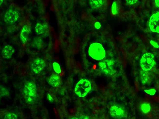
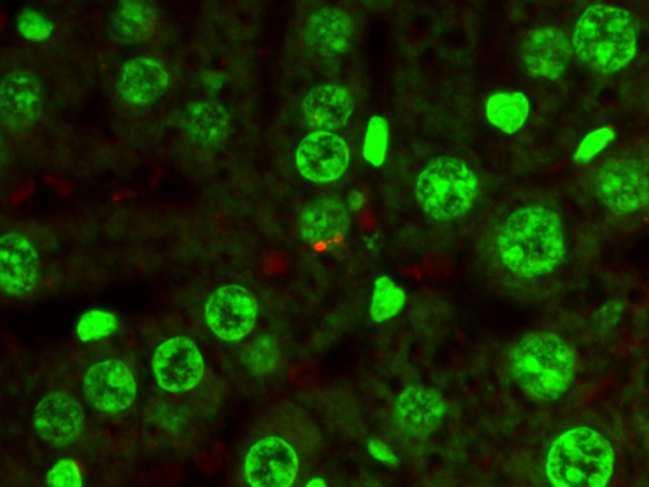
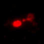
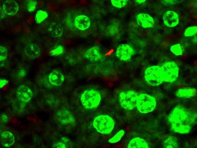
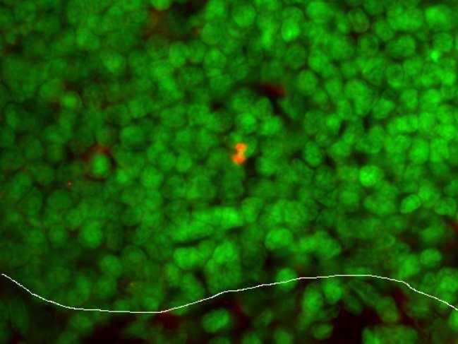
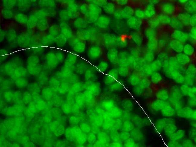
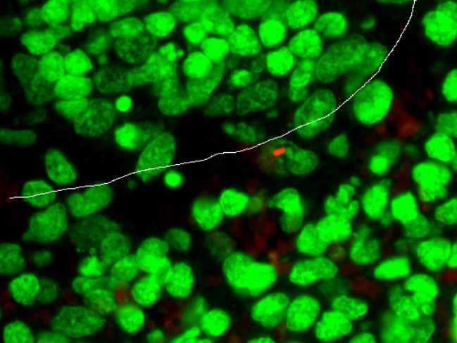
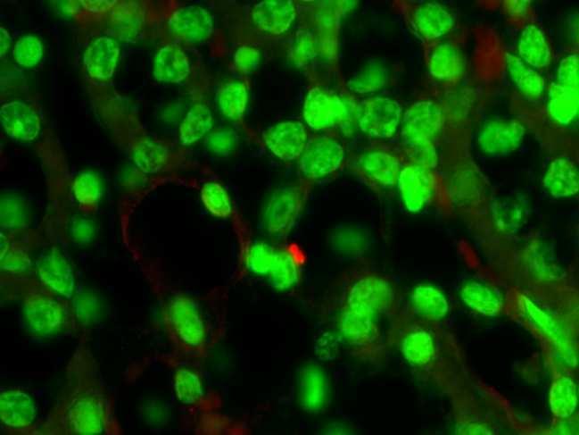
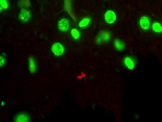
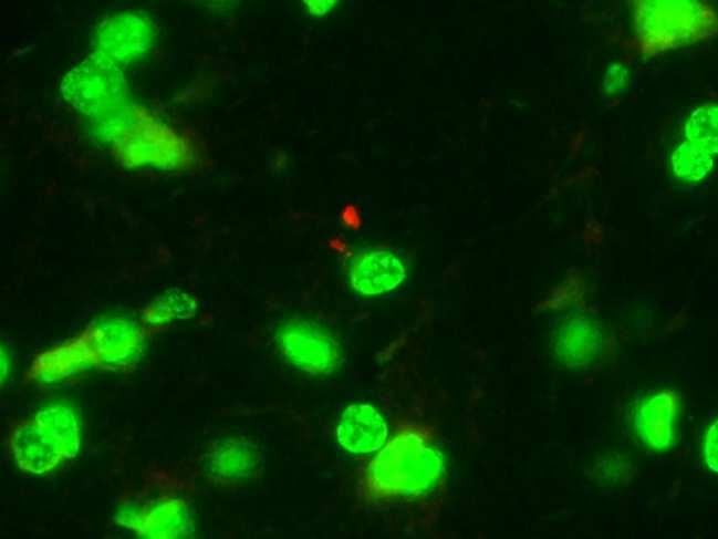
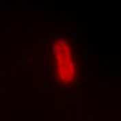
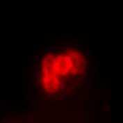
178 PAO1F CLJ1 +ExlA 20 µm wp wp mz wp mz mz Figure S3 Figure S3. Localisation of bacteria in infected organs Mice were subjected to lung infection with different bacterial strains ( ) as indicated. After 18 h.p.i., mice were euthanised and organs (indicated on the left) were paraffin-embedded. Sections were labelled with anti-lps antibody for bacteria staining (red) and with Yo-Pro (green) for nuclei straining. Bacteria (arrows) were detected in lung alveoli, hepatic lobules, white pulp (wp) and marginal zone (mz) of spleen, intertubular space of kidney and brain parenchyma.
 were incubated with WGA-alexa 647 (red) to label")

 indicates the cell shown in the next panels. Arrows show the edges of the ruptured plasma membrane. Figure S5 Figure S5.")

179 30 min 250 min 270 min 10 µm * Figure S4 Figure S4. Cell membrane rupture induced by CLJ1 Confluent A549-EGFP cells (green) were incubated with WGA-alexa 647 (red) to label the plasma membranes. Cells were infected with CLJ1 at MOI of 10 and were observed by confocal microscopy in real time. Images were acquired every 10 min. The figure shows representative images of one z-plane at indicated time points post-infection. (*) indicates the cell shown in the next panels. Arrows show the edges of the ruptured plasma membrane. Figure S5 Figure S5. ILβ expression and secretion in infected A549 cells A549 cells were either uninfected (NI) or incubated with PAO1F, CLJ1, +ExlA or -ExlA at MOI 10. Cell supernatants were harvested at 3 h.p.i. and cell lysates were prepared. All samples were analysed by Western blot to reveal IL1b and b-actin. Pro-ILβ/ β-actin signal ratios are shown below the lanes. Pro- IL1β signals were detected in the supernatants, indicating that some cells have lysed.
180
181 IX. ExlA induit le clivage des E- et VE-cadhérines des jonctions adhérentes
182
183 Résultats & Discussions Chapitre IX Résumé Dans le chapitre précédent, nous avons pu mettre en évidence l effet spectaculaire d ExlA sur les alvéoles pulmonaires, facilitant la dissémination bactérienne (Bouillot et al., 2017). A la suite de cette étude, nous avons cherché à comprendre, in vitro, les dommages causés par ExlA sur les cellules épithéliales et endothéliales. ExlA induit la mort cellulaire, mais pas seulement. Elsen et al. ont mis en évidence une dissociation des jonctions intercellulaires induite par CLJ1. Dans ce sens, nous nous sommes intéressés à comprendre plus en détails le rôle d ExlA sur les jonctions adhérentes des cellules épithéliales et endothéliales qui composent la barrière pulmonaire. Dans notre étude, nous avons pu observer, après infection, une disparition rapide des cadhérines dans ces deux types cellulaires, bien avant la rupture de la membrane plasmique. Nous avons mené notre étude en parallèle sur la toxine ShlA de Serratia marcescens, dont nous avons montré qu elle induisait également la dégradation des cadhérines. Comme ExlA et ShlA partagent 35% d identité, le but était de savoir s il existait un mécanisme commun dans l action de ces deux toxines sur les jonctions intercellulaires. Il était peu probable que ces toxines soient des protéases ; nous nous sommes donc orientés vers un mécanisme de subversion d une protéase eucaryote. Nous nous sommes intéressés à la protéase ADAM10. En effet, cette protéase a pour substrat les cadhérines et produit des fragments de clivage de même taille que ceux induits par ExlA. En outre, ADAM10 a déjà été impliquée dans le clivage des cadhérines induit par la PFT Hla de S. aureus. Hla est capable d interagir directement avec ADAM10, permettant à la fois l'insertion dans la membrane de la toxine et l'activation de la protéase (Wilke and Wardenburg, 2010). Par l utilisation d inhibiteurs pharmacologiques d ADAM10, de cellules épithéliales déficientes en ADAM10 et de cellules endothéliales dans lesquelles l expression d ADAM10 a été diminuée, nous avons pu démontrer le rôle d ADAM10 comme partenaire indirect du clivage des E- et VE-cadhérines dépendant d ExlA et ShlA. ADAM10 n est pas le récepteur protéique de ces deux toxines puisque son absence ne bloque pas la mort cellulaire induite par ExlA ou ShlA. Dans un second temps, nous avons cherché à comprendre le mécanisme d activation d ADAM10 par ExlA. ADAM10 est maintenue inactive par sa liaison avec la calmoduline, une
184 Résultats & Discussions Chapitre IX protéine ayant également une forte affinité pour le calcium. Lorsque le calcium intracellulaire est augmenté, la calmoduline se lie au calcium et se dissocie d ADAM10, qui peut donc être activée par la furine et exportée à la membrane où elle peut exercer son activité protéolytique. La plupart des pores formés par les PFTs provoquent un flux calcique entrant. Nous montrons que c est effectivement le cas pour ExlA et ShlA dans les cellules épithéliales et endothéliales. Les différentes expériences menées ont permis de montrer que les flux calciques provoqués par les pores formés par ExlA ou ShlA étaient à l origine de l activation d ADAM10 et par conséquent, provoquaient le clivage des cadhérines. Malgré de nombreux efforts de plusieurs membres de l équipe, ExlA et ShlA n ont pas pu être purifiées sous une forme active. Par conséquent, nous n avons pas pu faire de courbes doseréponse, ni affirmer que ces toxines n agissent pas conjointement à d autres facteurs bactériens. La toxicité sur les cadhérines de souches d E. coli, à priori dépourvues de facteurs de virulence, mais surexprimant ExlA, permet de conforter l hypothèse d une action seulement dépendante d ExlA. En conclusion, les souches bactériennes produisant ExlA ou ShlA détournent un mécanisme naturel de l'hôte pour induire la perte de l'intégrité tissulaire. La dissociation des jonctions et la rupture de la membrane plasmique induits par ExlA donnent une explication aux effets spectaculaires observés sur les alvéoles pulmonaires de souris lors de l infection : dégradation des cadhérines in vivo et lyse des pneumocytes et des cellules endothéliales (Bouillot et al., 2017) (Chapitre VIII). Cela explique la facilité des bactéries à transmigrer à travers le tissu pulmonaire pour coloniser d autres organes
185 Résultats & Discussions Chapitre IX Publication 1. Participation au projet : Dans cette étude, j ai produit l ensemble des résultats décrits, à l exception de la Figure 1E. Les expériences de flux calciques (Figures 4 & 6D, E, F, G) ont été réalisées avec l aide de Stéphanie Bouillot, technicienne au sein de l équipe. 2. Article
186 Résultats & Discussions Chapitre IX
187 RESEARCH ARTICLE Pseudomonas aeruginosa ExlA and Serratia marcescens ShlA trigger cadherin cleavage by promoting calcium influx and ADAM10 activation Emeline Reboud 1, Stéphanie Bouillot 1, Sabine Patot 2, Benoît Béganton 1, Ina Attrée 1, Philippe Huber 1 * a a a a a Université Grenoble Alpes, CNRS ERL5261, CEA BIG-BCI, INSERM UMR1036, Grenoble, France, 2 CIRI, Centre International de Recherche en Infectiologie, Inserm U1111, Université Lyon 1, Ecole Normale Supérieure de Lyon, CNRS UMR 5308, Lyon, France Current address: Institut de Recherche en Cancérologie de Montpellier, Montpellier, France * [email protected] Abstract OPEN ACCESS Citation: Reboud E, Bouillot S, Patot S, Béganton B, Attrée I, Huber P (2017) Pseudomonas aeruginosa ExlA and Serratia marcescens ShlA trigger cadherin cleavage by promoting calcium influx and ADAM10 activation. PLoS Pathog 13(8): e ppat Editor: Alice Prince, Columbia University, UNITED STATES Received: April 21, 2017 Accepted: August 12, 2017 Published: August 23, 2017 Copyright: 2017 Reboud et al. This is an open access article distributed under the terms of the Creative Commons Attribution License, which permits unrestricted use, distribution, and reproduction in any medium, provided the original author and source are credited. Data Availability Statement: All relevant data are within the paper and its Supporting Information files. Funding: This work was supported by institutional grants from the Commissariat à l Énergie Atomique ( the Institut National de la Santé Et de la Recherche Médicale ( inserm.fr/); the Université Grenoble-Alpes ( and the Centre National de la Recherche Scientifique ( Pore-forming toxins are potent virulence factors secreted by a large array of bacteria. Here, we deciphered the action of ExlA from Pseudomonas aeruginosa and ShlA from Serratia marcescens on host cell-cell junctions. ExlA and ShlA are two members of a unique family of pore-forming toxins secreted by a two-component secretion system. Bacteria secreting either toxin induced an ExlA- or ShlA-dependent rapid cleavage of E-cadherin and VE-cadherin in epithelial and endothelial cells, respectively. Cadherin proteolysis was executed by ADAM10, a host cell transmembrane metalloprotease. ADAM10 activation is controlled in the host cell by cytosolic Ca 2+ concentration. We show that Ca 2+ influx, induced by ExlA or ShlA pore formation in the plasma membrane, triggered ADAM10 activation, thereby leading to cadherin cleavage. Our data suggest that ADAM10 is not a cellular receptor for ExlA and ShlA, further confirming that ADAM10 activation occurred via Ca 2+ signalling. In conclusion, ExlA- and ShlA-secreting bacteria subvert a regulation mechanism of ADAM10 to activate cadherin shedding, inducing intercellular junction rupture, cell rounding and loss of tissue barrier integrity. Author summary Pore-forming toxins are the most widespread toxins delivered by pathogenic bacteria and are required for full virulence. Pore-forming toxins perforate membranes of host cells for intracellular delivery of bacterial factors, for bacterial escape from phagosomes or in order to kill cells. Loss of membrane integrity, especially the plasma membrane, has broad implications on cell and tissue physiology. Here, we show that two members of a unique family of pore-forming toxins, secreted by Pseudomonas aeruginosa and Serratia marcescens, have the capacity to disrupt cell-cell junctions of epithelial and endothelial cells, hence breaching two major tissue barriers. PLOS Pathogens August 23, / 20
188 ExlA and ShlA induce cadherin cleavage through ADAM10 activation cnrs.fr/), as well as the Laboratoire d Excellence GRAL [grant number ANR-10-LABX to ER] ( and Agence National de la Recherche [ANR-15-CE ] ( The funders had no role in study design, data collection and analysis, decision to publish, or preparation of the manuscript. Competing interests: The authors have declared that no competing interests exist. Introduction Multicellular organisms have developed barriers to protect their internal body from microbial invasion. Mucosae constituted of single-layered epithelial cells are the favoured tissue barriers that bacteria may cross. Pathogenic bacteria have engineered different weapons to traverse these borders, either going through the cells (transcellular route) or at cell-cell junctions (paracellular route). Opportunistic pathogens, like Pseudomonas aeruginosa or Serratia marcescens, can only cross epithelial barriers when tissues are damaged or proliferate after physical trauma or biological insults. Once across the epithelial layer, bacteria may cross the endothelium, translocate into the vascular system and disseminate into the body. Exolysin (ExlA hereafter) is a pore-forming toxin recently identified in a subset of strains from Pseudomonas aeruginosa species, generally called PA7-like strains, from the name of the first-identified strain of this category [1 11]. ExlA gene is located in the same locus as exlb, forming a Two-Partner Secretion System, in which ExlB is required for ExlA secretion in the extracellular milieu [12]. PA7-like strains have been isolated from patients with various infection types, such as acute or chronic pneumonia, urinary infections, burns, otitis, and recently from a patient suffering from hemorrhagic pneumonia. The PA7-like strains isolated so far do not possess a Type III secretion system (T3SS) and their virulence potential on cellular models is mainly correlated with the level of secreted ExlA [3, 13]. In the mouse lungs, ExlA-secreting strains induced major injuries of the alveolo-capillary barrier, leading to pulmonary hemorrhages and allowing bacterial dissemination in the body. Osmotic protection assays revealed that the inner diameter of the pore formed by ExlA in the host plasma membrane is approximately 1.6 nm, likely allowing the trafficking of small molecules[12]. Pore formation ultimately provokes cell death by plasma membrane rupture, as observed by microscopy, or monitored by lactate dehydrogenase (LDH) release and propidium iodide (PI) incorporation [3, 12]. Homologous ExlA proteins were identified in related Pseudomonas species, such as P. protegens, P. entomophila and P. putida [12]. Importantly, a 35% identity was found with ShlA pore-forming toxin from Serratia marcescens [3], which displays the same domain organization and a comparable size (172 kda for ExlA and 165 kda for ShlA), and shares a similar secretory pathway [14]. ShlA forms pores of 1 2 nm, is cytolytic for various cell types and induces hemorrhagic pneumonia in humans and in infected mouse lungs [15 17]. Hence, ShlA is related to ExlA in several structural and functional aspects. Other related toxins have been identified based on sequence homology and secretion pathway, including HpmA from Proteus mirabilis, HhdA from Haemophilus ducreyi, PhlA from Photorhabdus luminescens, EthA from Edwardsiella tarda and ChlA from Chromobacterium violaceum [14, 18 22], for which very little information is available. Altogether, these toxins constitute a unique family of pore-forming toxins, for which neither the 3-dimensional structure, nor the potential oligomerization and mechanisms of pore formation are known, and importantly, for which the mechanism of toxicity remains elusive. Here, we show that ExlA and ShlA have the capacity to disrupt the cell-cell junctions of epithelial and endothelial cells, using an indirect mechanism. The pore formed by either toxin in the host cell triggers a Ca 2+ influx, which activates A Disintegrin And Metalloproteinase domain-containing protein 10 (ADAM10), a transmembrane metalloprotease, whose natural substrates are transmembrane proteins, including some cadherins. ExlA- and ShlA-dependent ADAM10 activation rapidly leads to E- or VE-cadherin cleavage in epithelial or endothelial cells, respectively. As cadherins are major determinants of intercellular adhesion, cadherin cleavage induces cell-cell junction breakdown and loss of tissue integrity. PLOS Pathogens August 23, / 20
189 ExlA and ShlA induce cadherin cleavage through ADAM10 activation Results ExlA-secreting strains induce E- and VE-cadherin cleavage We previously reported that T3SS-positive P. aeruginosa strains incubated with endothelial cells induce VE-cadherin cleavage, which is mediated by a protease (LasB) released by the T2SS [23]. LasB cleaves VE-cadherin in the middle of its extracellular domain, preventing its adhesive activity. We further showed that E-cadherin, located at epithelial cell-cell junctions, was resistant to LasB proteolytic activity [23]. As cadherins are required for tissue integrity, we tested the ExlA-secreting isolate CLJ1 for its capacity to cleave E-cadherin by incubation with the human alveolar cell line A549 (Fig 1A, left). CLJ1 induced a rapid and dramatic decrease in full-length E-cadherin levels, which paralleled the onset of a C-terminal E-cadherin fragment of 30 kda. As previously reported [23], two ExlA-negative P. aeruginosa strains injecting ExoS, T and Y toxins through their T3SS, did not degrade E-cadherin, even at longer time points (Fig 1A, left). We also tested an ExlA-negative T3SS-positive strain injecting ExoU, a toxin endowed with phospholipase activity, known to induce membrane permeabilisation to nuclei dyes [24]. A549 cell incubation with the ExoU-positive strain did not induce E-cadherin cleavage (S1 Fig). Thus, membrane permeabilisation by a phospholipase is not sufficient to promote cadherin cleavage. In similar experiments, VE-cadherin was rapidly degraded after incubation of primary human endothelial cells (HUVECs) with CLJ1 (Fig 1A, right). The onset of a C-terminal VEcadherin fragment of 30 kda was also observed concomitantly to the decrease of full-length VE-cadherin. As previously noticed, VE-cadherin was partially degraded due to LasB activity [23], however at much longer time points (Fig 1A, right). To examine whether the rapid E- and VE-cadherin cleavage activity was dependent upon ExlA, we used the ExlA-secreting IHMA87 strain [12, 13] that can be manipulated genetically, as opposed to CLJ1. Hence, we tested the isogenic mutant IHMA87ΔexlA and its complemented counterpart IHMA87ΔexlA/exlA for their ability to degrade the cadherins (Fig 1B). IHMA87 similarly induced E-cadherin and VE-cadherin cleavages, albeit more slowly than CLJ1, while IHMA87ΔexlA did not, even at longer time points. The complemented strain recovered this ability. Thus, the cleavage of both types of cadherin is ExlAdependent. We previously reported that Type IV pili ( pili hereafter) facilitated the ExlA-dependent toxicity of IHMA87 towards A549 cells, probably by enhancing bacterial adhesion [12]. We thus tested the effect of a mutant devoid of pili (IHMA87ΔpilA) on cadherin cleavage (S2 Fig). The lack of pili had no effect on E-cadherin cleavage and a very partial effect on VE-cadherin. Next, we investigated whether other P. aeruginosa factors were needed for ExlA action on cadherins. As our attempts to purify a functional ExlA protein were unsuccessful, we used an Escherichia coli strain ectopically expressing ExlB-ExlA, and devoid of other P. aeruginosa factors. The E. coli-exlba strain induced the cleavage of both E- and VE-cadherins, suggesting that ExlA alone can induce cadherin proteolysis (Fig 1C). We then examined the fate of E-cadherin at cell-cell junctions by confocal videomicroscopy during infection using A549 expressing E-cadherin fused to GFP (Fig 1D). We observed a loss of E-cadherin-GFP labelling at cell-cell junctions when cells were incubated with CLJ1 or IHMA87, but not when incubated with IHMA87ΔexlA or PAO1F. This feature is in agreement with the above results by Western blot, except that the kinetics differ due to different experimental conditions. Taken together, the results show that ExlA-dependent E-cadherin cleavage promotes E-cadherin loss from cell-cell junctions. To assess cell membrane permeability, the non-permeant dye propidium iodide was added to the cell medium. Membrane disruption, as monitored by propidium iodide incorporation into cell nuclei, occurred much later than E- PLOS Pathogens August 23, / 20
.")
190 ExlA and ShlA induce cadherin cleavage through ADAM10 activation Fig 1. ExlA-dependent cleavage of E- and VE-cadherins. A. A549 cells (left) or HUVECs (right) were incubated with various P. aeruginosa strains: CHA, PAO1F or CLJ1, or were mock-infected with LB (NI). Cell extracts were prepared at different times postinfection, as indicated and analysed by Western blot using E-cadherin (left) or VE-cadherin (right) antibodies. In both cases, the fulllength (FL) and post-cleavage C-terminal (CTF) fragments are shown. β-actin was used as loading control. The experiment was performed 3 times for A549 and twice for HUVECs with similar results. B. A549 cells (left) and HUVECs (right) were incubated with PLOS Pathogens August 23, / 20
191 ExlA and ShlA induce cadherin cleavage through ADAM10 activation IHMA87, IHMA87ΔexlA or IHMA87ΔexlA/exlA strains, and cellular extracts were analysed as above. The experiment was performed 3 times for A549 and once for HUVECs. C. Similar experiments with A549 cells (left) or HUVECs (right) incubated with E. coli containing the empty vector (exlba -) or E-coli expressing ExlB-ExlA (exlba +). D. A549-E-cadherin-GFP cells were incubated with CLJ1, IHMA87, IHMA87ΔexlA or PAO1F bacteria. E-cadherin-GFP (green) as well as nuclei labelling by propidium iodide (red) were followed by confocal videomicroscopy. Times post-infection are shown as h:min. One z-position is represented. The experiment was performed twice, with 4 5 positions recorded each time. All films showed similar results. E. Mice (5 per condition) were infected by bacteria inhalation (2.5x10 6 ), using CLJ1 strain, or were mock-infected with PBS (NI). Mice were euthanized at 18 h.p.i.; protein extracts were prepared from lungs and were analysed by Western blot using E- and VE-cadherin antibodies (left). Histograms (right) show the E-cadherin/ β-actin and VE-cadherin/ β-actin ratios of band intensities, represented as means + s.d. Significance was calculated using Mann-Whitney s test, as variances were not equal. The experiment was repeated once, with similar results. cadherin loss (Fig 1D). Thus, ExlA effects on E-cadherin could be observed earlier than those leading to cell death. To definitely prove that ExlA induces barrier disruption, a bacterial transmigration assay was performed using Transwell filters. The bacterial transmigration across A549 monolayers were significantly decreased when IHMA87ΔexlA was used, compared to IHMA87 (S3 Fig). To investigate the action of ExlA-secreting bacteria on cadherin cleavage in vivo, we infected mice by inhalation of a suspension of CLJ1 bacteria. IHMA87 was not used in this assay, as it is not a potent infective agent in this model [13]. Mice were euthanized at 18 h.p.i. and lung extracts were analysed by Western blot (Fig 1E). The data revealed that CLJ1-infected lungs contained significantly lower amounts of E- and VE-cadherins than the mock-infected lungs. Likewise, CLJ1 induces the cleavage of both cadherins in vivo. These results also show that ExlA can promote cadherin cleavage from a different species. ADAM10 is the executer of ExlA-dependent cadherin cleavage ADAMs are transmembrane metalloproteases, modulating cell-cell and cell-matrix interactions. They are major ectodomain sheddases, releasing a variety of cell-surface proteins. ADAM10 is an ubiquitous member of this family, for which several protein substrates have been identified, including E- and VE-cadherins [25 28]. ADAM10 cleaves both cadherins in their extracellular membrane-proximal domains, releasing the extracellular domain in the supernatant. The transmembrane domain is then rapidly cleaved by the γ-secretase and the cytoplasmic domain is targeted to the proteasome for degradation [26, 28]. In our experiments, the transient C-terminal cleavage fragment (30 kda) induced by CLJ1 co-migrated with that produced by cell incubation with ionomycin, a potent inducer of ADAM10 activity [25, 29] (Fig 2A). ADAM17 activation by PMA did not produce a similar cleavage. Thus, these preliminary results suggested that ExlA could activate ADAM10, as has been reported for Staphylococcus aureus pore-forming toxin Hla [30, 31]. To further evaluate the role of ADAM10 in ExlA-dependent cadherin cleavage, we first used protease inhibitors: GM6001, a wide-spectrum Zn-metalloprotease inhibitor and GI254023X, a highly specific ADAM10 inhibitor [32]. Both inhibitors prevented E- and VEcadherin cleavages after infection with CLJ1 or IHMA87 (Fig 2B). To definitely prove the role of ADAM10 in this mechanism, we generated ADAM10-deficient A549 cells, using CRISPR/Cas9 technology. The absence of ADAM10 was verified by FACS analysis (Fig 2C, right). ADAM10-deficient A549 cells were resistant to E-cadherin cleavage after incubation with CLJ1 or IHMA87 (Fig 2C, left). To evaluate the VE-cadherin cleavage in primary cells, we knocked down ADAM10 in HUVECs using sirna, as it is not possible to generate gene deficiency in primary cells using the CRISPR/Cas9 system. ADAM10-surface expression was dramatically decreased in ADAM10 sirna-treated HUVECs (Fig 2D, right). VE- PLOS Pathogens August 23, / 20
192 ExlA and ShlA induce cadherin cleavage through ADAM10 activation Fig 2. ADAM10 requirement for ExlA-dependent cadherin cleavage. A. A549 cells were either treated with 0.1 μg/ml PMA or 5 μm ionomycin (Iono) for 30 min, or infected with CLJ1 (90 min), or left untreated/uninfected (NI). Cellular extracts were analysed by Western blot using E-cadherin and β-actin antibodies. FL, full-length; CTF, C-terminal fragment. The experiment was performed twice. B. A549 cells (left) or HUVECs (right) were pre-treated with DMSO, the general metalloprotease inhibitor GM6001 (10 μg/ml) or the specific ADAM10 inhibitor GI254023X (5 μm) and then incubated with CLJ1 or IHMA87 (90 min), or PLOS Pathogens August 23, / 20
.")
193 ExlA and ShlA induce cadherin cleavage through ADAM10 activation uninfected (NI). Cellular extracts were analysed as above. The experiment was performed twice for A549 and 3 times for HUVECs. C. A549 or ADAM10-deficient A549 (A549 ADAM10 -/- ) cells were incubated with either CLJ1 or IHMA87. Cellular extracts were prepared at different time points post-infection as indicated and analysed by Western blot (left). The right panel shows the FACS analysis of ADAM10 surface expression of both cell lines, as well as the negative control. The experiment was performed 3 times. D. Similar experiment with HUVECs, either transfected with ADAM10 sirna or untreated. The experiment was performed twice. cadherin cleavage was prevented in ADAM10 sirna-treated HUVECs infected with CLJ1 and IHMA87 (Fig 2D, left). Altogether, these findings demonstrate that ADAM10 is the executer of ExlA for cadherin cleavage and is absolutely required for this activity. ADAM10 is dispensable for the necrotic activity of ExlA To examine whether ADAM10 was required for ExlA-dependent plasma membrane rupture, we tested the necrotic activity of ExlA in ADAM10-deficient A549 cells (Fig 3). LDH release was independent of the presence of ADAM10, showing that ADAM10 is not involved in cell death and that ADAM10 is not the membrane receptor of ExlA. ExlA triggers intracellular calcium elevation As previously described, high cytosolic Ca 2+ concentrations induce cadherin cleavage through activation of ADAM10 [26, 33], which was further confirmed here using the calcium ionophore ionomycin (Fig 2A). Therefore, we tested the capacity of ExlA-secreting strains to induce intracellular Ca 2+ elevation in A549 cells and HUVECs. We used Fluo3-AM, a cell-permeant fluorescent probe, to monitor intracellular Ca 2+ together with Draq7, a non-permeant fluorescent probe, to visualize necrotic cells after plasma membrane rupture. Cell fluorescence were recorded on both channels by videomicroscopy and their intensities measured on Fig 3. ExlA necrotizing activity is preserved in ADAM10-deficient cells. Plasma membrane rupture was monitored by LDH release in the supernatant. A549 or A549ADAM10 -/- cells were incubated for 5 hours with IHMA87, IHMA87ΔexlA or IHMA87ΔexlA/exlA strains. The supernatants were the tested for LDH activity. The histograms show the mean ± s.d. of triplicates. The data are representative of 3 experiments. PLOS Pathogens August 23, / 20
194 ExlA and ShlA induce cadherin cleavage through ADAM10 activation individual cells. No Fluo3 signal was obtained in uninfected conditions and inconsistent signals were detected when cells were infected with PAO1F (Fig 4A 4D), as previously reported [34]. In contrast, sharp increases of Fluo3 intensities were observed when either cell types were infected with CLJ1 (Fig 4E and 4F); this increase takes place earlier in HUVECs. Similar observations were made with IHMA87 infection (Fig 4G and 4H), except that Ca 2+ rise was not consistent for each A549 cell and less marked in general. When IHMA87ΔexlA was used, no Ca 2+ elevation was noted (Fig 4I and 4J), while Fluo3 signal increases were observable within IHMA87ΔexlA::exlBA-infected cells (Fig 4K and 4L). Hence, ExlA triggers a massive Ca 2+ elevation in host cells, because of Ca 2+ entry through the pore formed in the plasma membrane. ADAM10 may thus be activated by this process. The increase of Fluo3 intensity was consistently followed by a sharp drop of fluorescence that was concomitant to Draq7 incorporation into the cells (Fig 4E 4H, 4K and 4L), suggesting that the Fluo3 diffused in the extracellular milieu when the plasma membrane was ruptured. ExlA-dependent calcium influx drives both ADAM10 activation and necrosis The inactive form of ADAM10 (pro-adam10), linked to its pro-domain, has been shown to interact with calmodulin, a cytosolic protein displaying high affinity for Ca 2+ [29, 35]. It was hypothesized that this interaction prevents ADAM10 pro-domain cleavage, and subsequent ADAM10 activation and export to the plasma membrane [35, 36]. The currently accepted model proposes that when cytosolic Ca 2+ concentration is increased, calmodulin and Ca 2+ interact, which dissociates calmodulin from pro-adam10, as its affinity is lower for the protease. ADAM10 hence becomes available for activation by furin and is eventually ready to process its own substrates [37, 38]. To test this hypothesis in our system, we first depleted Ca 2+ in the extracellular medium, but the absence of Ca 2+ triggered a striking over-secretion of ExlA by the bacteria (S4 Fig) that masked the results on ADAM10 activation. Therefore, we employed a chemical compound, trifluoperazine (TFP), that exhibits high affinity for calmodulin and dissociates calmodulin from its other interactants [29, 35]. TFP treatment of A549 resulted in a dose-dependent induction of E-cadherin cleavage, confirming that calmodulin prevents ADAM10 activation in uninfected A549 cells (Fig 5A). To further demonstrate the role of intracellular Ca 2+ in ADAM10 activation, we pre-incubated A549 cells with BAPTA-AM, a cell permeant Ca 2+ chelator. The chelating action of BAPTA-AM in these cells was tested by Ca 2+ imaging using Fluo3-AM probe (S5 Fig). Fluo-3 signal inhibition by BAPTA-AM was total in CLJ1-infected cells and almost complete when cells were infected with IHMA87. BAPTA-AM strongly decreased CLJ1-induced E-cadherin cleavage and its action was partial in IHMA87-infected (Fig 5B), consistent with the differential ability of BAPTA-AM to sequester intracellular Ca 2+ in the two conditions. In conclusion, cytosolic Ca 2+ is the messenger allowing ADAM10 activation in infected cells. Interestingly, BAPTA-AM also significantly diminished LDH release induced by CLJ1 or IHMA87 (Fig 5C), suggesting that Ca 2+ is also involved in ExlA-induced membrane permeability, independently of cadherin cleavage. Serratia marcescens ShlA toxin induces calcium influx, ADAM10 activation and cell death As mentioned above, the pore-forming toxin ShlA is homologous to ExlA in terms of sequence identity (35%), secretion pathway and domain organization [12]. We thus examined whether ShlA had the same capacity to promote cadherin cleavage through Ca 2+ influx and ADAM10 PLOS Pathogens August 23, / 20
195 ExlA and ShlA induce cadherin cleavage through ADAM10 activation Fig 4. Intracellular calcium elevation generated by ExlA-secreting bacteria. Intracellular Ca2+ content was analysed by videomicroscopy using the permeant Fluo3-AM fluorescent probe. Plasma membrane rupture was evaluated using the non-permeant Draq7 fluorescent probe. A549 cells (A,C,E,G,I,K) or HUVECs (B,D,F,H,J,L) were either non-infected (A,B) or infected with PAO1F (C,D), CLJ1 (E,F), IHMA87 (G, H), IHMA87ΔexlA (I,J) or IHMA87ΔexlA::exlBA (K,L). The fluorescence intensities of five cells were analysed PLOS Pathogens August 23, / 20
196 ExlA and ShlA induce cadherin cleavage through ADAM10 activation in each case; the Fluo3 intensities are represented by solid lines and the Draq7 intensities by dashed lines, using the same colour code for one cell. The data are representative of 4 7 experiments. Uninfected conditions were performed in each experiment. activation. To address this question, we used a ShlA-secreting strain of S. marcescens, Db11, and its isogenic shlb mutant, 21C4, which is impaired in ShlA secretion [39, 40]. Db11 rapidly cleaved E-cadherin with C-terminal cleavage fragment of similar size as ExlA, whereas 21C4 did not induce E-cadherin degradation (Fig 6A left). Similar data were obtained for VE-cadherin (Fig 6A right). Incubation of A549 ADAM10 -/- cells with Db11 did not induce E-cadherin cleavage (Fig 6B). E-cadherin degradation was only partially prevented in Db11-infected A549 cells by cell pre-incubation with BAPTA-AM (Fig 6C). However, BAPTA-AM incompletely chelated intracellular Ca 2+ (S5 Fig). Db11 increased intracellular Ca 2+ in A549 cells and Fig 5. Role of calcium influx in cadherin cleavage and necrosis. A. A549 cells were incubated with various concentrations of TFP, as indicated, to impede calmodulin interaction with ADAM10. Ionomycin was used as positive controls. E-cadherin cleavage was assessed by Western blot. The experiment was performed twice. B. Western blot analysis of A549 E-cadherin contents after infection with CLJ1 or IHMA87, in presence or absence of BAPTA-AM. Both experiments were performed 3 times. C. LDH release of A549 cells infected with either CLJ1 or IHMA87, in presence/ absence of BAPTA-AM. Student s t-test showed significance between the two treatments for both CLJ1 and IHMA87 data (p-values indicated above the bars). The experiment was performed 3 times. PLOS Pathogens August 23, / 20
197 ExlA and ShlA induce cadherin cleavage through ADAM10 activation Fig 6. Effects of S. marcescens ShlA on cadherin cleavage and calcium influx. A. A549 cells (left) or HUVECs (right) were incubated with the S. marcescens ShlA-secreting strain Db11, or with the non-shla-secreting mutant 21C4. Cellular extracts were analysed for their E- or VE-cadherin contents. The experiment was performed twice for the left panel and once for the right panel. B. Similar analysis using A549 ADAM10-/-. The experiment was performed once. C. Similar analysis using A549 cells, in presence/ absence of BAPTA-AM. D-G. Intracellular Ca2+ contents and plasma membrane permeability were measured using Fluo3-AM and PLOS Pathogens August 23, / 20
 and HUVECs (E,G) were infected with Db11 (D,E) or 21C4 (F,G) and fluorescence was recorded on both channels by videomicroscopy.")
198 ExlA and ShlA induce cadherin cleavage through ADAM10 activation Draq7 fluorescent probes, respectively. A549 cells (D,F) and HUVECs (E,G) were infected with Db11 (D,E) or 21C4 (F,G) and fluorescence was recorded on both channels by videomicroscopy. Five cells were analysed in each case; the Fluo3 intensities are represented by straight lines and the Draq7 intensities by dashed lines, using the same colour code for one cell. Data are representative of 8 and 5 independent experiments for A549 and HUVECs, respectively. HUVECs, followed by Draq7 incorporation (Fig 6D and 6E), while no Fluo3 or Draq7 signals were detected with 21C4 (Fig 6F and 6G). Therefore, we conclude that ShlA has the same capacity as ExlA to induce E-cadherin cleavage through Ca 2+ influx and ADAM10 activation. Discussion Cell-cell junctions of mucosal and vascular barriers play an essential role in impeding the spread of bacterial pathogens in the body (for a review on pore-forming toxin s action on tissue barriers, see [41]). Here, we employed ExlA- and ShlA-secreting bacteria, and their respective non-secreting mutants, to decipher how the two toxins disrupt adherens junctions. Our results demonstrate that one of the primary effects of the cytotoxic action of ExlA and ShlA is that they induce cleavage of E- and VE-cadherins, which disrupts epithelium and endothelium integrity. The massive Ca 2+ influx triggered by pore formation initiates a cascade of events leading to junction disruption. The model proposed in Fig 7 includes the following steps: in uninfected conditions, calmodulin is associated with pro-adam10, preventing its Fig 7. Mechanisms of ExlA/ShlA-induced cadherin cleavage. In uninfected cells, pro-adam10 is associated with calmodulin, preventing its maturation and export to the plasma membrane. Pore formation by ExlA or ShlA induces a massive Ca 2+ influx in host cells. Intracellular Ca 2+ interacts with the Ca 2+ -binding protein calmodulin, which detaches from pro-adam10, allowing its maturation to m-adam10. m-adam10 cleaves E- and VE-cadherin in epithelial and endothelial cells, respectively, provoking intercellular junction rupture. PLOS Pathogens August 23, / 20
199 ExlA and ShlA induce cadherin cleavage through ADAM10 activation maturation. Calmodulin has a high affinity for Ca 2+. When Ca 2+ enters the cell through the ExlA or ShlA pore, it interacts with calmodulin, which presumably alters the conformation of calmodulin and causes its dissociation from ADAM10. Then, the free pro-adam10 protein is activated by furin and translocates to the plasma membrane where it cleaves the cadherins. As cadherins are required for cell-cell adhesion, their cleavage is closely followed by cell retraction, owing to actin-cytoskeleton centripetal forces. Similar data were obtained with CLJ1 and IHMA87, except that the latter was less effective in most assays. The ExlA proteins in these two strains share 99.6% identity, suggesting that the milder effect of IHMA87 may be related to a reduced capacity to secrete ExlA [13]. Ca 2+ influx started earlier in HUVECs than in A549 cells, suggesting either that the pores are formed more rapidly or that a larger number of pores are formed in HUVECs. This effect could be explained by a higher expression of receptors for ExlA and ShlA at the surface of endothelial cells compared to epithelial cells. However, as no receptor has yet been identified for these two toxins, we cannot currently confirm this hypothesis. For ExlA, the receptor appears to be relatively ubiquitous as ExlA-secreting bacteria are toxic for most tested cell types except erythrocytes [13]. In contrast, ShlA targets erythrocytes, epithelial cells, fibroblasts [17] and endothelial cells (this study), but not myeloid cells. The somewhat different target host cell suggests that the cellular receptors are not identical for the two toxins. Interestingly, the LDH response was unmodified in the absence of ADAM10, when cells were exposed to ExlA- or ShlA-secreting bacteria. Thus, unlike for Hla [31], ADAM10 is not the receptor of either ExlA or ShlA, nor it is involved in ExlA-induced cell death. Unlike most bacterial pore-forming toxins, ExlA and ShlA are highly unstable in aqueous solution and are biologically inactive when purified. The instability of ExlA and ShlA and the fact that Type IV pili are required for ExlA-related toxicity of the IHMA87 strain [12] suggest that these toxins must be delivered close to host cells where they can rapidly insert into the plasma membrane. The mechanism of insertion into the membrane and the toxin s oligomerisation capacity remain to be determined. Because of their role in maintaining tissue barriers, cadherins are frequent targets for bacterial pathogens. Cadherins can be cleaved by ADAM10, which is activated by the calcium imbalance caused by the pore-forming action of the toxins, as previously demonstrated for Hla from S. aureus and pneumolysin from Streptococcus pneumoniae [30, 31, 42] and here for ExlA and ShlA. This mechanism of toxicity might also be used by other pore-forming toxins that are known to promote Ca 2+ influx, such as streptolysin O (SLO) fromstreptococcuspyogenes, α- toxin from Clostridium septicum, aerolysin from Aeromonas hydrophila, and listeriolysin O from Listeria monocytogenes [43 46]. Alternatively, E-cadherin can also be cleaved by HtrA bacterial proteases, released by Helicobacter pylori, enteropathogenic E. coli, Shigella flexneri and Campylobacter jejuni [47, 48]. As mentioned above, VE-cadherin is cleaved by P. aeruginosa LasB protease [23]. E-cadherin is also the target of Clostridium botulinum neurotoxin-a, preventing cadherin homotypic adhesion [49]. Finally, an intermediate mechanism was found for the metalloprotease BFT from Bacteroides fragilis, which activates the γ-secretase, promoting E-cadherin cleavage [50]. Thus, bacteria employ several independent but related pathways to target the junctions. Along the intercellular space, the cluster of E-cadherin molecules forming the adherens junctions, is located closer to the basal side of the cell layer than the tight junctions. The tight junctions seal intercellular gaps on the apical side to restrict the diffusion of molecules (e.g. proteases) present on the apical side of the cell layer. This restriction is less effective in endothelia, where the tight junctions tend to be less developed and where they are mixed with adherens junctions [51]. Bacterial proteases therefore have limited access to E-cadherin in most infection conditions, and the indirect action of pore-forming toxins on E-cadherin PLOS Pathogens August 23, / 20
200 ExlA and ShlA induce cadherin cleavage through ADAM10 activation appears to be an efficient alternative mechanism to the secretion of proteases directly targeting the junctions. Because ADAM10 can efficiently cleave adhesive and signalling receptors, its activity is tightly regulated in eukaryotic cells. The subversion mechanism of ADAM10 activation by ExlA and ShlA is probably shared by several pore-forming toxins. This may have functional consequences beyond cellular adhesion because of the numerous substrates of ADAM10, even when cell death is prevented by induction of the membrane repair machinery. Methods Ethical statement All protocols in this study were conducted in strict accordance with the French guidelines for the care and use of laboratory animals [52]. The protocols for mouse infection were approved by the animal research committee of the institute (CETEA, project number ) and the French Ministry of Research. Anesthesia was performed using a mixture of xylazine/ketamine and euthanasia by CO2 inhalation. P. aeruginosa, S. marcescens and E. coli strains and culture The strains and the plasmids used in this study are described in S1 File. Bacteria were grown in liquid LB medium at 37 C with agitation until the cultures reached an optical density value of 1.0 to be in the exponential growth phase. All conditions of infection were performed at MOI of 10, unless indicated in the legend. Cell culture HUVECs were prepared from anonymized human umbilical cords (coming from the Groupe Hospitalier Mutualiste de Grenoble) as previously described [53] and grown in supplemented EBM2 medium (Lonza). A549 (ATCC), A549-E-cadherin GFP and A549ADAM10 -/- cells (both generated in this study) were grown in DMEM medium, supplemented with 10% foetal calf serum (all from Life Technologies). Cells were tested for the presence of mycoplasma before freezing vials. Each experiment started from a mycoplasma-free frozen vial. A549 cells were tested for the presence of E-cadherin junctional labelling. A549 E-cadherin GFP cells were obtained after transfection with pcdna3.1-e-cadherin- GFP (Addgene) using Lipofectamine 2000 (Life Technologies). Positive cells were selected by antibiotic treatment and fluorescent cells were sorted by MoFlo flow cytometer (Beckman Coulter). When indicated, cells were pretreated 30 min before infection with TFP at indicated concentrations, 0.1 μg/ml phorbol 12-myristate 13-acetate (PMA), 5 μm ionomycin, 10 μg/ ml GM6001, 5μM GI254023X or 25μM BAPTA-AM (all from Sigma). BAPTA-AM was washed out before the infection step. For videomicroscopy experiments, cells were seeded at 150,000 cells per well on Labtek II 8-chambered (Thermo Fisher Scientific) coverslips and used 48-h later to obtain highly confluent monolayers. Medium was replaced with non-supplemented EBM-2 medium one hour before infection. Cells were infected and immediately observed by videomicroscopy. For sirna experiments, HUVECs were seeded at cells/well in P12 plate. The next day, cells were transfected with anti-sense ADAM10 sirna (UAACAUGACUGGAUAUCU GGG) using Lipofectamine RNAiMAX (Life Technologies). Three days after transfection, cells were analysed by FACScalibur flow cytometer (Becton Dickinson) after staining with ADAM10 antibody (R&D Systems) and anti-mouse-alexa 488 antibody (Molecular Probes). PLOS Pathogens August 23, / 20
201 ExlA and ShlA induce cadherin cleavage through ADAM10 activation CRISPR/Cas9-based ADAM10 gene knockout ADAM10 gene knockout was generated by using guide RNA oligos specifying the humanadam10 gene (forward: 5 -CACCGGATACCTCTCATATTTACAC; reverse: 5 - AAACGTGTAAATATGAGAGGTATCC). These oligos were designed by using the tool available at Oligos were obtained from Eurofins Genomics and then cloned into the pspcas9(bb)-2a-gfp vector (#48138, Addgene). The constructs or the empty vector were transfected into the A549 cells using the Neon Transfection System (Life Technologies). After 24 h, transfected cells were selected by flow cytometry using the GFP signal and were distributed directly in 96-well plates using a BD FACSAria flow cytometer. After 10 days of culture, cells were stained with ADAM10-phycoerythrin antibody (R&D Systems) for 30 min at room temperature. The samples were analysed using a BD Accuri C6 flow cytometer. ADAM10-negative clones were selected and theadam10 gene was sequenced. To further determine if the selected clones had mutations in both alleles, Guide-it sgrna in vitro Transcription coupled to Guide-it Genotype Confirmation Kits (Clontech Laboratories) were used following the manufacturer recommendations. One clone with both allelic mutations (A549ADAM10 -/- ) was used in this study and confirmed using BD FACScalibur flow cytometer after staining with ADAM10 antibody (R&D systems) and anti-mouse-alexa488 antibody (Molecular Probes) for 1 h at 4 C for each antibody. Mouse pulmonary infection Pathogen-free BALB/c female mice (8 10 weeks) were obtained from Harlan Laboratories and housed in the institute animal care facility. Bacteria from exponential growth (OD = 1.0) were centrifuged and resuspended in sterile PBS at 0.85x10 8 per ml. Mice were anesthetized by intraperitoneal administration of a mixture of xylazine (10 mg.kg -1 ) and ketamine (50 mg. Kg -1 ). Then, 30μL of bacterial suspension (2.5x10 6 ) were deposited into the mouse nostrils. Mice were euthanized by CO 2 inhalation at 18 h.p.i. and lungs were withdrawn and homogenized in 2 ml of PBS using a Polytron. The number of animals required for the study was deduced from previous work [23]. Randomization was performed by Harlan Laboratories. The experiment did not required blinding. Western blotting Cells and tissues were lysed in Triton X-100 and protein concentration of the lysates was determined with a Micro-BCA kit (Pierce) using BSA as standard. Protein extracts were then separated by SDS-PAGE, transferred onto Hybond ECL membrane (Amersham Biosciences) and revealed with E-cadherin-Cter (BD transduction laboratories), VE-cadherin (Santa Cruz) or β- actin (Sigma) antibodies. Luminescent signals were revealed using a ChemiDoc (BioRad). Only unsaturated signal intensities are presented. To examine ExlA secretion, the cell supernatants were collected at 2 h.p.i. and concentrated by trichloroacetic acid (TCA) precipitation. Briefly, 100 μl of 2% Na deoxycholate were added to 10 ml of cellular supernatant and incubated 30 min at 4 C. Then, 1 ml of TCA were added and the mixture was incubated overnight at 4 C. After centrifugation for 15 min, at 15,000 g and 4 C, the pellet was resuspended in 100 μl of electrophoresis loading buffer. The ExlA antibody [3] was used to reveal the western blots. PLOS Pathogens August 23, / 20
202 ExlA and ShlA induce cadherin cleavage through ADAM10 activation Lactate dehydrogenase release LDH release in the supernatant was measured using the Cytotoxicity Detection Kit from Roche Applied Science, following the recommended protocol. Briefly, cells were seeded at in 96-well plates two days before, and infected in non-supplemented EBM2. At different post-infection times, 30μL of supernatant were mixed with 100μL of reaction mix and OD was read at 492 nm. OD values were subtracted with that of uninfected cells and Triton-solubilized cells were used to determine the total LDH present in the cell culture. Confocal videomicroscopy For fluorescence time-lapse microscopy, cells were imaged using a confocal spinning-disk inverted microscope (Nikon TI-E Eclipse) equipped with an Evolve EMCCD camera. The optical sectioning was performed by a Yokogawa motorized confocal head CSUX1-A1. Images were acquired using an illumination system from Roper Scientific (ilaspulsed) with a CFI Plan Fluor oil immersion objective (40X, N.A. 1.3). Z-series were generated every 10 min using a motorized Z-piezo stage (ASI) by acquiring 25 z-plane images with a step size of 1μm. Microscope was controlled with MetaMorph software (Molecular Devices). Temperature, CO 2, and humidity control was performed using a chamlide TC system (TC-A, Quorum technologies). Solid-state 491 and 561 nm lasers (ilas, Roper Scientific) and ET 525/50M (Chroma) and FF01-605/54 (Semrock) emission filters were used for excitation and emission of EGFP and propidium iodide fluorescence, respectively. Calcium imaging by wide-field videomicroscopy Calcium imaging was performed using Fluo3-AM probe (Molecular probes). Cells were seeded at in Labtek 8-chambered (Thermo Fisher Scientific) one or two days before for A549 cells or HUVECs, respectively. Before experiments, cells were washed twice with PBS. Then cells were loaded with 3μM Fluo3-AM in PBS or EBM2 containing 2.5 mm probenecid and 40 mg/ml pluronic acid for 1.5 h at room temperature (A549) or 0.5 h at 37 C (HUVECs) in the dark. Cells were washed twice in EBM2. Infection was performed in EBM2 medium with 0.15 μm Draq7 (Abcam). Labtek chambers containing the infected cells were placed in an incubator equilibrated at 37 C and 5% CO2 located on a IX71 Olympus microscope controlled by the CellR Olympus system, automated in x, y, z axis and driven by Xcellence software (Olympus). Fluo3 was excited at 480/40 nm, and emission was collected at 535/50 nm. Draq7 dye was excited at 620/60 nm, and emission was collected at 700/75 nm. Images were captured with a Hammamatsu Orca-ER camera and a 40X (N.A. 1.35) oil objective. Acquisitions were generated every 5 min. The cells used for analysis were chosen at the initial step of recording. Statistics Data on lung cadherin content (Fig 1D) passed the normality test (Shapiro-Wilk s test), but not the equal variance test. Therefore, significance was evaluated on ranks, using a two-sided Mann-Whitney s test. LDH data (Fig 5C) passed the normality and equal variance tests; significance was thus analysed by a two-sided Student s t-test. Statistics were performed using Sigma- Plot software. Supporting information S1 File. Bacterial strains used and supplemental method. (PDF) PLOS Pathogens August 23, / 20
203 ExlA and ShlA induce cadherin cleavage through ADAM10 activation S1 Fig. Absence of E-cadherin cleavage in cells incubated with an ExoU-positive P. aeruginosa strain. (PDF) S2 Fig. Absence of pili did not prevent cadherin degradation. (PDF) S3 Fig. ExlA increases bacterial transmigration across A549 monolayers. (PDF) S4 Fig. Calcium depletion increases ExlA secretion. (PDF) S5 Fig. Visualisation of Ca 2+ titration by BAPTA-AM. (PDF) Acknowledgments We thank Thomas Henry for helpful discussions and Florian Marro for technical help. Author Contributions Conceptualization: Ina Attrée, Philippe Huber. Data curation: Emeline Reboud, Stéphanie Bouillot, Philippe Huber. Formal analysis: Ina Attrée. Funding acquisition: Ina Attrée, Philippe Huber. Investigation: Emeline Reboud, Stéphanie Bouillot, Sabine Patot, Benoît Béganton, Philippe Huber. Methodology: Emeline Reboud, Stéphanie Bouillot, Sabine Patot, Benoît Béganton. Project administration: Ina Attrée, Philippe Huber. Resources: Sabine Patot. Supervision: Philippe Huber. Validation: Ina Attrée, Philippe Huber. Visualization: Emeline Reboud, Stéphanie Bouillot. Writing original draft: Philippe Huber. Writing review & editing: Emeline Reboud, Stéphanie Bouillot, Sabine Patot, Benoît Béganton, Ina Attrée. References 1. Boukerb AM, Marti R, Cournoyer B. Genome Sequences of Three Strains of the Pseudomonas aeruginosa PA7 Clade. Genome Announc. 2015; 3(6). PMID: ;. 2. De Soyza A, Perry A, Hall AJ, Sunny SS, Walton KE, Mustafa N, et al. Molecular epidemiological analysis suggests cross-infection with Pseudomonas aeruginosa is rare in non-cystic fibrosis bronchiectasis. The European respiratory journal. 2014; 43(3): PMID: Elsen S, Huber P, Bouillot S, Coute Y, Fournier P, Dubois Y, et al. A type III secretion negative clinical strain of Pseudomonas aeruginosa employs a two-partner secreted exolysin to induce hemorrhagic PLOS Pathogens August 23, / 20
204 ExlA and ShlA induce cadherin cleavage through ADAM10 activation pneumonia. Cell Host Microbe. 2014; 15(2): PMID: Huber P, Basso P, Reboud E, Attree I. Pseudomonas aeruginosa renews its virulence factors. Environ Microbiol Rep PMID: King JD, Mulrooney EF, Vinogradov E, Kneidinger B, Mead K, Lam JS. lfna from Pseudomonas aeruginosa O12 and wbux from Escherichia coli O145 encode membrane-associated proteins and are required for expression of 2,6-dideoxy-2-acetamidino-L-galactose in lipopolysaccharide O antigen. Journal of bacteriology. 2008; 190(5): PMID: ;. 6. Kos VN, Deraspe M, McLaughlin RE, Whiteaker JD, Roy PH, Alm RA, et al. The resistome of Pseudomonas aeruginosa in relationship to phenotypic susceptibility. Antimicrobial agents and chemotherapy. 2015; 59(1): PMID: ;. 7. Mai-Prochnow A, Bradbury M, Murphy AB. Draft Genome Sequence of Pseudomonas aeruginosa ATCC 9027 (DSM 1128), an Important Rhamnolipid Surfactant Producer and Sterility Testing Strain. Genome Announc. 2015; 3(5). PMID: ;. 8. Pirnay JP, Bilocq F, Pot B, Cornelis P, Zizi M, Van Eldere J, et al. Pseudomonas aeruginosa population structure revisited. PloS one. 2009; 4(11):e PMID: ;. 9. Roy PH, Tetu SG, Larouche A, Elbourne L, Tremblay S, Ren Q, et al. Complete genome sequence of the multiresistant taxonomic outlier Pseudomonas aeruginosa PA7. PloS one. 2010; 5(1):e PMID: ;. 10. Toska J, Sun Y, Carbonell DA, Foster AN, Jacobs MR, Pearlman E, et al. Diversity of Virulence Phenotypes among Type III Secretion Negative Pseudomonas aeruginosa Clinical Isolates. PloS one. 2014; 9 (1):e PMID: van Belkum A, Soriaga LB, LaFave MC, Akella S, Veyrieras JB, Barbu EM, et al. Phylogenetic Distribution of CRISPR-Cas Systems in Antibiotic-Resistant Pseudomonas aeruginosa. MBio. 2015; 6(6): e PMID: ;. 12. Basso P, Ragno M, Elsen S, Reboud E, Golovkine G, Bouillot S, et al. Pseudomonas aeruginosa Pore- Forming Exolysin and Type IV Pili Cooperate To Induce Host Cell Lysis. MBio. 2017; 8(1). org/ /mbio PMID: ;. 13. Reboud E, Elsen S, Bouillot S, Golovkine G, Basso P, Jeannot K, et al. Phenotype and toxicity of the recently discovered exla-positive Pseudomonas aeruginosa strains collected worldwide. Environ Microbiol. 2016; 18(10): PMID: Hertle R. The family of Serratia type pore forming toxins. Current protein & peptide science. 2005; 6 (4): PMID: Goldstein JD, Godleski JJ, Balikian JP, Herman PG. Pathologic patterns of Serratia marcescens pneumonia. Hum Pathol. 1982; 13(5): PMID: Gonzalez-Juarbe N, Mares CA, Hinojosa CA, Medina JL, Cantwell A, Dube PH, et al. Requirement for Serratia marcescens cytolysin in a murine model of hemorrhagic pneumonia. Infection and immunity. 2015; 83(2): PMID: ;. 17. Hertle R, Hilger M, Weingardt-Kocher S, Walev I. Cytotoxic action of Serratia marcescens hemolysin on human epithelial cells. Infection and immunity. 1999; 67(2): PMID: ;. 18. Brillard J, Duchaud E, Boemare N, Kunst F, Givaudan A. The PhlA hemolysin from the entomopathogenic bacterium Photorhabdus luminescens belongs to the two-partner secretion family of hemolysins. Journal of bacteriology. 2002; 184(14): PMID: ;. 19. Brumbach KC, Eason BD, Anderson LK. The Serratia-type hemolysin of Chromobacterium violaceum. FEMS microbiology letters. 2007; 267(2): PMID: Hirono I, Tange N, Aoki T. Iron-regulated haemolysin gene from Edwardsiella tarda. Molecular microbiology. 1997; 24(4): PMID: Palmer KL, Munson RS Jr. Cloning and characterization of the genes encoding the hemolysin of Haemophilus ducreyi. Molecular microbiology. 1995; 18(5): PMID: Uphoff TS, Welch RA. Nucleotide sequencing of the Proteus mirabilis calcium-independent hemolysin genes (hpma and hpmb) reveals sequence similarity with the Serratia marcescens hemolysin genes (shla and shlb). Journal of bacteriology. 1990; 172(3): PMID: ;. 23. Golovkine G, Faudry E, Bouillot S, Voulhoux R, Attree I, Huber P. VE-cadherin cleavage by LasB protease from Pseudomonas aeruginosa facilitates type III secretion system toxicity in endothelial cells. PLoS pathogens. 2014; 10(3):e PMID: ;. PLOS Pathogens August 23, / 20
205 ExlA and ShlA induce cadherin cleavage through ADAM10 activation 24. Hauser AR. The type III secretion system of Pseudomonas aeruginosa: infection by injection. Nature reviews. 2009; 7(9): PMID: Dreymueller D, Pruessmeyer J, Groth E, Ludwig A. The role of ADAM-mediated shedding in vascular biology. European journal of cell biology. 2012; 91(6 7): PMID: Maretzky T, Reiss K, Ludwig A, Buchholz J, Scholz F, Proksch E, et al. ADAM10 mediates E-cadherin shedding and regulates epithelial cell-cell adhesion, migration, and beta-catenin translocation. Proceedings of the National Academy of Sciences of the United States of America. 2005; 102(26): PMID: ;. 27. Pruessmeyer J, Ludwig A. The good, the bad and the ugly substrates for ADAM10 and ADAM17 in brain pathology, inflammation and cancer. Semin Cell Dev Biol. 2009; 20(2): /j.semcdb PMID: Schulz B, Pruessmeyer J, Maretzky T, Ludwig A, Blobel CP, Saftig P, et al. ADAM10 regulates endothelial permeability and T-Cell transmigration by proteolysis of vascular endothelial cadherin. Circulation research. 2008; 102(10): PMID: Horiuchi K, Le Gall S, Schulte M, Yamaguchi T, Reiss K, Murphy G, et al. Substrate selectivity of epidermal growth factor-receptor ligand sheddases and their regulation by phorbol esters and calcium influx. Molecular biology of the cell. 2007; 18(1): PMID: ;. 30. Powers ME, Kim HK, Wang Y, Bubeck Wardenburg J. ADAM10 mediates vascular injury induced by Staphylococcus aureus alpha-hemolysin. The Journal of infectious diseases. 2012; 206(3): PMID: ;. 31. Wilke GA, Bubeck Wardenburg J. Role of a disintegrin and metalloprotease 10 in Staphylococcus aureus alpha-hemolysin-mediated cellular injury. Proceedings of the National Academy of Sciences of the United States of America. 2010; 107(30): PMID: ;. 32. Ludwig A, Hundhausen C, Lambert MH, Broadway N, Andrews RC, Bickett DM, et al. Metalloproteinase inhibitors for the disintegrin-like metalloproteinases ADAM10 and ADAM17 that differentially block constitutive and phorbol ester-inducible shedding of cell surface molecules. Comb Chem High Throughput Screen. 2005; 8(2): PMID: Marambaud P, Shioi J, Serban G, Georgakopoulos A, Sarner S, Nagy V, et al. A presenilin-1/gammasecretase cleavage releases the E-cadherin intracellular domain and regulates disassembly of adherens junctions. The EMBO journal. 2002; 21(8): PMID: ;. 34. Jacob T, Lee RJ, Engel JN, Machen TE. Modulation of cytosolic Ca(2+) concentration in airway epithelial cells by Pseudomonas aeruginosa. Infection and immunity. 2002; 70(11): /IAI PMID: ;. 35. Nagano O, Murakami D, Hartmann D, De Strooper B, Saftig P, Iwatsubo T, et al. Cell-matrix interaction via CD44 is independently regulated by different metalloproteinases activated in response to extracellular Ca(2+) influx and PKC activation. The Journal of cell biology. 2004; 165(6): /jcb PMID: ;. 36. Ponnuchamy B, Khalil RA. Role of ADAMs in endothelial cell permeability: cadherin shedding and leukocyte rolling. Circulation research. 2008; 102(10): PMID: ;. 37. Ebsen H, Lettau M, Kabelitz D, Janssen O. Identification of SH3 domain proteins interacting with the cytoplasmic tail of the a disintegrin and metalloprotease 10 (ADAM10). PloS one. 2014; 9(7):e PMID: ;. 38. Seals DF, Courtneidge SA. The ADAMs family of metalloproteases: multidomain proteins with multiple functions. Genes & development. 2003; 17(1): PMID: Flyg C, Kenne K, Boman HG. Insect pathogenic properties of Serratia marcescens: phage-resistant mutants with a decreased resistance to Cecropia immunity and a decreased virulence to Drosophila. J Gen Microbiol. 1980; 120(1): PMID: Kurz CL, Chauvet S, Andres E, Aurouze M, Vallet I, Michel GP, et al. Virulence factors of the human opportunistic pathogen Serratia marcescens identified by in vivo screening. The EMBO journal. 2003; 22(7): PMID: ;. 41. Los FC, Randis TM, Aroian RV, Ratner AJ. Role of pore-forming toxins in bacterial infectious diseases. Microbiol Mol Biol Rev. 2013; 77(2): PMID: ;. PLOS Pathogens August 23, / 20
206 ExlA and ShlA induce cadherin cleavage through ADAM10 activation 42. Inoshima I, Inoshima N, Wilke GA, Powers ME, Frank KM, Wang Y, et al. A Staphylococcus aureus pore-forming toxin subverts the activity of ADAM10 to cause lethal infection in mice. Nature medicine. 2011; 17(10): PMID: Cywes Bentley C, Hakansson A, Christianson J, Wessels MR. Extracellular group A Streptococcus induces keratinocyte apoptosis by dysregulating calcium signalling. Cellular microbiology. 2005; 7 (7): PMID: Gekara NO, Groebe L, Viegas N, Weiss S. Listeria monocytogenes desensitizes immune cells to subsequent Ca2+ signaling via listeriolysin O-induced depletion of intracellular Ca2+ stores. Infection and immunity. 2008; 76(2): PMID: ;. 45. Kennedy CL, Smith DJ, Lyras D, Chakravorty A, Rood JI. Programmed cellular necrosis mediated by the pore-forming alpha-toxin from Clostridium septicum. PLoS pathogens. 2009; 5(7):e PMID: ;. 46. Nelson KL, Brodsky RA, Buckley JT. Channels formed by subnanomolar concentrations of the toxin aerolysin trigger apoptosis of T lymphomas. Cellular microbiology. 1999; 1(1): PMID: Hoy B, Geppert T, Boehm M, Reisen F, Plattner P, Gadermaier G, et al. Distinct roles of secreted HtrA proteases from gram-negative pathogens in cleaving the junctional protein and tumor suppressor E- cadherin. The Journal of biological chemistry. 2012; 287(13): C PMID: ;. 48. Hoy B, Lower M, Weydig C, Carra G, Tegtmeyer N, Geppert T, et al. Helicobacter pylori HtrA is a new secreted virulence factor that cleaves E-cadherin to disrupt intercellular adhesion. EMBO Rep. 2010; 11 (10): PMID: ;. 49. Lee K, Zhong X, Gu S, Kruel AM, Dorner MB, Perry K, et al. Molecular basis for disruption of E-cadherin adhesion by botulinum neurotoxin A complex. Science (New York, NY. 2014; 344(6190): PMID: ;. 50. Wu S, Rhee KJ, Zhang M, Franco A, Sears CL. Bacteroides fragilis toxin stimulates intestinal epithelial cell shedding and gamma-secretase-dependent E-cadherin cleavage. Journal of cell science. 2007; 120(Pt 11): PMID: ;. 51. Wallez Y, Huber P. Endothelial adherens and tight junctions in vascular homeostasis, inflammation and angiogenesis. Biochimica et biophysica acta. 2008; 1778(3): bbamem PMID: Council NR. Guide for the Care and Use of Laboratory Animals French Version. Washington, DC: The National Academies Press; p. 53. Huber P, Bouillot S, Elsen S, Attree I. Sequential inactivation of Rho GTPases and Lim kinase by Pseudomonas aeruginosa toxins ExoS and ExoT leads to endothelial monolayer breakdown. Cell Mol Life Sci. 2014; 71(10): PMID: PLOS Pathogens August 23, / 20
207 SUPPLEMENTAL INFORMATIONS S1 Table: P. aeruginosa, S. marcescens and E. coli strains used in this work Strains Description Abbreviations Refs Pseudomonas aeruginosa PAO1F (RP1831) Wild-type laboratory strain T3SS+ PAO1F [1] (exos,t,y) CHA Wild-type laboratory strain T3SS+ CHA [2] (exos,t,y) PA14 Wild-type laboratory strain T3SS+ PA14 [3] (exou,t,y) CLJ1 Clinical strain T3SS-, ExlA+ CLJ1 [4] IHMA Clinical strain T3SS-, ExlA+ IHMA87 [5] IHMA87 exla IHMA87 with exla inactivated IHMA87 exla [6] IHMA87 exla/exla IHMA87 exla with plasmidic expression of exla IHMA87 exla/exla This work IHMA87 exla::exlba IHMA87 exla with chromosomal insertion of exlba IHMA87 exla::exlba [6] IHMA87 pila IHMA87 with pila deletion IHMA87 pila [6] Serratia marcescens Db11 Clinical strain expressing ShlA [7] 21C4 Escherichia coli shlb mutant of Db11 with no secretion of ShlA [8] TOP10 psw196 TOP10 psw196exlba Strain containing psw196 vector E. coli-vector [4] only Strain expressing the exlba operon E. coli-exlba [4] REFERENCES 1. Cisz M, Lee PC, Rietsch A (2008) ExoS controls the cell contact-mediated switch to effector secretion in Pseudomonas aeruginosa. J Bacteriol 190:
208 2. Toussaint B, Delic-Attree I, Vignais PM (1993) Pseudomonas aeruginosa contains an IHFlike protein that binds to the algd promoter. Biochem Biophys Res Commun 196: Berthelot P, Attree I, Plesiat P, Chabert J, de Bentzmann S, et al. (2003) Genotypic and phenotypic analysis of type III secretion system in a cohort of Pseudomonas aeruginosa bacteremia isolates: evidence for a possible association between O serotypes and exo genes. J Infect Dis 188: Elsen S, Huber P, Bouillot S, Coute Y, Fournier P, et al. (2014) A type III secretion negative clinical strain of Pseudomonas aeruginosa employs a two-partner secreted exolysin to induce hemorrhagic pneumonia. Cell Host Microbe 15: Kos VN, Deraspe M, McLaughlin RE, Whiteaker JD, Roy PH, et al. (2015) The resistome of Pseudomonas aeruginosa in relationship to phenotypic susceptibility. Antimicrob Agents Chemother 59: Basso P, Ragno M, Elsen S, Reboud E, Golovkine G, et al. (2017) Pseudomonas aeruginosa Pore-Forming Exolysin and Type IV Pili Cooperate To Induce Host Cell Lysis. MBio Flyg C, Kenne K, Boman HG (1980) Insect pathogenic properties of Serratia marcescens: phage-resistant mutants with a decreased resistance to Cecropia immunity and a decreased virulence to Drosophila. J Gen Microbiol 120: Kurz CL, Chauvet S, Andres E, Aurouze M, Vallet I, et al. (2003) Virulence factors of the human opportunistic pathogen Serratia marcescens identified by in vivo screening. EMBO J 22: Bacterial transmigration across Transwell devices Three days before infection, A549 cells ( ) were seeded in DMEM medium on Transwell filters (Greiner ThinCert 24 well, 3.0 µm). At day 0, the medium was replaced and bacteria (MOI 10) were added in the upper compartment. At 4 h.p.i., 100 µl of medium in the lower compartment were withdrawn, serially diluted and spotted onto LB-agar plates to determine the number of transmigrated bacteria.
 or were mock-infected with LB. Cellular extracts were analysed for their E-cadherin contents.")
 or HUVECs (right) were incubated with IHMA87, IHMA87DexlA or IHMA87DpilA")
209 Figure S1 S1 Figure: Absence of E-cadherin cleavage in cells incubated with an ExoU-positive P. aeruginosa strain. A549 cells were incubated with CLJ1, PA14 (ExoU+) or were mock-infected with LB. Cellular extracts were analysed for their E-cadherin contents. The experiment was performed twice with similar results. Figure S2 S2 Figure: Absence of pili did not prevent cadherin degradation A549 cells (left) or HUVECs (right) were incubated with IHMA87, IHMA87DexlA or IHMA87DpilA bacteria. Cellular extracts were analysed for their E- or VE-cadherin contents after 2 h.p.i.. The results are representative of three experiments for A549 cells and two experiments for HUVECs.

210 1200 p= IHMA87 IHMA87 exla Figure S3 S3 Figure: ExlA increases bacterial transmigration across A549 monolayers A549 cells were seeded onto Transwell filters and grown until confluency. IHMA87 or IHMA87DexlA bacteria were added to the upper compartment and bacteria were collected at 4 h.p.i. in the lower compartment. CFU were counted by plating onto LB plates. The graph represents individual triplicates from two independent experiments. The red bars show the medians. Significance was calculated using Mann-Whitney s test. Figure S4 S4 Figure: Calcium depletion increases ExlA secretion A549 cells were incubated with CLJ1 or IHMA87 bacteria in Tyrode buffer with various Ca2+ concentrations. The supernatants were harvested after 2 hours, TCA-precipitated and analysed by Western blot for their content in ExlA. The results are representative of three experiments.
211 A CLJ1 B CLJ1 + BAPTA-AM C IHMA87 D IHMA87 + BAPTA-AM E Db11 F Db11 + BAPTA-AM Figure S5 S5 Figure: Visualisation of Ca2+ titration by BAPTA-AM A549 cells were pre-incubated with BAPTA-AM and Fluo3-AM and incubated with CLJ1, IHMA87 or Db11. Fluo3 signals were followed by videomicroscopy. Each coloured line represent the signal for a single cell. BAPTA-AM totally or partially inhibited the available cytosolic Ca2+ depending on the strain. The data are representative of three independent experiments.
212

213 Résultats & Discussions Chapitre IX Résultats complémentaires & discussion 1. Les flux calciques sont médiés par le pore d ExlA L augmentation du calcium intracellulaire, visible au cours de l infection, peut provenir du milieu extracellulaire (à travers le pore formé par ExlA ou après activation de canaux calciques) ou de stocks intracellulaires (après activation de signalisations cellulaires). Afin d établir le rôle du calcium extracellulaire dans le clivage des cadhérines, nous avons bloqué le relargage de calcium des stocks intracellulaires en employant la drogue 2-APB. Cette drogue bloque les flux calciques du réticulum endoplasmique et de l appareil de Golgi. Nous pouvons voir, en condition infectée par CLJ1, que le 2-APB n a aucun effet d inhibition sur le clivage des cadhérines (Figure 23). Des résultats similaires ont été obtenus avec la souche IHMA87 (données non montrées). Nous pouvons en conclure qu ExlA agit en faisant pénétrer le calcium extracellulaire dans la cellule. Figure 23 : Les stocks intracellulaires de calcium n interviennent pas dans le clivage de la E- cadhérine dépendante d ExlA. Les cellules épithéliales (A549) sont traitées avec le 2-APB puis infectées par CLJ1 à MOI 10. Les extraits cellulaires sont analysés par western blot en utilisant des anticorps anti E-cadhérine et β-actine (données non publiées). Afin d éliminer l hypothèse d une activation des canaux calciques qui pourrait être à l origine des flux calciques observés, nous avons utilisé deux métaux connus pour inhiber ces canaux : le lanthane et le gadolinium. Aucun de ces métaux ne bloque la toxicité d ExlA sur les cellules épithéliales (données non montrées). Cela confirme bien que les flux calciques observés ont lieu à travers le pore d ExlA et non par activation de canaux calciques
, une toxine de la famille des CDC, sur les jonctions")
214 Résultats & Discussions Chapitre IX 2. La Streptolysine O induit le clivage des cadhérines Suite à notre étude, nous nous sommes intéressés à l effet de la Streptolysine O (SLO), une toxine de la famille des CDC, sur les jonctions adhérentes. Nous avons, pour cela, utilisé plusieurs doses de la protéine purifiée. A partir de 500 unités de toxines, nous observons une dégradation totale de la VE-cadhérine. SLO produit des fragments de clivage de la VEcadhérine de mêmes tailles que ceux induits par ExlA, ce qui semble indiquer l implication de la métalloprotéase ADAM10 (Figure 24). Cependant, cette hypothèse n a pas été vérifiée. En revanche, la E-cadhérine est beaucoup plus résistante à l action de SLO et une dose de 1000 unités ne provoque qu une très faible dégradation de la E-cadhérine (Figure 24). Nous savons que SLO active des mécanismes d élimination du pore, arrêtant de ce fait l influx calcique. Nous pouvons donc supposer que ces mécanismes différents entre les cellules endothéliales (HUVECs) et les cellules épithéliales (A549) expliquent les différences de résultats. Une autre explication pourrait être la formation d un plus grand nombre de pores dans les cellules endothéliales. Figure 24 : Clivage de la VE-cadhérine par la Streptolysine O Les cellules endothéliales (HUVECs) et épithéliales (A549) ont été incubées avec différentes unités de la Streptolysine O purifée. Les lysats cellulaires ont été produits après 2 h d incubation avec la toxine puis analysés par Western blot en utilisant un anticorps anti-ve-cadhérine, anti-e-cadhérine et anti-βactine. La taille de la E- ou la VE-cadhérine non dégradée (FL) et le fragment C-terminal clivé (CTF) sont représentés
215 Résultats & Discussions Chapitre IX 3. Les jonctions intercellulaires : une cible pour les pathogènes i. Effets des PFTs A travers notre étude, nous avons mis en évidence l effet d ExlA et ShlA sur les jonctions adhérentes par l intermédiaire de la métalloprotéase eucaryote ADAM10. Nous avons également montré la dégradation de la VE-cadhérine par SLO. Ce mécanisme a également été décrit pour Hla de S. aureus et la pneumolysine (PLY) de Streptococcus pneumoniae (Inoshima et al., 2011). Dans la pathogénicité induite par Hla, ADAM10 a deux rôles. Il est le récepteur d Hla et il est essentiel au clivage des cadhérines. Cependant ce sont les flux calciques engendrés par la formation du pore qui semblent nécessaires au clivage des cadhérines et non pas la seule interaction protéique, car le clivage n a pas lieu en absence de calcium extracellulaire (Inoshima et al., 2011). Contrairement à Hla, ADAM10 n est pas le récepteur de la toxine PLY. En effet, PLY est une cytolysine utilisant le cholestérol comme récepteur. Cependant, elle est également capable d activer ADAM10 par l intermédiaire de la formation du pore (Inoshima et al., 2011; Powers et al., 2012). Ces travaux précédents et notre étude montrent qu ADAM10 peut être potentiellement activée par de nombreuses pore-forming toxins appartenant à différentes familles, induisant un flux calcique. D autres mécanismes moléculaires induits par des PFTs bactériennes et ciblant les jonctions ont été décrits : - Les PFTs de la famille des aérolysines ciblent les jonctions serrées. L aérolysine d Aeromonas hydrophila induit une redistribution des protéines des jonctions serrées entrainant une altération de l épithélium intestinal. Ce phénomène est médié par les flux calciques qui active la MLC kinase (moysin light chain kinase) entrainant la contraction de l actomyosine (Bucker et al., 2011). L aérolysine affecte l ensemble des claudines qui composent les jonctions serrées. - L entérotoxine de Clostridium perfringens cible principalement les claudines-3, 4, 6, 8 et 14 qui sont essentielles à la formation du pore. L interaction avec les claudines perturbe l'intégrité des jonctions serrées (Freedman et al., 2016; Eichner et al., 2017)
216 Résultats & Discussions Chapitre IX ii. La E-cadhérine, une cible pour d autres toxines que les PFT Comme décrit dans le chapitre III, les bactéries mettent à profit d autres facteurs de virulence que des PFTs pour dégrader les protéines des jonctions serrées ou des jonctions adhérentes (E ou VE-cadhérine). Helicobacter pylori sécrète la protéase HtrA (high temperature requirement A) qui clive la E- cadhérine des cellules épithéliales à des sites spécifiques, ce qui lui permet de transmigrer à travers l épithélium gastro-intestinal. HtrA a été retrouvée chez d autres pathogènes à gram négatif, tels que E. coli, Campylobacter jejuni et Shigella flexneri. (Hoy et al., 2010; Hoy et al., 2012; Schmidt et al., 2016a; Schmidt et al., 2016b). La E-cadhérine est également la cible de la neurotoxine-a de Clostridium botulinium, une entérobactérie. La toxine interagit avec la protéine jonctionnelle avec une spécificité élevée compromettant ainsi le maintien des jonctions adhérentes et facilitant la transmigration paracellulaire de la bactérie (Lee et al., 2014). Et enfin, un autre mécanisme de dégradation de la E-cadhérine a été décrit pour la metalloprotease BFT de Bacteroides fragilis. Cette protéine est capable d activer la γ-sécrétase, une protéine qui clive la E-cadhérine au niveau de son domaine transmembranaire (Wu et al., 2007). Cibler la E-cadhérine peut également faciliter l internalisation de la bactérie par les cellules non phagocytaires et permettre une migration transcellulaire à travers les barrières tissulaires. C est le cas du pathogène Listeria monocytogenes. La bactérie sécrète l internaline InlA qui est capable interagir spécifiquement avec la E-cadhérine des cellules épithéliales. Cette interaction induit le recrutement de facteurs d endocytose et un remodelage du cytosquelette d actine permettant l entrée de la bactérie dans la cellule. La E-cadhérine étant inaccessible du fait de la présence des jonctions serrées, L. monocytogenes profite de zones de faiblesse de la paroi intestinale (Mengaud et al., 1996; Pizarro-Cerda et al., 2012; Ribet and Cossart, 2015). Très peu de pathogènes sont capables de cibler spécifiquement la VE-cadhérine. Il a été décrit dans le chapitre III, l effet de LasB de P. aeruginosa sur la VE-cadhérine (Beaufort et al., 2013; Golovkine et al., 2014). Neisseria meningitidis, un pathogène provoquant des méningites, induit, par son interaction avec le domaine apical des cellules endothéliales, une délocalisation des protéines jonctionnelles, dont la VE-cadhérine. Cela a pour conséquence un affaiblissement
217 Résultats & Discussions Chapitre IX des jonctions, qui sont très développées dans la barrière hémato-encéphalique, permettant ainsi au pathogène le franchissement paracellulaire de cette barrière (Coureuil et al., 2009). 4. Les flux calciques et rupture des jonctions intercellulaires Comme nous avons pu le voir précédemment, de nombreuses PFTs altèrent les jonctions intercellulaires (Tableau 7). Toutes provoquent des flux calciques, et comme cela a été montré pour certaines toxines de la famille des hémolysines et des CDCs, cela entraine un clivage des cadhérines. En revanche, les toxines de la famille des aérolysines ciblent les jonctions serrées, par des mécanismes qui restent peu décrits, notamment pour l implication du calcium. (Tableau 7). Cependant, les flux calciques très importants provoqués par les CDCs activent des mécanismes de réparation de la membrane plasmique, contrairement aux hémolysines et aux autres PFTs formant des petits pores, ce qui modifie leur impact sur la mortalité cellulaire (Chapitre IV). Par conséquent, ces PFTs induisent des réponses différentes, qui doivent être étudiées individuellement
218 Résultats & Discussions Chapitre IX PFTs organismes Famille de Flux Activation Effets sur Références PFTs Calcique d ADAM10 les jonctions Streptolysine Streptococcus CDC* X? Jonctions Cette étude O (SLO) pyogenes adhérentes (Idone et al., 2008) Pneumolysine Streptococcus CDC* X X Jonctions (Inoshima (PLY) pneumoniae adhérentes et al., 2011) Aérolysine Aeromonas Aérolysine X? Jonctions (Bucker et hydrophila serrées al., 2011) CPE Clostridium Aérolysine X? Jonctions (Eichner et perfringens serrées al., 2017) Hla Staphylococc Hémolysine X X Jonctions (Inoshima us aureus adhérentes et al., 2011) ExlA Pseudomonas Hémolysine X X Jonctions Cette étude aeruginosa adhérentes ShlA Serratia Hémolysine X X Jonctions Cette étude marcescens adhérentes Tableau 7 : Les flux calciques induits par les PFTs ciblent les jonctions intercellulaires. *CDC : Cholesterol-dependent cytolysin
219 Conclusions
220
221 En 2014, la caractérisation de la souche CLJ1 a permis à l équipe PB&RC de mettre en évidence un nouveau facteur de virulence : la pore-forming toxin ExlA (Elsen et al., 2014). L étude des souches PA7-like et de la toxine ExlA est devenue un des axes de recherche majeurs au sein de l équipe. PA7-like : une nouvelle famille de souches de P. aeruginosa L étude des souches «marginales» de type PA7 a montré la diversité et la complexité du génome de P. aeruginosa. Ces souches forment une nouvelle famille au sein de l espèce P. aeruginosa, très peu étudiée auparavant. Il sera intéressant de voir s il existe des souches intermédiaires entre les souches classiques et ces souches marginales, et en particulier, s il existe des souches possédant à la fois le SST3 et ExlA. L étude du génome de CLJ1 est en cours et permettra éventuellement de mettre en évidence d autres caractéristiques génomiques de cette nouvelle famille de souche de P. aeruginosa. Grâce à différentes collaborations, une trentaine de souches PA7-like ont été identifiées. D autres souches environnementales de ce type sont recherchées par l équipe de Benoit Cournoyer à Lyon, et de nouvelles souches ont été ainsi isolées et identifiées par des tests génétiques. Cependant, aucun test clinique ne permet à l heure actuelle d identifier ces souches émergeantes à l hôpital, afin de connaitre la prévalence d ExlA par rapport au SST3 en milieu médical. Il est particulièrement important de détecter les souches exla-positives en milieu hospitalier, puisque ExlA est responsable de dommages alvéolaires sévères, facilitant la dissémination bactérienne dans l organisme. La mise au point d un système de détection rapide des souches exla-positives est en projet au sein de l équipe. L évaluation de la prévalence de ces souches en milieu hospitalier permettra de déterminer si un traitement thérapeutique spécifique, antibiotique ou symptomatique, doit être mis en place pour ce type d infection
222 ExlA : une pore-forming toxin aux multiples facettes Les différentes études menées au sein de l équipe, ont permis de comprendre le mécanisme de sécrétion d ExlA par ExlB à travers l identification d un système TPS. Ce système est régulé par la voie de régulation Vfr/AMPc (Chapitre V & Figure 25), mais d autres facteurs de transcription ont été récemment identifiés et sont en cours de validation. ExlA est une protéine de 172 kda, formant des pores de 1,6 nm dans la membrane plasmique de plusieurs types de cellules, à l'exception des érythrocytes. Le pore provoque la rétraction des cellules hôtes et finit par induire la mort des cellules. ExlA est homologue à d'autres toxines, y compris ShlA de S. marcescens. Ces toxines constituent une famille particulière de PFTs possédant des caractéristiques spécifiques. Dans les poumons de souris infectés, ExlA confère aux bactéries la capacité à proliférer dans les poumons et à disséminer dans les organes secondaires, de façon équivalente aux bactéries possédant un SST3. Mais, contrairement aux bactéries SST3 positives, ExlA induit des lésions alvéolaires majeures. In vivo et in vitro, ExlA induit le clivage de la E- et de la VE-cadhérine, deux protéines adhésives requises pour l'intégrité tissulaire. Nous avons démontré que la formation de pores par ExlA dans les membranes eucaryotes induisait une entrée massive et rapide de calcium dans le cytosol. Cet afflux de calcium permet la maturation et l'activation d'adam10, une protéase eucaryote située à la membrane cellulaire. L'activation d'adam10 induit le clivage de ses substrats naturels : les E- et VE-cadhérines, entraînant la perturbation des jonctions adhérentes (Figure 25). En conclusion, ExlA induit une pneumonie hémorragique en provoquant la rupture de la barrière alvéolo-vasculaire. A l heure actuelle, il n est pas possible de déterminer laquelle de ses deux actions, sur les jonctions ou sur la mort cellulaire, est prépondérante dans la dissémination bactérienne. Il faudrait, pour cela, utiliser des charges bactériennes plus faibles, n entrainant pas de nécrose aussi forte, ou des inhibiteurs pharmacologiques de l une ou l autre de ces deux actions
223 1. ExlA une PFT de la famille des two-partner secretion systems ExlA appartient à une sous-famille spécifique de pore-forming toxins, dont font également partie ShlA, HpmA, ChlA (Chapitre V). Ces toxines sont caractérisées par leur taille importante, leur mode de sécrétion par TPS, et leur homologie de séquence. Malheureusement ces toxines ont été peu étudiées, et leurs caractéristiques biochimiques, leur récepteur, leur mécanisme d insertion dans la membrane de l hôte et leur mode de toxicité sont inconnus ou très partiellement connus. Il est probable que ShlA fonctionne de façon similaire à ExlA, au vu des lésions induites par ShlA dans les poumons de souris (Gonzalez-Juarbe et al., 2015) et de son action sur les jonctions. 2. Récepteur cellulaire et formation du pore d ExlA Pour l instant, le récepteur cellulaire d ExlA, impliqué dans la formation du pore, est inconnu. Cependant, pour comprendre le mécanisme de formation du pore il est primordial de le déterminer. L étude par protéomique des partenaires membranaires d ExlA n a pas été concluante. Une nouvelle stratégie a été mise en place dans l équipe : l étude de la résistance à la mort cellulaire d une librairie de mutants aléatoires de macrophages obtenue par CRISPR/cas9, suite à une infection par IHMA87. Cette technique a pour but d identifier les facteurs cellulaires impliqués dans la toxicité d ExlA. L exploitation des résultats est en cours au sein de l équipe. Néanmoins, il est possible que la toxicité induite par ExlA sur les macrophages et les cellules épithéliales ne soit pas la même. Il serait donc intéressant de cribler par ce test une librairie CRISPR/cas9 de cellules épithéliales, afin de comparer les facteurs requis pour la mort cellulaire, qui pourront différer entre ces deux types cellulaires. La détermination de la structure d ExlA, qui est menée en collaboration avec l Institut de Biologie Structurale de Grenoble, nous permettra également de mieux appréhender le mécanisme d insertion dans la membrane et de formation du pore par ExlA. Après une compréhension globale sur la formation du pore par ExlA, il sera intéressant d identifier un mécanisme d inhibition de la formation du pore. Une des possibilités serait l utilisation d anticorps spécifique ciblant ExlA afin de bloquer la formation du pore. L utilisation de la cyclodextrine semble également prometteuse. En effet il a été montré que
224 des dérivés de la cyclodextrine pouvaient inhiber la formation du pore d Hla de S. aureus et de la toxine anthrax de Bacillus anthracis (Karginov et al., 2007). 3. Effets sur le système immunitaire Pauline Basso, doctorante au laboratoire, a étudié l effet d ExlA (via l infection par IHMA87) sur un modèle de macrophages primaires murins issus de la moelle osseuse. Elle a démontré que la formation d un pore dans la membrane plasmique des macrophages entrainait des efflux potassiques activant la protéine NLRP3. L activation de cette voie canonique de l inflammasome permet la production d IL-1β à partir de pro-il-1β et induit la mort par pyroptose des macrophages (Figure 25) (Basso et al., 2017a). Dans ce contexte, la surproduction de pro-il-1β par les macrophages est induite par l activation des TLR5 et TLR4 par le flagelle et les LPS, respectivement. Comme nous l avons vu précédemment, CLJ1 n induit pas de production d IL-1β. Il est donc probable que cette souche n entraine pas les mêmes effets sur les macrophages. Ce mécanisme nous montre la multiplicité des cibles d ExlA puisque la toxine est capable d induire la mort cellulaire de cellules épithéliales et endothéliales mais également des cellules du système immunitaire telles que des macrophages. L élimination des macrophages par ExlA donne à la bactérie un réel avantage dans la colonisation et la dissémination des tissus


 ainsi que la mort des cellules.")
225 Figure 25 : Mécanisme d action d ExlA sur les cellules épithéliales et endothéliales et sur les macrophages. La voie de régulation vfr/ampc permet l activation de la transcription de l opéron exlba. ExlB et ExlA forme un TPS où ExlB est un transporteur permettant la sécrétion d ExlA à travers la membrane externe de la bactérie. Les flux calciques engendrés par le pore formé par ExlA dans les cellules eucaryotes, activent la protéine ADAM10 des cellules épithéliales et endothéliales entrainant le clivage des jonctions adhérentes (E- et VE-cadhérines) ainsi que la mort des cellules. Les flux potassiques médiés par le pore d ExlA activent la voie NLRP3 de l inflammasome entrainant la mort par pyroptose des macrophages et la production d IL1ß
226 En conclusion, ExlA et ShlA sont des PFTs sécrétées par un système TPS dont le mécanisme d action envers les jonctions adhérentes, a été décrit pour la première fois dans notre étude. Les effets spectaculaires d ExlA et ShlA sur les alvéoles pulmonaires, montrent l importance de poursuivre l étude et de déterminer les mécanismes de formation du pore et de mort cellulaire induits par ces toxines. De plus, ExlA est sécrétée par une nouvelle famille de souche de P. aeruginosa qui possède une grande divergence de génome avec les souches classiques, ce qui pose des questions intéressantes sur le plan phylogénétique. Il est également essentiel de déterminer la prévalence de ces souches en milieu hospitalier et de mieux comprendre la pathogénicité et l adaptabilité des souches produisant ExlA, afin de prévenir leur dispersion et éventuellement mettre en place des thérapies adaptées
227 Annexes
228
229 Annexes Annexe 1 : Revue sur les souches PA7-like Pseudomonas aeruginosa renews its virulence factors Philippe Huber 1,2,3,4,*, Pauline Basso 1,2,3,4, Emeline Reboud 1,2,3,4 and Ina Attree 1,2,3,4 1Univ. Grenoble Alpes, Grenoble, France. 2CNRS, ERL5261, Grenoble, France. 3CEA, irtsv-bci, Grenoble, France. 4INSERM, U1036, Grenoble, France. Environmental Microbiology Report, doi: / Participation au projet : J ai participé à la synthèse des résultats publiés dans l article (Reboud et al., 2016) ainsi qu à la relecture du manuscrit. Summary Highly divergent strains of the major human opportunistic pathogen Pseudomonas aeruginosa have been isolated around the world by different research laboratories. They came from patients with various types of infectious diseases or from the environment. These strains are devoid of the major virulence factor used by classical strains, the Type III secretion system, but possess additional putative virulence factors, including a novel two-partner secretion system, ExlBA, responsible for the hypervirulent behavior of some clinical isolates. Here, we review the genetic and phenotypic characteristics of these recently-discovered P. aeruginosa outliers
230 Annexes
231 Environmental Microbiology Reports (2016) 8(5), doi: / Minireview Pseudomonas aeruginosa renews its virulence factors Philippe Huber, 1,2,3,4 * Pauline Basso, 1,2,3,4 Emeline Reboud 1,2,3,4 and Ina Attree 1,2,3,4 1 University of Grenoble Alpes, Grenoble 38000, France. 2 CNRS, ERL5261, Grenoble 38000, France. 3 CEA, BIG-BCI, Grenoble, 38000, France. 4 INSERM, U1036, Grenoble 38000, France Summary Highly divergent strains of the major human opportunistic pathogen Pseudomonas aeruginosa have been isolated around the world by different research laboratories. They came from patients with various types of infectious diseases or from the environment. These strains are devoid of the major virulence factor used by classical strains, the Type III secretion system, but possess additional putative virulence factors, including a novel two-partner secretion system, ExlBA, responsible for the hypervirulent behavior of some clinical isolates. Here, we review the genetic and phenotypic characteristics of these recentlydiscovered P. aeruginosa outliers. Introduction The Gram-negative bacillus Pseudomonas aeruginosa is a wide-spread bacterium that inhabits soil and aquatic environments. Its metabolic versatility, adaptive regulatory systems and high intrinsic antibiotic resistance favor its persistence in different habitats, notably in hospital settings. P. aeruginosa is recognized as a major human opportunistic pathogen responsible for acute lifethreatening infections in individuals with diverse underlying affection, such as burns, cancer, HIV infections, or patients with surgical wounds, carrying catheters or submitted to bone marrow and organ transplantations which results in bacteremia, urinary tract infections and pneumonia. P. aeruginosa is also capable to provoke persistent, chronic infections, notably in patients with cystic fibrosis and chronic obstructive pulmonary disease (Williams et al., 2010; Gellatly and Hancock, 2013). Received 6 June, 2016; accepted 13 July, *For correspondence. [email protected]; Tel ; Fax Access to genome sequences of several hundreds of P. aeruginosa isolates, together with several reports on the phenotypic characteristics of individual clones, suggested a highly diverse population structure. Indeed, detailed phylogenic analysis based on single nucleotide polymorphisms of the core genomes allowed to divide P. aeruginosa population in four major clades, with the most divergent clade populated by clonal outliers related to the PA7 strain (Thrane et al., 2015). Genomics of P. aeruginosa outliers In 2010, the teams of Roy and Paulsen (Roy et al., 2010), reported the whole-genome sequence of a P. aeruginosa wound isolate from Argentina, PA7, showing a highly divergent genome from all other classical strains, represented by two in-laboratory used strains: PAO1 and PA14. With on average 93.5% of pairwise identity, the PA7 strain is characterized by two major differences comparing to classical strains (i) the absence of the whole type III secretion system (T3SS) loci comprising five operons and 36 genes and (ii) the absence of all genes encoding the T3SS toxins (exos, exot, exoy and exou). In addition, the PA7 genome possesses 18 genomic islands inserted in regions of genomic plasticity (RGP) that are absent in PAO1 and PA14. Since this first publication in 2010 and the advances in nextgeneration sequencing technology, the genomes of 14 PA7-like strains (Dingemans et al., 2014; Boukerb et al., 2015; Kos et al., 2015; Mai-Prochnow et al., 2015; van Belkum et al., 2015) and of five related strains (Dingemans et al., 2014; Boukerb et al., 2015; van Belkum et al., 2015) have been fully sequenced, deposited in NCBI and made available at Pseudomonas genome project website ( (Table 1). Isolation of a hypervirulent PA7-like strain, named CLJ1, from a patient with hemorrhagic pneumonia in Grenoble University Hospital, France, allowed the identification and characterization of a novel two partner secretion (TPS) system exporting a 172-kDa protein, called Exolysin, belonging to the hemaglutinin/hemolysin family (Elsen et al., 2014). The identified TPS, encoded by a two-gene operon PSPA_4641 PSPA_4642, dubbed exlb-exla, was found directly related to the hyper-virulence of strains secreting ExlA in various VC 2016 Society for Applied Microbiology and John Wiley & Sons Ltd
232 VC 2016 Society for Applied Microbiology and John Wiley & Sons Ltd, Environmental Microbiology Reports, 8, Table 1. Genomic characteristics of exla1 strains. Strains Synonyms Sequence types Countries References Biosamples toxa txc system operon Mutated mvfr cbpe tfpw Group A PA Argentina (Roy et al., 2010) SAMN JT USA (Toska et al., 2014) CPHL USA (Pirnay et al., 2009) CLJ France (Elsen et al., 2014) IHMA AZPAE China (Kos et al., 2015) SAMN IHMA AZPAE Germany (Kos et al., 2015) SAMN IHMA AZPAE India (Kos et al., 2015) SAMN ATCC IMG (King et al., 2008) SAMN BL USA na na 2 na na na na EML (Boukerb et al., 2015) SAMN DSM1128 ATCC USA (Mai-Prochnow SAMN et al., 2015) Zw Germany na na na na na na LMG5031 WH-SGI-V Puerto Rico (Pirnay et al., 2009) SAMN WH-SGI-V Br Belgium (van Belkum et al., 2015) SAMN WH-SGI-V France (van Belkum et al., 2015) SAMN WH-SGI-V USA (van Belkum et al., 2015) SAMN WH-SGI-V USA (van Belkum et al., 2015) SAMN WH-SGI-V France (van Belkum et al., 2015) SAMN WH-SGI-V USA (van Belkum et al., 2015) SAMN WH-SGI-V USA (van Belkum et al., 2015) SAMN Group B CF_PA Belgium (Dingemans et al., 2014) SAMN PA UK (De Soyza et al., 2014) na na na na na na na PA UK (De Soyza et al., 2014) na na na na na na na DVL Belgium (Pirnay et al., 2009) na na na na na na na TA Australia (Pirnay et al., 2009) na na na na na na na Can UK (Pirnay et al., 2009) na na na na na na na Group C EML Germany (Boukerb et al., 2015) SAMN EML528 nd Germany (Boukerb et al., 2015) SAMN WH-SGI-V USA (van Belkum et al., 2015) SAMN WH-SGI-V France (van Belkum et al., 2015) SAMN na, genome sequence not available; nd, not determined. pyocin S6 opra Pseudomonas aeruginosa renews its virulence factors 565
.")
233 566 P. Huber, P. Basso, E. Reboud and I. Attree Fig. 1. Phylogenetic tree of exla1 P. aeruginosa strains. Maximal-likelihood tree based on single nucleotide polymorphisms of seven housekeeping genes (asca, aroe, guaa, mutl, nuod, ppsa and trpe). The gene sequences were concatenated and aligned using CLC bio software. The classical strains are represented by PAO1 and PA14 strains. See Table 1 for the references of the genome sequences. models of infection (see below). When comparing with the PAO1 genome organization, exlb-exla locus is present between genes corresponding to PA0874 and PA0873, and this organization is conserved in all PA7- like strains sequenced to date ((Reboud et al., 2016) and Basso, unpublished results). Therefore, exlb-exla could be considered as a novel RGP, potentially specific to PA7-like strains. Interestingly, a recent systematic search for strains lacking the T3SS genes, but possessing exla, revealed that, in addition to the PA7-like clonal outliers, exlb-exla could be found in strains that are closer phylogenetically (using the divergence of seven housekeeping genes) to PAO1/PA14 than to PA7 (Fig. 1 and Table 1). We named those strains Group B, in comparison to the Group A formed by the PA7-like strains (Reboud et al., 2016). Notably, Group A and Group B strains, while all lacking T3SS toxins, differ by the extent of the deletion occurring at the T3SS loci, suggesting that exlb-exla locus has been acquired before exclusion of the T3SS. Finally, a phylogenetic analysis of recently published T3SS-negative, exlb-exlapositive strains (Boukerb et al., 2015; van Belkum et al., 2015) revealed the existence of a third group of strains that we propose to call Group C (Fig. 1 and Table 1). Strains from Group C possess the same type of T3SS deletion as in Group A and the exlb-exla locus is inserted in the same genetic environment. Until now, no P. aeruginosa strain with both exlb-exla and T3SS genes has been identified. The novel hypervirulent ExlA-ExlB system Exolysin (ExlA) was identified in the secretomes of various Group A and Group B strains and displays 35% identity with ShlA hemolysin from Serratia marcescens (Elsen et al., 2014). ExlB, encoding an outer membrane porin which is required for ExlA secretion, has 34% identity with ShlB from S. marcescens and belongs to the TPS family of OM transporters. Although not found in other P. aeruginosa classical clades, highly homologous exlb-exla loci are present in P. putida, P. fluorescens and P. entomophila, strongly suggesting that this locus was acquired by horizontal transfer from other Pseudomonas species (Basso et al., unpublished). ExlA rapidly induces the formation of a pore in eukaryotic host plasma membranes, eventually leading to plasma membrane rupture and cell death (Basso et al. unpublished). The secreted ExlA protein has no toxic VC 2016 Society for Applied Microbiology and John Wiley & Sons Ltd, Environmental Microbiology Reports, 8,
234 Pseudomonas aeruginosa renews its virulence factors 567 Table 2. Phenotypes of group A and B strains. Virulence factors Motility Biological activity Name Origin Serotype ExlA Secretion HCN LasB Chicory activity Swarming Cytotoxicity a Mice b leaves b Group A PA7 Burn O12 1/ nd 6 JT87 Urinary O12 1/2 11 1/ CPHL11451 Unknown O12 1/ CLJ1 Expec. (COPD) O IHMA Peritoneal O nd 11 IHMA Urinary O11_O IHMA Abscess/Pus O11_O nd 19 ATCC Burn O nd 2 BL043 Bacteremia O nd 1 EML548 Otitis O1 1/ nd 15 DSM1128 Otitis O nd 19 Zw26 Expec. (CF) O11 1/ nd 8 LMG5031 Aglaonema nt commutatum Group B CF_PA39 Expec. (CF) O /2 11 nd 3 PA213 Expec. (Non-CF PA nd 5 Bronchiectasis) PA70 Expec. (Non-CF PA nd 7 Bronchiectasis) DVL1758 Pond water nt TA19 Urinary nt 1/2 1/ nd 12 Can5 Unknown (canine) O a. Sum of rank number of the cytotoxicity in six cell types (epithelial, endothelial, fibroblastic, myeloid and lymphoid), from less to most toxic (min: 6, max: 114). b. Toxicity range from less to most toxic in acute pneumonia (out of 7) and chicory leave (out of 19) models. CF, cystic fibrosis; Expec., Expectoration; COPD, Chronic obstructive pulmonary disease; nt, not typable; nd, not determined. Derived from Reboud and colleagues (2016). activity on nucleated cells, either because the protein is unstable in the medium or more likely (but not exclusively) because the bacterium has to be in the vicinity of the host, as shown in experiments where host cells and bacteria are filter-separated. This latter hypothesis is further supported by the requirement of Type IV pili proteins for toxicity, suggesting an involvement of pili in the toxic behavior of ExlA (Basso et al., unpublished). ExlA is present in purified host membranes after infection, yet the mechanisms of toxin insertion into host membranes are totally unknown. In particular, the putative receptor for ExlA on host cells is unidentified. ExlA1 strains secrete variable amounts of ExlA (Table 2), although their exlba locus sequences are highly homologous (> 85% identity)(reboud et al., 2016), suggesting the existence of differential regulatory pathways for ExlA and ExlB expressions. The more detailed pangenomic analysis of exla1 strains together with genomewide mutagenesis on selected strains for loss or gain of ExlA secretion could give a hint in the regulation of ExlAdependent virulence. Interestingly, the majority of the Group B strains neither express nor secrete the Exolysin, and in consequence are poorly cytotoxic. Among the Group A strains, a direct correlation was observed between the secreted ExlA levels and cell lysis activity (Table 2)(Reboud et al., 2016). Target cells can be epithelial, endothelial, myeloid and fibroblastic cells from human, dog or mouse origins. Human erythrocytes are weakly lysed by exla1 strains, unless ExlA is overexpressed, suggesting that high affinity receptors for ExlA are lacking at the surface of erythrocytes. Similar observations were made on mouse erythrocytes in vivo. Therefore, ExlA has to be considered as a cytolysin rather than an hemolysin. ExlA-secreting strains displayed virulence in the mouse pneumonia model, but their toxicity were not completely correlated with their cytolytic properties (Reboud et al., 2016). Data on strain toxicity towards neutrophils and macrophages, as well as the capability of the inflammatory cells to phagocyte exla1 strains are missing to better understand the behavior of these strains in vivo. As for the classical strains, the capacity of exla1 strains to infect plants is heterogeneous and there is a correlation neither with ExlA secretion levels, nor with their lytic activity on mammalian cells (Table 2) (Reboud et al., 2016). Therefore, it is likely that these bacteria use different virulence mechanisms to infect mammals and plants, and that ExlA is not required for bacterial toxicity on plants. It seems thus that exla1 strains behave much like T3SS1 strains concerning global VC 2016 Society for Applied Microbiology and John Wiley & Sons Ltd, Environmental Microbiology Reports, 8,
235 568 P. Huber, P. Basso, E. Reboud and I. Attree pathogenicity profiles (Hilker et al., 2015), meaning they show unequal level of virulence that could not be attributed only to one toxin, ExlA. A third type II secretion system Type II secretion is a two-stage process: effectors are first translocated into the periplasm through the Sec or Tat translocons, and then excreted in the extracellular milieu using macromolecular complexes called secretons. Two types of T2SS secretons have been identified in classical P. aeruginosa strains: the Xcp complex, which secretes a broad range of proteins, and the Hxc complex, which is only active under phosphate-limiting conditions and specialized in alkaline phosphatase secretion. These systems have been reviewed previously (Bleves et al., 2010; Filloux, 2011). Xcp and Hxc complexes are present and functional in PA7 (Cadoret et al., 2014). The main effector of Xcp is exotoxin A (ToxA), a potent inhibitor of protein translation in the host. The gene encoding ToxA is lacking in PA7 genome (Roy et al., 2010), as well as in most genomes of Group A and Group C strains, except for LMG5031 and WH-SGI-V-07050, while toxa is present in the only reported Group B genome, CF_PA39 (Table 1). Other major Xcp substrates are proteases, including LasB that targets several extracellular proteins and host cell receptors (Hong and Ghebrehiwet, 1992; Mariencheck et al., 2003; Schmidtchen et al., 2003; Dulon et al., 2005; Leduc et al., 2007; Beaufort et al., 2011; Kuang et al., 2011; Golovkine et al., 2014). Most exla1 strains display caseinolytic activity, demonstrating a general proteolytic activity, but only a few of them exhibit a prototypical LasB activity (Table 2), while this activity is present in most classical strain secretomes (Coin et al., 1997; Schaber et al., 2004). The lasb gene was found in all strains at same position and with sequence identity ranging 91 98% from the lasb PAO1 sequence. LasB expression is under the control of the quorum sensing (QS), and lack of the QS genes might affect lasb expression. The QS genes: vqsm vqsr, lasr, lasi, rhlr, rhli are all present at identical locations in the genomes of all strains, and their sequences display 98% identity, except for strain PA39 (92%). However, an internal frameshift mutation in the mvfr gene, coding for an important regulator of the QS, was noted in PA7 genome (Roy et al., 2010). This mutation is also present in mvfr genes of WH-SGI-V and Group C strains (Table 1), but is absent in most Group A strains lacking LasB activity (Table 2). Thus, reported QS and lasb sequences cannot simply explain the absence of LasB activity in most of these strains. The lack of these two major virulence factors, ToxA and LasB, in most Group A and C strains, in addition to the absence of T3SS, indicate that the toxic behavior of the exla1 strains in vivo is likely totally different from that of classical strains. A third T2SS has been identified in PA7, called Txc for third homolog for Xcp (Cadoret et al., 2014). A cluster of 14 genes, including 11 core genes (txcp to txcz or PSPA7_1417 to PSPA7_1407), a secreted substrate (cbpe or PA7_1419) and a regulatory protein (ttss or PSPA7_1420) have been identified in a region of genome plasticity (RGP69). CbpE is a chitin-binding protein that can only be secreted by the Txc system, and whose secretion is regulated by the two-component system TtsR/TtsS, in which TtsR is the regulator and TtsS the sensor. The Txc locus is a hallmark of strains from Group A, B and C, for which genomic data are available (Table 1), suggesting that these strains are adapted to grow on insect carapaces. Interestingly, Txc systems are also found in two other Pseudomonas species: P. fluorescens and P. putida, as well as in Yersinia enterocolitica. However, multiple genetic evidences are in favor of a loss of Txc locus in classical strains, rather than an acquisition in PA7-like strains (Cadoret et al., 2014). Serotype O12 is prevalent in exla1 strains Detailed phenotypic analysis of exla1 strains established in our laboratory also revealed that the majority of strains are of O12 serotype (Table 2), as already reported for PA7 (Reboud et al., 2016). In recent years, O12 serotype isolates have attracted a lot of attention due to several related outbreaks and high antibiotic resistance (Maatallah et al., 2011; Witney et al., 2014). Wide comparative genomic approach performed on 83 isolates pointed out the horizontal transfer of the so called serotype island between PA7 and non-pa7 strains that confers to the majority of isolates both O12 serotype and resistance to fluoroquinolone, notably due to mutation in gyra gene located in the island (PSPA7_1944 to PSPA7_2005). Intriguingly, serotype islands in different O12 isolates may vary in size from 62 to 185 kbp (Thrane et al., 2015) possibly carrying other strain-specific genes of unknown function. The link between exla and O12 serotype islands still needs to be investigated. Motility is nearly normal Pseudomonas aeruginosa uses a polar flagellum for swimming in liquid medium and Type IV pili for surface motility, a motility type generally referred to as twitching. A third motility behavior, called swarming, has been identified, which may use the flagellum, Type IV pili and bacterium-produced surfactants in different possible associations (Josenhans and Suerbaum, 2002; Burrows, 2012). Swarming allows bacterial motion on soft substrates. As for classical strains, all possible combinations exist for exla1 strains, and the proportions of VC 2016 Society for Applied Microbiology and John Wiley & Sons Ltd, Environmental Microbiology Reports, 8,
236 Pseudomonas aeruginosa renews its virulence factors 569 swimming or twitching strains are similar in classical and exla1 strains (Table 2) (Murray et al., 2010; Reboud et al., 2016). However, it was noticed that the proportion of strains able to engage swarming motility is lower in exla1 strains compared to classical strains (21% and 63% respectively) (Murray et al., 2010; Reboud et al., 2016). Swarming may be considered as a transition step between attachment and biofilm formation, a process involved in tissue or material colonization, and promoting chronic infections. However, two exla1 strains isolated from patients with chronic infections were unable to swarm (Reboud et al., 2016), suggesting that swarming deficiency does not preclude colonization. Type IV pili are composed of monomeric subunits of pilin encoded by the pila gene. In the majority of strains studied so far, the pilins are glycosylated at Ser148 (Comer et al., 2002). An oligosaccharide transferase (TfpO) attaches an O-antigen subunit of membrane lipopolysaccharide to this single amino-acid. Therefore, pilins of classical strains have a glycan moiety that matches the serotype of the strain. In P. aeruginosa as well as in other bacteria, roles for pilin glycosylation have been demonstrated in adhesion, protection from proteolytic cleavage, solubility, antigenic variation and protective immunity (Doig et al., 1988; Kuo et al., 1996; Marceau and Nassif, 1999; Szymanski et al., 2002). However, in some strains, including PA7, the pilin is glycosylated at multiple sites with unusual a1,5-linked D-arabino-furanose (a1,5-d- Araf) glycans, identical to those found in the cell wall of Mycobacterium spp (Voisin et al., 2007). In PA7, D-Araf biosynthetic pathway requires three genes: PSPA_6246, 6248 and 6249 that are sufficient for D-Araf synthesis in heterologous system (Harvey et al., 2011). The transfer on pili is operated by an arabinosyltransferase, TfpW, specific of these strains (Kus et al., 2008). TfpW deficiency leads to fewer surface pili and reduced motility, indicating that arabinosylation modifies pili assembly or dynamics. The various genes encoding the D-Araf pathway were identified in all exla1 strains, except WH-SGI and CF_PA39 (Table 1), indicating that this feature can be used as a general marker of these strains. Hydrogen cyanide and pyocins Hydrogen cyanide (HCN) is released as a volatile compound by most P. aeruginosa strains, and is relatively specific to P. aeruginosa species (Gilchrist et al., 2011; Smith et al., 2013). HCN is a poison inhibiting mitochondrial cytochrome c oxidase, hence halting cellular respiration of eukaryotic cells. HCN belongs to the arsenal of P. aeruginosa factors participating to its pathogenicity. Interestingly, exla1 strains are much more heterogeneous regarding HCN production than classical strains, again suggesting that the entire repertoire of toxins, independent of the T3SS, is different in these strains (Table 2) (Reboud et al., 2016). Another example is a specific bacteriocin, pyocin S6, released by the exla1 strain CF_PA39 (Dingemans et al., 2014). Bacteriocins, called pyocins when secreted by P. aeruginosa, are produced as antagonistic molecules, usually in stress conditions, to compete with closely related bacteria (Michel-Briand and Baysse, 2002; Ghequire and De Mot, 2014). Pyocin S6 is a soluble pyocin (S-type), endowed with 16S RNA cleavage activity, that kills bacteria lacking the immunity protein Imm6. The pyocin S6 gene is shared by other exla strains (61% of reported genomes, Table 1). Thus, exla1 strains developed (or retained) the ability to compete with other bacteria using a specific weapon. Antibiotic resistance In general, treatment of P. aeruginosa infections has become extremely difficult due to inefficiency of available antibiotics. The PA7 strain has been reported as multiresistant (Roy et al., 2010). Typical mutations in genes responsible for fluoroquinolone resistance, gyra and parc, and to tobramycin, aaca4, have been identified in PA7 genome (Roy et al., 2010). In addition, the resistance to aminoglycosides (including, amikacin, gentamicin and tobramycin) can be attributed to the presence of an additional outer-membrane protein of the Opr family, OprA, encoded by a gene located in the operon of the major P. aeruginosa efflux pump, MexXY. While the deletion of the whole operon makes the strain sensitive to aminoglycosides, the deletion of opra alone is not sufficient to modify resistant phenotype, suggesting that OprA could be replaced by other OMPs (Morita et al., 2012). Based on available genomes, the presence of opra gene is a general feature of exla1 strains, regardless of the phylogenetic group (Table 1). Moreover, Morita and colleagues 2015 identified in PA7 a novel variant of a regulatory gene, mexs, responsible for upregulation of another multidrug efflux pump, MexEF-OprN, contributing further to multi-antibiotic resistance of this strain. Although the integrated data of antibiograms are lacking for the majority of the exla1 strains, we noted that the strain CLJ3, isolated from the same patient as CLJ1 but after the antibiotic treatment, has increased resistance to numerous hospital-used antibiotics (Elsen et al., 2014). Genetic determinants that are responsible for this adaptation and multiresistance are under investigation. Conclusion The discovery of these atypical strains demonstrates that P. aeruginosa species is not restricted to PAO1- and PA14-like strains, but encompasses a much larger and diverse bacterial family than previously thought. VC 2016 Society for Applied Microbiology and John Wiley & Sons Ltd, Environmental Microbiology Reports, 8,
237 570 P. Huber, P. Basso, E. Reboud and I. Attree Accession to bacterial whole genomes together with the characterization of virulence strategies of clinical strains allowed to re-classify P. aeruginosa members. Several strains lacking T3SS, but employing a novel virulence strategy to intoxicate host cells have been collected and analysed. Their presence in hospital units, together with reports on multi-drug resistance, calls for more research on their distribution, as well as their diversity and pathogenicity in order to foresee anti-infective interventions. In particular, as exla1 strains use virulence modes highly different from classical P. aeruginosa strains, it will be important to diagnose these strains in hospital units, which is easy to perform using PCR or mass spectrometry approaches. Clearly, accumulation of genomic data and raising collections of phenotypically characterized exla1 strains should give in the future a more global picture of evolution and occurrence of those strains in hospital settings. Acknowledgements This study was performed thanks to institutional grants from CEA, Inserm, University Grenoble-Alpes and CNRS, and from ANR grants (ANR-10-LABX and ANR-15- CE ). References Beaufort, N., Corvazier, E., Hervieu, A., Choqueux, C., Dussiot, M., Louedec, L., et al. (2011) The thermolysinlike metalloproteinase and virulence factor LasB from pathogenic pseudomonas aeruginosa induces anoikis of human vascular cells. Cell Microbiol 13: van Belkum, A., Soriaga, L.B., LaFave, M.C., Akella, S., Veyrieras, J.B., Barbu, E.M., et al. (2015) Phylogenetic distribution of CRISPR-Cas systems in antibiotic-resistant pseudomonas aeruginosa. MBio 6: e01796 e Bleves, S., Viarre, V., Salacha, R., Michel, G.P., Filloux, A., and Voulhoux, R. (2010) Protein secretion systems in Pseudomonas aeruginosa: a wealth of pathogenic weapons. Int J Med Microbiol 300: Boukerb, A.M., Marti, R., and Cournoyer, B. (2015) Genome sequences of three strains of the Pseudomonas aeruginosa PA7 Clade. Genome Announc 3: e Burrows, L.L. (2012) Pseudomonas aeruginosa twitching motility: type IV pili in action. Annu Rev Microbiol 66: Cadoret, F., Ball, G., Douzi, B., and Voulhoux, R. (2014) Txc, a new type II secretion system of pseudomonas aeruginosa strain PA7, is regulated by the TtsS/TtsR twocomponent system and directs specific secretion of the CbpE chitin-binding protein. J Bacteriol 196: Coin, D., Louis, D., Bernillon, J., Guinand, M., and Wallach, J. (1997) LasA, alkaline protease and elastase in clinical strains of Pseudomonas aeruginosa: quantification by immunochemical methods. FEMS Immunol Med Microbiol 18: Comer, J.E., Marshall, M.A., Blanch, V.J., Deal, C.D., and Castric, P. (2002) Identification of the Pseudomonas aeruginosa 1244 pilin glycosylation site. Infect Immun 70: De Soyza, A., Perry, A., Hall, A.J., Sunny, S.S., Walton, K.E., Mustafa, N., et al. (2014) Molecular epidemiological analysis suggests cross-infection with Pseudomonas aeruginosa is rare in non-cystic fibrosis bronchiectasis. Eur Respir J 43: Dingemans, J., Ye, L., Hildebrand, F., Tontodonati, F., Craggs, M., Bilocq, F., et al. (2014) The deletion of TonB-dependent receptor genes is part of the genome reduction process that occurs during adaptation of Pseudomonas aeruginosa tothecysticfibrosislung. Pathog Dis 71: Doig, P., Todd, T., Sastry, P.A., Lee, K.K., Hodges, R.S., Paranchych, W., and Irvin, R.T. (1988) Role of pili in adhesion of Pseudomonas aeruginosa to human respiratory epithelial cells. Infect Immun 56: Dulon, S., Leduc, D., Cottrell, G.S., D alayer, J., Hansen, K.K., Bunnett, N.W., et al. (2005) Pseudomonas aeruginosa elastase disables proteinase-activated receptor 2 in respiratory epithelial cells. Am J Respir Cell Mol Biol 32: Elsen, S., Huber, P., Bouillot, S., Coute, Y., Fournier, P., Dubois, Y., et al. (2014) A type III secretion negative clinical strain of Pseudomonas aeruginosa employs a two-partner secreted exolysin to induce hemorrhagic pneumonia. Cell Host Microbe 15: Filloux, A. (2011) Protein secretion systems in Pseudomonas aeruginosa: an essay on diversity, evolution, and Function. Front Microbiol 2: 155. Gellatly, S.L., and Hancock, R.E. (2013) Pseudomonas aeruginosa: new insights into pathogenesis and host defenses. Pathog Dis 67: Ghequire, M.G., and De Mot, R. (2014) Ribosomally encoded antibacterial proteins and peptides from Pseudomonas. FEMS Microbiol Rev 38: Gilchrist, F.J., Alcock, A., Belcher, J., Brady, M., Jones, A., Smith, D., et al. (2011) Variation in hydrogen cyanide production between different strains of Pseudomonas aeruginosa. Eur Respir J 38: Golovkine, G., Faudry, E., Bouillot, S., Voulhoux, R., Attree, I., and Huber, P. (2014) VE-cadherin cleavage by LasB protease from Pseudomonas aeruginosa facilitates type III secretion system toxicity in endothelial cells. PLoS Pathog 10: e Harvey, H., Kus, J.V., Tessier, L., Kelly, J., and Burrows, L.L. (2011) Pseudomonas aeruginosa D-arabinofuranose biosynthetic pathway and its role in type IV pilus assembly. J Biol Chem 286: Hilker, R., Munder, A., Klockgether, J., Losada, P.M., Chouvarine, P., Cramer, N., et al. (2015) Interclonal gradient of virulence in the pseudomonas aeruginosa pangenome from disease and environment. Environ Microbiol 17: Hong, Y.Q., and Ghebrehiwet, B. (1992) Effect of Pseudomonas aeruginosa elastase and alkaline protease on serum complement and isolated components C1q and C3. Clin Immunol Immunopathol 62: Josenhans, C., and Suerbaum, S. (2002) The role of motility as a virulence factor in bacteria. Int J Med Microbiol 291: VC 2016 Society for Applied Microbiology and John Wiley & Sons Ltd, Environmental Microbiology Reports, 8,
238 Pseudomonas aeruginosa renews its virulence factors 571 King, J.D., Mulrooney, E.F., Vinogradov, E., Kneidinger, B., Mead, K., and Lam, J.S. (2008) lfna from Pseudomonas aeruginosa O12 and wbux from Escherichia coli O145 encode membrane-associated proteins and are required for expression of 2,6-dideoxy-2-acetamidino-L-galactose in lipopolysaccharide O antigen. J Bacteriol 190: Kos, V.N., Deraspe, M., McLaughlin, R.E., Whiteaker, J.D., Roy, P.H., Alm, R.A., et al. (2015) The resistome of Pseudomonas aeruginosa in relationship to phenotypic susceptibility. Antimicrob Agents Chemother 59: Kuang, Z., Hao, Y., Walling, B.E., Jeffries, J.L., Ohman, D.E., and Lau, G.W. (2011) Pseudomonas aeruginosa elastase provides an escape from phagocytosis by degrading the pulmonary surfactant protein-a. PLoS One 6: e Kuo, C., Takahashi, N., Swanson, A.F., Ozeki, Y., and Hakomori, S. (1996) An N-linked high-mannose type oligosaccharide, expressed at the major outer membrane protein of Chlamydia trachomatis, mediates attachment and infectivity of the microorganism to HeLa cells. J Clin Invest 98: Kus, J.V., Kelly, J., Tessier, L., Harvey, H., Cvitkovitch, D.G., and Burrows, L.L. (2008) Modification of Pseudomonas aeruginosa Pa5196 type IV Pilins at multiple sites with D- Araf by a novel GT-C family Arabinosyltransferase, TfpW. J Bacteriol 190: Leduc, D., Beaufort, N., de Bentzmann, S., Rousselle, J.C., Namane, A., Chignard, M., and Pidard, D. (2007) The Pseudomonas aeruginosa LasB metalloproteinase regulates the human urokinase-type plasminogen activator receptor through domain-specific endoproteolysis. Infect Immun 75: Maatallah, M., Cheriaa, J., Backhrouf, A., Iversen, A., Grundmann, H., Do, T., et al. (2011) Population structure of Pseudomonas aeruginosa from five Mediterranean countries: evidence for frequent recombination and epidemic occurrence of CC235. PLoS One 6: e Mai-Prochnow, A., Bradbury, M., and Murphy, A.B. (2015) Draft genome sequence of Pseudomonas aeruginosa ATCC 9027 (DSM 1128), an important rhamnolipid surfactant producer and sterility testing strain. Genome Announc 3: e Marceau, M., and Nassif, X. (1999) Role of glycosylation at Ser63 in production of soluble pilin in pathogenic Neisseria. J Bacteriol 181: Mariencheck, W.I., Alcorn, J.F., Palmer, S.M., and Wright, J.R. (2003) Pseudomonas aeruginosa elastase degrades surfactant proteins a and D. Am J Respir Cell Mol Biol 28: Michel-Briand, Y., and Baysse, C. (2002) The pyocins of Pseudomonas aeruginosa. Biochimie 84: Morita, Y., Tomida, J., and Kawamura, Y. (2012) Primary mechanisms mediating aminoglycoside resistance in the multidrug-resistant pseudomonas aeruginosa clinical isolate PA7. Microbiology 158: Morita, Y., Tomida, J., and Kawamura, Y. (2015) Effluxmediated fluoroquinolone resistance in the multidrugresistant Pseudomonas aeruginosa clinical isolate PA7: identification of a novel MexS variant involved in upregulation of the mexef-oprn multidrug efflux operon. Front Microbiol 6: 8. Murray, T.S., Ledizet, M., and Kazmierczak, B.I. (2010) Swarming motility, secretion of type 3 effectors and biofilm formation phenotypes exhibited within a large cohort of Pseudomonas aeruginosa clinical isolates. J Med Microbiol 59: Pirnay, J.P., Bilocq, F., Pot, B., Cornelis, P., Zizi, M., Van Eldere, J., et al. (2009) Pseudomonas aeruginosa population structure revisited. PLoS One 4: e7740. Reboud, E., Elsen, S., Bouillot, S., Golovkine, G., Basso, P., Jeannot, K., et al. (2016) Phenotype and toxicity of the recently discovered exla-positive Pseudomonas aeruginosa strains collected worldwide. Environ Microbiol doi: / Roy, P.H., Tetu, S.G., Larouche, A., Elbourne, L., Tremblay, S., Ren, Q., et al. (2010) Complete genome sequence of the multiresistant taxonomic outlier Pseudomonas aeruginosa PA7. PLoS One 5: e8842. Schaber, J.A., Carty, N.L., McDonald, N.A., Graham, E.D., Cheluvappa, R., Griswold, J.A., and Hamood, A.N. (2004) Analysis of quorum sensing-deficient clinical isolates of Pseudomonas aeruginosa. J Med Microbiol 53: Schmidtchen, A., Holst, E., Tapper, H., and Bjorck, L. (2003) Elastase-producing Pseudomonas aeruginosa degrade plasma proteins and extracellular products of human skin and fibroblasts, and inhibit fibroblast growth. Microb Pathog 34: Smith, D., Spanel, P., Gilchrist, F.J., and Lenney, W. (2013) Hydrogen cyanide, a volatile biomarker of Pseudomonas aeruginosa infection. J Breath Res 7: Szymanski, C.M., Burr, D.H., and Guerry, P. (2002) Campylobacter protein glycosylation affects host cell interactions. Infect Immun 70: Thrane, S.W., Taylor, V.L., Freschi, L., Kukavica-Ibrulj, I., Boyle, B., Laroche, J., et al. (2015) The widespread multidrug-resistant serotype O12 Pseudomonas aeruginosa clone emerged through concomitant horizontal transfer of serotype antigen and antibiotic resistance gene clusters. MBio 6: e01396 e Toska, J., Sun, Y., Carbonell, D.A., Foster, A.N., Jacobs, M.R., Pearlman, E., and Rietsch, A. (2014) Diversity of virulence phenotypes among type III secretion negative Pseudomonas aeruginosa clinical isolates. PLoS One 9: e Voisin, S., Kus, J.V., Houliston, S., St-Michael, F., Watson, D., Cvitkovitch, D.G., et al. (2007) Glycosylation of Pseudomonas aeruginosa strain Pa5196 type IV pilins with mycobacterium-like alpha-1,5-linked d-araf oligosaccharides. J Bacteriol 189: Williams, B.J., Dehnbostel, J., and Blackwell, T.S. (2010) Pseudomonas aeruginosa: host defence in lung diseases. Respirology 15: Witney, A.A., Gould, K.A., Pope, C.F., Bolt, F., Stoker, N.G., Cubbon, M.D., et al. (2014) Genome sequencing and characterization of an extensively drug-resistant sequence type 111 serotype O12 hospital outbreak strain of Pseudomonas aeruginosa. Clin Microbiol Infect 20: O609 O618. VC 2016 Society for Applied Microbiology and John Wiley & Sons Ltd, Environmental Microbiology Reports, 8,


239 Annexes Annexe 2 : Lettre scientifique de BIG N 50 La lettre scientifique de l institut des Biosciences et des biotechnologies de Grenoble est une lettre électronique bimensuelle qui a pour but de communiquer à l ensemble de l institut les nouvelles découvertes réalisées par chaque laboratoire. L article publié dans Environmental microbiology (Reboud et al., 2016) a fait l objet d une rubrique dans la lettre n 50, publiée en mai
 l objet d un communiqué sur le site du «CEA Sciences», en juin 2016.")
240 Annexes Annexe 3 : CEA Sciences L article publié dans Environmental Microbiology (Reboud et al., 2016) l objet d un communiqué sur le site du «CEA Sciences», en juin a également fait
241 Annexes Annexe 4 : Poster présenté lors du congrès «Young Microbiology symposium» à Dundee (Ecosse) du 29 au 30 Juin Behavior and toxicity of novel hyper-virulent strains of Pseudomonas aeruginosa Emeline Reboud 1, Sylvie Elsen 1, Stéphanie Bouillot 1, Guillaume Golovkine 1, Pauline Basso 1, Katy Jeannot 2, Ina Attrée 1, Philippe Huber 1 1 INSERM, U1036, Biology of Cancer and Infection, Grenoble, France / CNRS, ERL 5261, Bacterial Pathogenesis and Cellular Responses, Grenoble, France / Université Grenoble-Alpes, France / CEA, DRF, BIG, Grenoble, France 2 University hospital of Besançon, Besançon, France Pseudomonas aeruginosa, is an opportunist pathogen, responsible for chronic and acute infections. The virulence of the most aggressive strains mainly relies on a type III secretion system (T3SS), injecting various toxins directly into the cytoplasm of target cells. We have recently isolated a novel hyper-toxic clinical strain of P. aeruginosa, named CLJ1, that does not possess this secretion system, but instead releases a membrane-permeabilizing toxin called ExlA. CLJ1 belongs to a recently discovered and highly divergent clade of P. aeruginosa, whose members possess an exla gene instead of the T3SS. To further describe the virulence potential of exla-positive strains, we collected 18 exla-positive strains around the world, obtained from various origins, e.g. human infections and environmental samples. We examined the capacity of these strains to display virulenceassociated phenotypes in different models. In addition to membrane toxicity, exla-positive strains demonstrated high proteolytic activities targeting VE and E-cadherins, two intercellular-junction adhesive proteins required for endothelium and epithelium integrity. We investigated the molecular mechanisms of E- cadherin cleavage by ExlA. Our preliminary data show that VE/E-cadherin cleavage is rapidly induced by ExlA and mediated by ADAM10, an eukaryotic protease located at the cell membrane. Ce poster a également été présenté lors du congrès régional «48h GRAL», le 14 et 15 mars

242 Annexes
243 Annexes Annexe 5 : Communication orale lors du congrès régional «G-RREMI» à Grenoble le 24 mars Behavior and toxicity of novel hyper-virulent strains of Pseudomonas aeruginosa Emeline Reboud 1, Sylvie Elsen 1, Stéphanie Bouillot 1, Guillaume Golovkine 1, Pauline Basso 1, Katy Jeannot 2, Ina Attrée 1, Philippe Huber 1 1 INSERM, U1036, Biology of Cancer and Infection, Grenoble, France / CNRS, ERL 5261, Bacterial Pathogenesis and Cellular Responses, Grenoble, France / Université Grenoble-Alpes, France / CEA, DRF, BIG, Grenoble, France 2 Hôpital Universitaire de Besançon, Besançon, France Pseudomonas aeruginosa, is an opportunist pathogen, responsible for chronic and acute infections. The virulence of the most aggressive strains mainly relies on a type III secretion system (T3SS), injecting various toxins directly into the cytoplasm of target cells. We have recently isolated a novel hyper-toxic clinical strain of P. aeruginosa, named CLJ1, that does not possess this secretion system, but instead releases a membrane-permeabilizing toxin called ExlA. CLJ1 belongs to a recently discovered and highly divergent clade of P. aeruginosa, whose members possess an exla gene instead of the T3SS. To further describe the virulence potential of exla-positive strains, we collected 18 exla-positive strains around the world, obtained from various origins, e.g. human infections and environmental samples. We examined the capacity of these strains to display virulenceassociated phenotypes in different models. In addition to membrane toxicity, exla-positive strains demonstrated high proteolytic activities targeting VE and E-cadherins, two intercellular-junction adhesive proteins required for endothelium and epithelium integrity. We investigated the molecular mechanisms of E- cadherin cleavage by ExlA. Our preliminary data show that E-cadherin cleavage is rapidly induced by ExlA and mediated by ADAM10, an eukaryotic protease located at the cell membrane
244 Annexes Annexe 6 : Communication orale lors du congrès régional «Bacto-GRE» à Grenoble le 30 mars Pseudomonas aeruginosa ExlA targets adherens cell junctions Emeline Reboud 1,2,3,4, Stéphanie Bouillot 1,2,3,4, Sabine Patot 5, Benoît Béganton 1,2,3,4, Ina Attrée 1,2,3,4 and Philippe Huber 1,2,3,4,* 1 Université Grenoble Alpes, F Grenoble, France 2 CNRS, ERL5261, F Grenoble, France 3 CEA, BIG-BCI, F Grenoble, France 4 INSERM, U1036, F Grenoble, France 5 CIRI, Inserm U1111, Université Lyon 1, Ecole Normale Supérieure de Lyon, CNRS UMR 5308, Lyon, France. Pseudomonas aeruginosa is a gram-negative bacterium. This opportunistic pathogen is the third agent responsible for nosocomial diseases. It provokes acute or chronic infections due to several virulence factors acting in concert. The most aggressive strains possess a Type III Secretion System (T3SS), injecting toxins directly into the cytoplasm of eukaryotic cells thanks to a nano-needle. Recently, a hyper-virulent clinical strain, called CLJ1, was isolated from a patient suffering of hemorrhagic pulmonary infection, at the intensive care unit of Grenoble University Hospital. This strain lacks a T3SS but secretes a pore-forming toxin, ExlA, not previously identified. ExlA causes the retraction of host cells, a disorganization of the actin cytoskeleton and a permeabilisation of the plasma membrane. We showed that CLJ1 belongs to a recently-discovered and highly divergent clade of P. aeruginosa, whose members possess the exla gene instead of the genes coding for the T3SS and its effectors. The strains we collected worldwide originate from human infections and environmental samples. Their cytotoxicity on various human cells and mouse models of infection was correlated with ExlA secretion levels. In addition to membrane toxicity, exla-positive strains displayed high proteolytic activities targeting VE and E-cadherins, two intercellular-junction adhesive proteins required for endothelium and epithelium integrity. We thus investigated the mechanisms of ExlAinduced cadherin cleavage. We demonstrated that ExlA pore formation in the eukaryotic membrane induces a massive and rapid entry of calcium into the cytosol. This calcium influx enables the maturation and activation of ADAM10, an eukaryotic protease located at the cell membrane. ADAM10 activation induces the cleavage of its natural substrates: VE/E-cadherins. Some homologous pore-forming toxins are present in other pathogenic bacteria, such as ShlA from Serratia marcescens, which shares 35% of identity with ExlA. We demonstrated that ShlA uses the same mechanism as ExlA to induce the cleavage of the cadherins. In conclusion, exla- and shla-positive strains hijack a natural mechanism of the host to induce the loss of tissue integrity
245 Annexes Annexe 7 : Poster présenté lors du congrès européen «ETOX18» à Paris du 26 au 30 juin Pseudomonas aeruginosa Exolysin induces alveolar damage and bacterial dissemination in mice by targeting cell-cell junctions and provoking cell death Emeline Reboud 1*, Stéphanie Bouillot 1, Patrick Munro 2, Benoit Gallet 3, François Cretin 1, Sabine Patot 4, Guillaume Golovkine 1, Ina Attrée 1, Emmanuel Lemichez 2, and Philippe Huber 1* 1 CEA, INSERM U1036, CNRS ERL5261, Université Grenoble Alpes, Grenoble, France 2 INSERM U1065, Université de Nice-Sophia-Antipolis, France 3 CEA, IBS, CNRS UMR5075, Université Grenoble Alpes, Grenoble, France 4 CIRI, Inserm U1111, Université Lyon 1, Ecole Normale Supérieure de Lyon, CNRS UMR 5308, Lyon, France. Pseudomonas aeruginosa is a gram-negative bacterium and the third agent responsible for nosocomial diseases. It provokes acute or chronic infections due to several virulence factors acting in concert. The most aggressive strains possess a Type III Secretion System (T3SS), injecting toxins directly into the cytoplasm of eukaryotic cells thanks to a nano-needle. Recently, a hyper-virulent clinical strain, called CLJ1, was isolated from a patient suffering of hemorrhagic pulmonary infection. This strain lacks a T3SS but secretes a pore-forming toxin, called Exolysin or ExlA, not previously described. CLJ1 belongs to a growing family of a P. aeruginosa subtype, whose members are identified worldwide. ExlA is a 172-kDa protein, forming 1.6-nm pores in the plasma membrane of several cell types, except erythrocytes. The pore causes the retraction of host cells and eventually induces necrotic cell death. ExlA is related to other toxins, including ShlA from Serratia marcescens, and altogether they constitute a family of pore-forming toxins with unique properties. In mouse infected lungs, ExlA conferred on bacteria the capacity to proliferate in lungs and to disseminate in secondary organs, similar to bacteria possessing a functional T3SS. But, as opposed to T3SS-positive bacteria, CLJ1 induced major injuries in the alveoli wall. CLJ1 did not promote a strong neutrophil infiltration in the alveoli, owing to the weak proinflammatory cytokine reaction engendered by the strain. However, CLJ1 was rapidly eliminated from the blood in a bacteremia model, suggesting that it can be promptly phagocytosed by immune cells. In addition, we showed in vivo and in vitro that CLJ1 induces the cleavage of cadherins, two adhesive proteins required for tissue integrity. We demonstrated that ExlA pore formation in eukaryotic membranes induces a massive and rapid entry of calcium into the cytosol. This calcium influx enables the maturation and activation of ADAM10, an eukaryotic protease located at the cell membrane. ADAM10 activation induces the cleavage of its natural substrates: VE- and E-cadherins, leading to junction disruption. In conclusion, ExlA induces hemorrhagic pneumonia by provoking the disruption of alveolo-vascular barrier. This occurs via cell-cell dissociation and activation of cell death pathways. In addition, CLJ1 can thrive in the lung without exacerbation of the inflammatory response. Ce poster a reçu le prix du meilleur poster, parmi trois autres

246 Annexes
247 Bibliographies
248
249 Bibliographies -A- Adamo, R., Sokol, S., Soong, G., Gomez, M.I., and Prince, A. (2004) Pseudomonas aeruginosa flagella activate airway epithelial cells through asialogm1 and toll-like receptor 2 as well as toll-like receptor 5. Am J Respir Cell Mol Biol 30: Aijaz, S., Balda, M.S., and Matter, K. (2006) Tight junctions: molecular architecture and function. Int Rev Cytol 248: Aird, W.C. (2007) Phenotypic heterogeneity of the endothelium: I. Structure, function, and mechanisms. Circ Res 100: Aird, W.C. (2012) Endothelial cell heterogeneity. Cold Spring Harb Perspect Med 2: a Allingham, M.J., van Buul, J.D., and Burridge, K. (2007) ICAM-1-mediated, Src- and Pyk2- dependent vascular endothelial cadherin tyrosine phosphorylation is required for leukocyte transendothelial migration. Journal of Immunology 179: Allport, J.R., Muller, W.A., and Luscinskas, F.W. (2000) Monocytes induce reversible focal changes in vascular endothelial cadherin complex during transendothelial migration under flow. Journal of Cell Biology 148: Alouf, J.E. (2003) Molecular features of the cytolytic pore-forming bacterial protein toxins. Folia Microbiologica 48: Angus, A.A., Evans, D.J., Barbieri, J.T., and Fleiszig, S.M.J. (2010) The ADP-Ribosylation Domain of Pseudomonas aeruginosa ExoS Is Required for Membrane Bleb Niche Formation and Bacterial Survival within Epithelial Cells. Infect Immun 78: B- Balczon, R., Prasain, N., Ochoa, C., Prater, J., Zhu, B., Alexeyev, M. et al. (2013) Pseudomonas aeruginosa Exotoxin Y-Mediated Tau Hyperphosphorylation Impairs Microtubule Assembly in Pulmonary Microvascular Endothelial Cells. PLoS One 8. Ball, G., Durand, E., Lazdunski, A., and Filloux, A. (2002) A novel type II secretion system in Pseudomonas aeruginosa. Mol Microbiol 43: Bardoel, B.W., van Kessel, K.P.M., van Strijp, J.A.G., and Milder, F.J. (2012) Inhibition of Pseudomonas aeruginosa Virulence: Characterization of the AprA AprI Interface and Species Selectivity. J Mol Biol 415: Basso, P., Wallet, P., Elsen, S., Soleilhac, E., Henry, T., Faudry, F., and Attrée, I. (2017a) Multiple Pseudomonas species secrete Exolysin-like toxins and provoke Caspase-1-dependent macrophage death. Environ Microbiol. Basso, P., Ragno, M., Elsen, S., Reboud, E., Golovkine, G., Bouillot, S. et al. (2017b) Pseudomonas aeruginosa Pore-Forming Exolysin and Type IV Pili Cooperate To Induce Host Cell Lysis. MBio
250 Bibliographies Beaufort, N., Corvazier, E., Mlanaoindrou, S., de Bentzmann, S., and Pidard, D. (2013) Disruption of the Endothelial Barrier by Proteases from the Bacterial Pathogen Pseudomonas aeruginosa: Implication of Matrilysis and Receptor Cleavage. PLoS One 8. Beckert, U., Wolter, S., Hartwig, C., Bahre, H., Kaever, V., Ladant, D. et al. (2014) ExoY from Pseudomonas aeruginosa is a nucleotidyl cyclase with preference for cgmp and cump formation. Biochem Biophys Res Commun 450: Belyy, A., Raoux-Barbot, D., Saveanu, C., Namane, A., Ogryzko, V., Worpenberg, L. et al. (2016) Actin activates Pseudomonas aeruginosa ExoY nucleotidyl cyclase toxin and ExoY-like effector domains from MARTX toxins. Nat Commun 7. Ben Haj Khalifa, A., Moissenet, D., Vu Thien, H., and Khedher, M. (2011) [Virulence factors in Pseudomonas aeruginosa: mechanisms and modes of regulation]. Ann Biol Clin (Paris) 69: Bernstein, H.D. (2010) Type V Secretion: the Autotransporter and Two-Partner Secretion Pathways. EcoSal Plus 4. Berube, B.J., Rangel, S.M., and Hauser, A.R. (2016) Pseudomonas aeruginosa: breaking down barriers. Curr Genet 62: Birmingham, C.L., Canadien, V., Kaniuk, N.A., Steinberg, B.E., Higgins, D.E., and Brumell, J.H. (2008) Listeriolysin O allows Listeria monocytogenes replication in macrophage vacuoles. Nature 451: Bischofberger, M., Iacovache, I., and van der Goot, F.G. (2012) Pathogenic Pore-Forming Proteins: Function and Host Response. Cell Host Microbe 12: Biswas, K.H., and Zaidel-Bar, R. (2017) Early events in the assembly of E-cadherin adhesions. Exp Cell Res. Bleves, S., Viarre, V., Salacha, R., Michel, G.P., Filloux, A., and Voulhoux, R. (2010) Protein secretion systems in Pseudomonas aeruginosa: A wealth of pathogenic weapons. Int J Med Microbiol 300: Bouillot, S., Munro, P., Gallet, B., Reboud, E., Cretin, F., Golovkine, G. et al. (2017) Pseudomonas aeruginosa Exolysin promotes bacterial growth in lungs, alveolar damage and bacterial dissemination. Sci Rep 7: Breidenstein, E.B.M., de la Fuente-Nunez, C., and Hancock, R.E.W. (2011) Pseudomonas aeruginosa: all roads lead to resistance. Trends in Microbiology 19: Broadley, S.P., Plaumann, A., Coletti, R., Lehmann, C., Wanisch, A., Seidlmeier, A. et al. (2016) Dual-Track Clearance of Circulating Bacteria Balances Rapid Restoration of Blood Sterility with Induction of Adaptive Immunity. Cell Host Microbe 20: Bruzaud, J., Tarrade, J., Coudreuse, A., Canette, A., Herry, J.M., de Givenchy, E.T. et al. (2015) Flagella but not type IV pili are involved in the initial adhesion of Pseudomonas aeruginosa PAO1 to hydrophobic or superhydrophobic surfaces. Colloids and Surfaces B-Biointerfaces 131:
251 Bibliographies Bucior, I., Pielage, J.F., and Engel, J.N. (2012) Pseudomonas aeruginosa pili and flagella mediate distinct binding and signaling events at the apical and basolateral surface of airway epithelium. PLoS Pathog 8: e Bucker, R., Krug, S.M., Rosenthal, R., Gunzel, D., Fromm, A., Zeitz, M. et al. (2011) Aerolysin From Aeromonas hydrophila Perturbs Tight Junction Integrity and Cell Lesion Repair in Intestinal Epithelial HT-29/B6 Cells. Journal of Infectious Diseases 204: Burrows, L.L. (2012) Pseudomonas aeruginosa twitching motility: type IV pili in action. Annu Rev Microbiol 66: C- Cadoret, F., Ball, G., Douzi, B., and Voulhoux, R. (2014) Txc, a new type II secretion system of Pseudomonas aeruginosa strain PA7, is regulated by the TtsS/TtsR two-component system and directs specific secretion of the CbpE chitin-binding protein. J Bacteriol 196: Cadwell, C.M., Su, W.J., and Kowalczyk, A.P. (2016) Cadherin tales: Regulation of cadherin function by endocytic membrane trafficking. Traffic 17: Campbell, H.K., Maiers, J.L., and DeMali, K.A. (2017) Interplay between tight junctions & adherens junctions. Exp Cell Res. Carayol, N., and Nhieu, G.T.V. (2013) Tips and tricks about Shigella invasion of epithelial cells. Current Opinion in Microbiology 16: Carden, D., Xiao, F., Moak, C., Willis, B.H., Robinson-Jackson, S., and Alexander, S. (1998) Neutrophil elastase promotes lung microvascular injury and proteolysis of endothelial cadherins. American Journal of Physiology-Heart and Circulatory Physiology 275: H385- H392. Cassidy, S.K.B., and O'Riordan, M.X.D. (2013) More Than a Pore: The Cellular Response to Cholesterol-Dependent Cytolysins. Toxins 5: Cereijido, M., Contreras, R.G., and Shoshani, L. (2004) Cell adhesion, polarity, and epithelia in the dawn of metazoans. Physiological Reviews 84: Chervin-Petinot, A., Courcon, M., Almagro, S., Nicolas, A., Grichine, A., Grunwald, D. et al. (2012) Epithelial protein lost in neoplasm (EPLIN) interacts with alpha-catenin and actin filaments in endothelial cells and stabilizes vascular capillary network in vitro. J Biol Chem 287: Chi, E., Mehl, T., Nunn, D., and Lory, S. (1991) Interaction of Pseudomonas aeruginosa with A549 pneumocyte cells. Infect Immun 59: Cirl, C., Wieser, A., Yadav, M., Duerr, S., Schubert, S., Fischer, H. et al. (2008) Subversion of Toll-like receptor signaling by a unique family of bacterial Toll/interleukin-1 receptor domaincontaining proteins. Nat Med 14:
252 Bibliographies Clantin, B., Delattre, A.S., Rucktooa, P., Saint, N., Meli, A.C., Locht, C. et al. (2007) Structure of the membrane protein FhaC: A member of the Omp85-TpsB transporter superfamily. Science 317: Cohen, T.S., Hilliard, J.J., Jones-Nelson, O., Keller, A.E., O'Day, T., Tkaczyk, C. et al. (2016) Staphylococcus aureus alpha toxin potentiates opportunistic bacterial lung infections. Science Translational Medicine 8. Comolli, J.C., Waite, L.L., Mostov, K.E., and Engel, J.N. (1999a) Pili binding to asialo-gm1 on epithelial cells can mediate cytotoxicity or bacterial internalization by Pseudomonas aeruginosa. Infect Immun 67: Comolli, J.C., Hauser, A.R., Waite, L., Whitchurch, C.B., Mattick, J.S., and Engel, J.N. (1999b) Pseudomonas aeruginosa gene products PilT and PilU are required for cytotoxicity in vitro and virulence in a mouse model of acute pneumonia. Infect Immun 67: Conrad, J.C., Gibiansky, M.L., Jin, F., Gordon, V.D., Motto, D.A., Mathewson, M.A. et al. (2011) Flagella and pili-mediated near-surface single-cell motility mechanisms in P. aeruginosa. Biophys J 100: Coopman, P., and Djiane, A. (2016) Adherens Junction and E-Cadherin complex regulation by epithelial polarity. Cellular and Molecular Life Sciences 73: Cordes, T.J., Worzalla, G.A., Ginster, A.M., and Forest, K.T. (2011) Crystal structure of the Pseudomonas aeruginosa virulence factor regulator. J Bacteriol 193: Cosgrove, G.P., Schwarz, M.I., Geraci, M.W., Brown, K.K., and Worthen, G.S. (2002) Overexpression of matrix metalloproteinase-7 in pulmonary fibrosis. Chest 121: 25s-26s. Costa, T.R., Felisberto-Rodrigues, C., Meir, A., Prevost, M.S., Redzej, A., Trokter, M., and Waksman, G. (2015) Secretion systems in Gram-negative bacteria: structural and mechanistic insights. Nat Rev Microbiol 13: Coureuil, M., Mikaty, G., Miller, F., Lecuyer, H., Bernard, C., Bourdoulous, S. et al. (2009) Meningococcal type IV pili recruit the polarity complex to cross the brain endothelium. Science 325: Craven, R.R., Gao, X., Allen, I.C., Gris, D., Bubeck Wardenburg, J., McElvania-Tekippe, E. et al. (2009) Staphylococcus aureus alpha-hemolysin activates the NLRP3-inflammasome in human and mouse monocytic cells. PLoS One 4: e7446. Cullen, L., and McClean, S. (2015) Bacterial Adaptation during Chronic Respiratory Infections. Pathogens 4:
253 Bibliographies -D- da Mata Madeira, P.V., Zouhir, S., Basso, P., Neves, D., Laubier, A., Salacha, R. et al. (2016) Structural Basis of Lipid Targeting and Destruction by the Type V Secretion System of Pseudomonas aeruginosa. J Mol Biol 428: Dal Peraro, M., and van der Goot, F.G. (2016) Pore-forming toxins: ancient, but never really out of fashion. Nature Reviews Microbiology 14: Darling, K.E., Dewar, A., and Evans, T.J. (2004) Role of the cystic fibrosis transmembrane conductance regulator in internalization of Pseudomonas aeruginosa by polarized respiratory epithelial cells. Cell Microbiol 6: Davey, M.E., Caiazza, N.C., and O'Toole, G.A. (2003) Rhamnolipid surfactant production affects biofilm architecture in Pseudomonas aeruginosa PAO1. J Bacteriol 185: Deng, Q., and Barbieri, J.T. (2008) Modulation of Host Cell Endocytosis by the Type III Cytotoxin, Pseudomonas ExoS. Traffic 9: Diep, B.A., Chan, L., Tattevin, P., Kajikawa, O., Martin, T.R., Basuino, L. et al. (2010) Polymorphonuclear leukocytes mediate Staphylococcus aureus Panton-Valentine leukocidininduced lung inflammation and injury. Proc Natl Acad Sci U S A 107: Dimasi, D., Sun, W.Y., and Bonder, C.S. (2013) Neutrophil interactions with the vascular endothelium. International Immunopharmacology 17: Doran, K.S., Banerjee, A., Disson, O., and Lecuit, M. (2013) Concepts and mechanisms: crossing host barriers. Cold Spring Harb Perspect Med 3. Dreymueller, D., Pruessmeyer, J., Groth, E., and Ludwig, A. (2012) The role of ADAMmediated shedding in vascular biology. Eur J Cell Biol 91: Duong, F., Bonnet, E., Geli, V., Lazdunski, A., Murgier, M., and Filloux, A. (2001) The AprX protein of Pseudomonas aeruginosa: a new substrate for the Apr type I secretion system. Gene 262: E- Edwards, D.R., Handsley, M.M., and Pennington, C.J. (2008) The ADAM metalloproteinases. Molecular Aspects of Medicine 29: Eichner, M., Protze, J., Piontek, A., Krause, G., and Piontek, J. (2017) Targeting and alteration of tight junctions by bacteria and their virulence factors such as Clostridium perfringens enterotoxin. Pflugers Archiv-European Journal of Physiology 469: Eiffler, I., Behnke, J., Ziesemer, S., Muller, C., and Hildebrandt, J.P. (2016) Staphylococcus aureus alpha-toxin-mediated cation entry depolarizes membrane potential and activates p38 MAP kinase in airway epithelial cells. American Journal of Physiology-Lung Cellular and Molecular Physiology 311: L676-L
254 Bibliographies El-Solh, A.A., Hattemer, A., Hauser, A.R., Alhajhusain, A., and Vora, H. (2012) Clinical outcomes of type III Pseudomonas aeruginosa bacteremia. Crit Care Med 40: Elsen, S., Huber, P., Bouillot, S., Coute, Y., Fournier, P., Dubois, Y. et al. (2014) A type III secretion negative clinical strain of Pseudomonas aeruginosa employs a two-partner secreted exolysin to induce hemorrhagic pneumonia. Cell Host Microbe 15: Endres, K., and Fahrenholz, F. (2010) Upregulation of the alpha-secretase ADAM10-risk or reason for hope? Febs Journal 277: Engel, J., and Eran, Y. (2011) Subversion of mucosal barrier polarity by pseudomonas aeruginosa. Front Microbiol 2: 114. Essmann, F., Bantel, H., Totzke, G., Engels, I.H., Sinha, B., Schulze-Osthoff, K., and Janicke, R.U. (2003) Staphylococcus aureus alpha-toxin-induced cell death: predominant necrosis despite apoptotic caspase activation. Cell Death Differ 10: Eum, S.Y., Jaraki, D., Bertrand, L., Andras, I.E., and Toborek, M. (2014) Disruption of epithelial barrier by quorum-sensing N-3-(oxododecanoyl)-homoserine lactone is mediated by matrix metalloproteinases. American Journal of Physiology-Gastrointestinal and Liver Physiology 306: G992-G F- Fahie, M., Romano, F.B., Chisholm, C., Heuck, A.P., Zbinden, M., and Chen, M. (2013) A non-classical assembly pathway of Escherichia coli pore-forming toxin cytolysin A. J Biol Chem 288: Fan, E., Chauhan, N., Udatha, D.B., Leo, J.C., and Linke, D. (2016) Type V Secretion Systems in Bacteria. Microbiol Spectr 4. Farquhar, M.G., and Palade, G.E. (1963) Junctional Complexes in Various Epithelia. Journal of Cell Biology 17: 375-&. Faure, L.M., Garvis, S., de Bentzmann, S., and Bigot, S. (2014) Characterization of a novel two-partner secretion system implicated in the virulence of Pseudomonas aeruginosa. Microbiology 160: Feldman, M., Bryan, R., Rajan, S., Scheffler, L., Brunnert, S., Tang, H., and Prince, A. (1998) Role of flagella in pathogenesis of Pseudomonas aeruginosa pulmonary infection. Infect Immun 66: Ferber, E.C., Kajita, M., Wadlow, A., Tobiansky, L., Niessen, C., Ariga, H. et al. (2008) A role for the cleaved cytoplasmic domain of E-cadherin in the nucleus. Journal of Biological Chemistry 283: Filloux, A. (2011) Protein Secretion Systems in Pseudomonas aeruginosa: An Essay on Diversity, Evolution, and Function. Front Microbiol 2:
255 Bibliographies Filloux, A., and Vallet, I. (2003) Biofilm: positioning and organisation of bacterial communities. M S-Medecine Sciences 19: Filloux, A., and Ventre, I. (2006) Two sensors to control bacterial life style: the choice between chronic or acute infection. M S-Medecine Sciences 22: Folkesson, A., Jelsbak, L., Yang, L., Johansen, H.K., Ciofu, O., Hoiby, N., and Molin, S. (2012) Adaptation of Pseudomonas aeruginosa to the cystic fibrosis airway: an evolutionary perspective. Nat Rev Microbiol 10: Freedman, J.C., Shrestha, A., and McClane, B.A. (2016) Clostridium perfringens Enterotoxin: Action, Genetics, and Translational Applications. Toxins 8. -G- Gallant, C., Raivio, T., Olson, J., Woods, D., and Storey, D. (2000) Pseudomonas aeruginosa cystic fibrosis clinical isolates produce exotoxin A with altered ADP-ribosyltransferase activity and cytotoxicity. Microbiology Ganash, M., Phung, D., Sedelnikova, S.E., Lindback, T., Granum, P.E., and Artymiuk, P.J. (2013) Structure of the NheA component of the Nhe toxin from Bacillus cereus: implications for function. PLoS One 8: e Ganter, M.T., Roux, J., Su, G., Lynch, S.V., Deutschman, C.S., Weiss, Y.G. et al. (2009) Role of Small GTPases and alpha v beta 5 Integrin in Pseudomonas aeruginosa-induced Increase in Lung Endothelial Permeability. Am J Respir Cell Mol Biol 40: Garcia-Suarez Mdel, M., Florez, N., Astudillo, A., Vazquez, F., Villaverde, R., Fabrizio, K. et al. (2007) The role of pneumolysin in mediating lung damage in a lethal pneumococcal pneumonia murine model. Respir Res 8: 3. Garrity-Ryan, L., Kazmierczak, B., Kowal, R., Comolli, J., Hauser, A., and Engel, J.N. (2000) The arginine finger domain of ExoT contributes to actin cytoskeleton disruption and inhibition of internalization of Pseudomonas aeruginosa by epithelial cells and macrophages. Infect Immun 68: Gavard, J., and Gutkind, J.S. (2006) VEGF controls endothelial-cell permeability by promoting the beta-arrestin-dependent endocytosis of VE-cadherin. Nature Cell Biology 8: 1223-U1217. Gellatly, S.L., and Hancock, R.E. (2013) Pseudomonas aeruginosa: new insights into pathogenesis and host defenses. Pathog Dis 67: Gendrin, C., Contreras-Martel, C., Bouillot, S., Elsen, S., Lemaire, D., Skoufias, D.A. et al. (2012) Structural basis of cytotoxicity mediated by the type III secretion toxin ExoU from Pseudomonas aeruginosa. PLoS Pathog 8: e Gentle, I.E., Burri, L., and Lithgow, T. (2005) Molecular architecture and function of the Omp85 family of proteins. Mol Microbiol 58:
256 Bibliographies Gilchrist, F.J., Alcock, A., Belcher, J., Brady, M., Jones, A., Smith, D. et al. (2011) Variation in hydrogen cyanide production between different strains of Pseudomonas aeruginosa. Eur Respir J 38: Goldmann, O., Sastalla, I., Wos-Oxley, M., Rohde, M., and Medina, E. (2009) Streptococcus pyogenes induces oncosis in macrophages through the activation of an inflammatory programmed cell death pathway. Cell Microbiol 11: Golovkine, G., Faudry, E., Bouillot, S., Voulhoux, R., Attree, I., and Huber, P. (2014) VEcadherin cleavage by LasB protease from Pseudomonas aeruginosa facilitates type III secretion system toxicity in endothelial cells. PLoS Pathog 10: e Golovkine, G., Faudry, E., Bouillot, S., Elsen, S., Attree, I., and Huber, P. (2016a) Pseudomonas aeruginosa Transmigrates at Epithelial Cell-Cell Junctions, Exploiting Sites of Cell Division and Senescent Cell Extrusion. PLoS Pathog 12: e Golovkine, G., Lemelle, L., Burny, C., Vaillant, C., Palierne, J.F., Place, C., and Huber, P. (2016b) Host cell surfaces induce a Type IV pili-dependent alteration of bacterial swimming. Sci Rep 6: Gonzalez-Juarbe, N., Mares, C.A., Hinojosa, C.A., Medina, J.L., Cantwell, A., Dube, P.H. et al. (2015) Requirement for Serratia marcescens cytolysin in a murine model of hemorrhagic pneumonia. Infect Immun 83: Gonzalez, M.R., Bischofberger, M., Pernot, L., van der Goot, F.G., and Freche, B. (2008) Bacterial pore-forming toxins: The (w)hole story? Cellular and Molecular Life Sciences 65: Goodenough, D.A., and Paul, D.L. (2009) Gap junctions. Cold Spring Harb Perspect Biol 1: a Green, E.R., and Mecsas, J. (2016) Bacterial Secretion Systems: An Overview. Microbiol Spectr 4. Green, S.K., Schroth, M.N., Cho, J.J., Kominos, S.D., and Vitanzajack, V.B. (1974) Agricultural Plants and Soil as a Reservoir for Pseudomonas-Aeruginosa. Appl Microbiol 28: Groulx, N., McGuire, H., Laprade, R., Schwartz, J.L., and Blunck, R. (2011) Single molecule fluorescence study of the Bacillus thuringiensis toxin Cry1Aa reveals tetramerization. J Biol Chem 286: Guérin, J., Bigot, S., Schneider, R., Buchanan, S.K., and Jacob-Dubuisson, F. (2017) Two- Partner Secretion: Combining Efficiency and Simplicity in the Secretion of Large Proteins for Bacteria-Host and Bacteria-Bacteria Interactions. Front Cell Infect Microbiol 7. Gul, I.S., Hulpiau, P., Saeys, Y., and van Roy, F. (2017) Evolution and diversity of cadherins and catenins. Exp Cell Res. Guttman, J.A., and Finlay, B.B. (2009) Tight junctions as targets of infectious agents. Biochimica Et Biophysica Acta-Biomembranes 1788:
257 Bibliographies Guzzo, J., Pages, J.M., Duong, F., Lazdunski, A., and Murgier, M. (1991) Pseudomonas aeruginosa alkaline protease: evidence for secretion genes and study of secretion mechanism. J Bacteriol 173: H- Haefliger, J.A., Nicod, P., and Meda, P. (2004) Contribution of connexins to the function of the vascular wall. Cardiovascular Research 62: Hahn, H.P. (1997) The type-4 pilus is the major virulence-associated adhesin of Pseudomonas aeruginosa--a review. Gene 192: Harrison, O.J., Jin, X.S., Hong, S.J., Bahna, F., Ahlsen, G., Brasch, J. et al. (2011) The Extracellular Architecture of Adherens Junctions Revealed by Crystal Structures of Type I Cadherins. Structure 19: Hatzfeld, M., Keil, R., and Magin, T.M. (2017) Desmosomes and Intermediate Filaments: Their Consequences for Tissue Mechanics. Cold Spring Harb Perspect Biol. Hauser, A.R. (2009) The type III secretion system of Pseudomonas aeruginosa: infection by injection. Nat Rev Microbiol 7: Hauser, A.R., Fleiszig, S., Kang, P.J., Mostov, K., and Engel, J.N. (1998) Defects in type III secretion correlate with internalization of Pseudomonas aeruginosa by epithelial cells. Infect Immun 66: Hauser, A.R., Cobb, E., Bodi, M., Mariscal, D., Valles, J., Engel, J.N., and Rello, J. (2002) Type III protein secretion is associated with poor clinical outcomes in patients with ventilatorassociated pneumonia caused by Pseudomonas aeruginosa. Crit Care Med 30: Hayashi, N., Nishizawa, H., Kitao, S., Deguchi, S., Nakamura, T., Fujimoto, A. et al. (2015) Pseudomonas aeruginosa injects type III effector ExoS into epithelial cells through the function of type IV pili. Febs Letters 589: Heimer, S.R., Evans, D.J., Stern, M.E., Barbieri, J.T., Yahr, T., and Fleiszig, S.M.J. (2013) Pseudomonas aeruginosa Utilizes the Type III Secreted Toxin ExoS to Avoid Acidified Compartments within Epithelial Cells. PLoS One 8. Heiniger, R.W., Winther-Larsen, H.C., Pickles, R.J., Koomey, M., and Wolfgang, M.C. (2010) Infection of human mucosal tissue by Pseudomonas aeruginosa requires sequential and mutually dependent virulence factors and a novel pilus-associated adhesin. Cell Microbiol 12: Hermant, B., Bibert, S., Concord, E., Dublet, B., Weidenhaupt, M., Vernet, T., and Gulino- Debrac, D. (2003) Identification of proteases involved in the proteolysis of vascular endothelium cadherin during neutrophil transmigration. Journal of Biological Chemistry 278: Hertle, R. (2000) Serratia Type Pore Forming Toxins. Current Protein & Peptide Science 1:
258 Bibliographies Hertle, R. (2002) Serratia marcescens hemolysin (ShIA) binds artificial membranes and forms pores in a receptor-independent manner. Journal of Membrane Biology 189: Hertle, R. (2005) The family of Serratia type pore forming toxins. Current Protein & Peptide Science 6: Hilliam, Y., Moore, M.P., Lamont, I.L., Bilton, D., Haworth, C.S., Foweraker, J. et al. (2017) Pseudomonas aeruginosa adaptation and diversification in the non-cystic fibrosis bronchiectasis lung. Eur Respir J 49. Hoiby, N., Ciofu, O., and Bjarnsholt, T. (2010) Pseudomonas aeruginosa biofilms in cystic fibrosis. Future Microbiol 5: Horng, T., Barton, G.M., and Medzhitov, R. (2001) TIRAP: an adapter molecule in the Toll signaling pathway. Nat Immunol 2: Hotze, E.M., and Tweten, R.K. (2012) Membrane assembly of the cholesterol-dependent cytolysin pore complex. Biochimica Et Biophysica Acta-Biomembranes 1818: Hoy, B., Lower, M., Weydig, C., Carra, G., Tegtmeyer, N., Geppert, T. et al. (2010) Helicobacter pylori HtrA is a new secreted virulence factor that cleaves E-cadherin to disrupt intercellular adhesion. Embo Reports 11: Hoy, B., Geppert, T., Boehm, M., Reisen, F., Plattner, P., Gadermaier, G. et al. (2012) Distinct roles of secreted HtrA proteases from gram-negative pathogens in cleaving the junctional protein and tumor suppressor E-cadherin. J Biol Chem 287: Huber, P., Bouillot, S., Elsen, S., and Attree, I. (2014) Sequential inactivation of Rho GTPases and Lim kinase by Pseudomonas aeruginosa toxins ExoS and ExoT leads to endothelial monolayer breakdown. Cell Mol Life Sci 71: Huber, P., Basso, P., Reboud, E., and Attree, I. (2016) Pseudomonas aeruginosa renews its virulence factors. Environ Microbiol Rep. Hunt, S., Green, J., and Artymiuk, P.J. (2010) Hemolysin E (HlyE, ClyA, SheA) and related toxins. Adv Exp Med Biol 677: I- Iacovache, I., Bischofberger, M., and van der Goot, F.G. (2010) Structure and assembly of poreforming proteins. Current Opinion in Structural Biology 20: Idone, V., Tam, C., Goss, J.W., Toomre, D., Pypaert, M., and Andrews, N.W. (2008) Repair of injured plasma membrane by rapid Ca2+-dependent endocytosis. J Cell Biol 180: Imbert, P.R., Louche, A., Luizet, J.B., Grandjean, T., Bigot, S., Wood, T.E. et al. (2017) A Pseudomonas aeruginosa TIR effector mediates immune evasion by targeting UBAP1 and TLR adaptors. EMBO J. 164
259 Bibliographies Inoshima, I., Inoshima, N., Wilke, G.A., Powers, M.E., Frank, K.M., Wang, Y., and Wardenburg, J.B. (2011) A Staphylococcus aureus pore-forming toxin subverts the activity of ADAM10 to cause lethal infection in mice. Nat Med 17: 1310-U J- Jacob-Dubuisson, F., Fernandez, R., and Coutte, L. (2004) Protein secretion through autotransporter and two-partner pathways. Biochimica Et Biophysica Acta-Molecular Cell Research 1694: Jacob-Dubuisson, F., Guerin, J., Baelen, S., and Clantin, B. (2013) Two-partner secretion: as simple as it sounds? Research in Microbiology 164: Johnson, M.D., Garrett, C.K., Bond, J.E., Coggan, K.A., Wolfgang, M.C., and Redinbo, M.R. (2011) Pseudomonas aeruginosa PilY1 binds integrin in an RGD- and calcium-dependent manner. PLoS One 6: e Jorgensen, I., Rayamajhi, M., and Miao, E.A. (2017) Programmed cell death as a defence against infection. Nature Reviews Immunology 17: Juan, C., Pena, C., and Oliver, A. (2017) Host and Pathogen Biomarkers for Severe Pseudomonas aeruginosa Infections. Journal of Infectious Diseases 215: S44-S51. -K- Kafsack, B.F.C., Pena, J.D.O., Coppens, I., Ravindran, S., Boothroyd, J.C., and Carruthers, V.B. (2009) Rapid Membrane Disruption by a Perforin-Like Protein Facilitates Parasite Exit from Host Cells. Science 323: Kanack, K.J., Runyen-Janecky, L.J., Ferrell, E.P., Suh, S.J., and West, S.E. (2006) Characterization of DNA-binding specificity and analysis of binding sites of the Pseudomonas aeruginosa global regulator, Vfr, a homologue of the Escherichia coli camp receptor protein. Microbiology 152: Karginov, V.A., Nestorovich, E.M., Schmidtmann, F., Robinson, T.M., Yohannes, A., Fahmi, N.E. et al. (2007) Inhibition of S. aureus alpha-hemolysin and B. anthracis lethal toxin by betacyclodextrin derivatives. Bioorg Med Chem 15: Kazmierczak, B.I., and Engel, J.N. (2002) Pseudomonas aeruginosa ExoT acts in vivo as a GTPase-activating protein for RhoA, Rac1, and Cdc42. Infect Immun 70: Kipnis, E., Sawa, T., and Wiener-Kronish, J. (2006) Targeting mechanisms of Pseudomonas aeruginosa pathogenesis. Med Mal Infect 36: Kirmusaoglu, S., Yurdugul, S., Metin, A., and Vehid, S. (2017) The Effect of Urinary Catheters on Microbial Biofilms and Catheter Associated Urinary Tract Infections. Urol J 14:
260 Bibliographies Kondos, S.C., Hatfaludi, T., Voskoboinik, I., Trapani, J.A., Law, R.H., Whisstock, J.C., and Dunstone, M.A. (2010) The structure and function of mammalian membrane-attack complex/perforin-like proteins. Tissue Antigens 76: Kos, V.N., Deraspe, M., McLaughlin, R.E., Whiteaker, J.D., Roy, P.H., Alm, R.A. et al. (2015) The resistome of Pseudomonas aeruginosa in relationship to phenotypic susceptibility. Antimicrob Agents Chemother 59: Kowalczyk, A.P., and Green, K.J. (2013) Structure, function, and regulation of desmosomes. Prog Mol Biol Transl Sci 116: Kristan, K.C., Viero, G., Dalla Serra, M., Macek, P., and Anderluh, G. (2009) Molecular mechanism of pore formation by actinoporins. Toxicon 54: Kroon, J., Daniel, A.E., Hoogenboezem, M., and van Buul, J.D. (2014) Real-time Imaging of Endothelial Cell-cell Junctions During Neutrophil Transmigration Under Physiological Flow. Jove-Journal of Visualized Experiments. Kumar, Y., and Valdivia, R.H. (2009) Leading a Sheltered Life: Intracellular Pathogens and Maintenance of Vacuolar Compartments. Cell Host Microbe 5: Kung, V.L., Ozer, E.A., and Hauser, A.R. (2010) The accessory genome of Pseudomonas aeruginosa. Microbiol Mol Biol Rev 74: Kwak, Y.K., Vikstrom, E., Magnusson, K.E., Vecsey-Semjen, B., Colque-Navarro, P., and Mollby, R. (2012) The Staphylococcus aureus Alpha-Toxin Perturbs the Barrier Function in Caco-2 Epithelial Cell Monolayers by Altering Junctional Integrity. Infect Immun 80: L- Laarman, A.J., Bardoel, B.W., Ruyken, M., Fernie, J., Milder, F.J., van Strijp, J.A., and Rooijakkers, S.H. (2012) Pseudomonas aeruginosa alkaline protease blocks complement activation via the classical and lectin pathways. J Immunol 188: Lam, J.S., Taylor, V.L., Islam, S.T., Hao, Y.A., and Kocincova, D. (2011) Genetic and functional diversity of Pseudomonas aeruginosa lipopolysaccharide. Front Microbiol 2. Law, R.H.P., Lukoyanova, N., Voskoboinik, I., Caradoc-Davies, T.T., Baran, K., Dunstone, M.A. et al. (2010) The structural basis for membrane binding and pore formation by lymphocyte perforin. Nature 468: 447-U277. Le Berre, R., Faure, K., Nguyen, S., Pierre, M., Ader, F., and Guery, B. (2006) [Quorum sensing: a new clinical target for Pseudomonas aeruginosa?]. Med Mal Infect 36: Le Berre, R., Nguyen, S., Nowak, E., Kipnis, E., Pierre, M., Quenee, L. et al. (2011) Relative contribution of three main virulence factors in Pseudomonas aeruginosa pneumonia. Crit Care Med 39:
261 Bibliographies Lee, K., Zhong, X., Gu, S., Kruel, A.M., Dorner, M.B., Perry, K. et al. (2014) Molecular basis for disruption of E-cadherin adhesion by botulinum neurotoxin A complex. Science 344: Legakis, N.J., Aliferopoulou, M., Papavassiliou, J., and Papapetropoulou, M. (1982) Serotypes of Pseudomonas aeruginosa in clinical specimens in relation to antibiotic susceptibility. J Clin Microbiol 16: Lepanto, P., Lecumberry, F., Rossello, J., and Kierbel, A. (2013) A confocal microscopy image analysis method to measure adhesion and internalization of Pseudomonas aeruginosa multicellular structures into epithelial cells. Mol Cell Probes. Lepanto, P., Bryant, D.M., Rossello, J., Datta, A., Mostov, K.E., and Kierbel, A. (2011) Pseudomonas aeruginosa interacts with epithelial cells rapidly forming aggregates that are internalized by a Lyn-dependent mechanism. Cell Microbiol 13: Letoffe, S., Redeker, V., and Wandersman, C. (1998) Isolation and characterization of an extracellular haem-binding protein from Pseudomonas aeruginosa that shares function and sequence similarities with the Serratia marcescens HasA haemophore. Mol Microbiol 28: Lickert, H., Bauer, A., Kemler, R., and Stappert, J. (2000) Casein kinase II phosphorylation of E-cadherin increases E-cadherin/beta-catenin interaction and strengthens cell-cell adhesion. J Biol Chem 275: Lister, P.D., Wolter, D.J., and Hanson, N.D. (2009) Antibacterial-resistant Pseudomonas aeruginosa: clinical impact and complex regulation of chromosomally encoded resistance mechanisms. Clin Microbiol Rev 22: Liu, K., Wang, X., Sha, K., Zhang, F., Xiong, F., Chen, J. et al. (2017) Nuclear protein HMGN2 attenuates pyocyanin-induced oxidative stress via Nrf2 signaling and inhibits Pseudomonas aeruginosa internalization in A549 cells. Free Radic Biol Med 108: Los, F.C.O., Randis, T.M., Aroian, R.V., and Ratner, A.J. (2013) Role of Pore-Forming Toxins in Bacterial Infectious Diseases. Microbiology and Molecular Biology Reviews 77: Lu, Q., Eggimann, P., Luyt, C.E., Wolff, M., Tamm, M., Francois, B. et al. (2014) Pseudomonas aeruginosa serotypes in nosocomial pneumonia: prevalence and clinical outcomes. Crit Care 18: R17. Lyczak, J.B., Cannon, C.L., and Pier, G.B. (2000) Establishment of Pseudomonas aeruginosa infection: lessons from a versatile opportunist. Microbes Infect 2: M- Maddugoda, M.P., Stefani, C., Gonzalez-Rodriguez, D., Saarikangas, J., Torrino, S., Janel, S. et al. (2011) camp Signaling by Anthrax Edema Toxin Induces Transendothelial Cell Tunnels, which Are Resealed by MIM via Arp2/3-Driven Actin Polymerization. Cell Host Microbe 10:
262 Bibliographies Madegowda, M., Eswaramoorthy, S., Burley, S.K., and Swaminathan, S. (2008) X-ray crystal structure of the B component of Hemolysin BL from Bacillus cereus. Proteins-Structure Function and Bioinformatics 71: Mahenthiralingam, E., Campbell, M.E., and Speert, D.P. (1994) Nonmotility and Phagocytic Resistance of Pseudomonas-Aeruginosa Isolates from Chronically Colonized Patients with Cystic-Fibrosis. Infect Immun 62: Marambaud, P., Shioi, J., Serban, G., Georgakopoulos, A., Sarner, S., Nagy, V. et al. (2002) A presenilin-1/gamma-secretase cleavage releases the E-cadherin intracellular domain and regulates disassembly of adherens junctions. Embo Journal 21: Marchiando, A.M., Graham, W.V., and Turner, J.R. (2010) Epithelial Barriers in Homeostasis and Disease. Annual Review of Pathology-Mechanisms of Disease 5: Marsden, A.E., Intile, P.J., Schulmeyer, K.H., Simmons-Patterson, E.R., Urbanowski, M.L., Wolfgang, M.C., and Yahr, T.L. (2016) Vfr Directly Activates exsa Transcription To Regulate Expression of the Pseudomonas aeruginosa Type III Secretion System. J Bacteriol 198: McClure, C.D., and Schiller, N.L. (1996) Inhibition of macrophage phagocytosis by Pseudomonas aeruginosa rhamnolipids in vitro and in vivo. Curr Microbiol 33: McGuire, J.K., Li, Q.L., and Parks, W.C. (2003) Matrilysin (matrix metalloproteinase-7) mediates E-cadherin ectodomain shedding in injured lung epithelium. American Journal of Pathology 162: Meng, W., and Takeichi, M. (2009) Adherens junction: molecular architecture and regulation. Cold Spring Harb Perspect Biol 1: a Mengaud, J., Ohayon, H., Gounon, P., Mege, R.M., and Cossart, P. (1996) E-cadherin is the receptor for internalin, a surface protein required for entry of L-monocytogenes into epithelial cells. Cell 84: Mercy, C., Ize, B., Salcedo, S.P., de Bentzmann, S., and Bigot, S. (2016) Functional Characterization of Pseudomonas Contact Dependent Growth Inhibition (CDI) Systems. PLoS One 11: e Minandri, F., Imperi, F., Frangipani, E., Bonchi, C., Visaggio, D., Facchini, M. et al. (2016) Role of Iron Uptake Systems in Pseudomonas aeruginosa Virulence and Airway Infection. Infect Immun 84: Mittal, R., Grati, M., Gerring, R., Blackwelder, P., Yan, D., Li, J.D., and Liu, X.Z. (2014) In Vitro Interaction of Pseudomonas aeruginosa with Human Middle Ear Epithelial Cells. PLoS One 9. Moradali, M.F., Ghods, S., and Rehm, B.H.A. (2017) Pseudomonas aeruginosa Lifestyle: A Paradigm for Adaptation, Survival, and Persistence. Front Cell Infect Microbiol 7. Morita, Y., Tomida, J., and Kawamura, Y. (2012) Primary mechanisms mediating aminoglycoside resistance in the multidrug-resistant Pseudomonas aeruginosa clinical isolate PA7. Microbiology-Sgm 158:
263 Bibliographies Morita, Y., Tomida, J., and Kawamura, Y. (2015) Efflux-mediated fluoroquinolone resistance in the multidrug-resistant Pseudomonas aeruginosa clinical isolate PA7: identification of a novel MexS variant involved in upregulation of the mexef-oprn multidrug efflux operon. Front Microbiol 6. Mougous, J.D., Cuff, M.E., Raunser, S., Shen, A., Zhou, M., Gifford, C.A. et al. (2006) A virulence locus of Pseudomonas aeruginosa encodes a protein secretion apparatus. Science 312: Mueller, M., Grauschopf, U., Maier, T., Glockshuber, R., and Ban, N. (2009) The structure of a cytolytic alpha-helical toxin pore reveals its assembly mechanism. Nature 459: 726-U135. Mulcahy, L.R., Isabella, V.M., and Lewis, K. (2014) Pseudomonas aeruginosa biofilms in disease. Microb Ecol 68: Muller, A.J., Kaiser, P., Dittmar, K.E.J., Weber, T.C., Haueter, S., Endt, K. et al. (2012) Salmonella Gut Invasion Involves TTSS-2-Dependent Epithelial Traversal, Basolateral Exit, and Uptake by Epithelium-Sampling Lamina Propria Phagocytes. Cell Host Microbe 11: Munro, P., Benchetrit, M., Nahori, M.A., Stefani, C., Clement, R., Michiels, J.F. et al. (2010) The Staphylococcus aureus Epidermal Cell Differentiation Inhibitor Toxin Promotes Formation of Infection Foci in a Mouse Model of Bacteremia. Infect Immun 78: N- Nekrasova, O., and Green, K.J. (2013) Desmosome assembly and dynamics. Trends Cell Biol 23: Newman, R.M., Salunkhe, P., Godzik, A., and Reed, J.C. (2006) Identification and characterization of a novel bacterial virulence factor that shares homology with mammalian Toll/interleukin-1 receptor family proteins. Infect Immun 74: Niedergang, F., Didierlaurent, A., Kraehenbuhl, J.P., and Sirard, J.C. (2004) Dendritic cells: the host Achille's heel for mucosal pathogens? Trends in Microbiology 12: Noe, V., Fingleton, B., Jacobs, K., Crawford, H.C., Vermeulen, S., Steelant, W. et al. (2001) Release of an invasion promoter E-cadherin fragment by matrilysin and stromelysin-1. J Cell Sci 114: Nomura, K., Obata, K., Keira, T., Miyata, R., Hirakawa, S., Takano, K. et al. (2014) Pseudomonas aeruginosa elastase causes transient disruption of tight junctions and downregulation of PAR-2 in human nasal epithelial cells. Respir Res 15: 21. Nygaard, T.K., Pallister, K.B., DuMont, A.L., DeWald, M., Watkins, R.L., Pallister, E.Q. et al. (2012) Alpha-toxin induces programmed cell death of human T cells, B cells, and monocytes during USA300 infection. PLoS One 7: e
264 Bibliographies -P- Pastor, A., Chabert, J., Louwagie, M., Garin, J., and Attree, I. (2005) PscF is a major component of the Pseudomonas aeruginosa type III secretion needle. FEMS Microbiol Lett 253: Penaloza, H.F., Nieto, P.A., Munoz-Durango, N., Salazar-Echegarai, F.J., Torres, J., Parga, M.J. et al. (2015) Interleukin-10 plays a key role in the modulation of neutrophils recruitment and lung inflammation during infection by Streptococcus pneumoniae. Immunology 146: Persat, A., Inclan, Y.F., Engel, J.N., Stone, H.A., and Gitai, Z. (2015) Type IV pili mechanochemically regulate virulence factors in Pseudomonas aeruginosa. Proc Natl Acad Sci U S A 112: Pertz, O., Bozic, D., Koch, A.W., Fauser, C., Brancaccio, A., and Engel, J. (1999) A new crystal structure, Ca2+ dependence and mutational analysis reveal molecular details of E-cadherin homoassociation. Embo Journal 18: Pizarro-Cerda, J., Kuhbacher, A., and Cossart, P. (2012) Entry of Listeria monocytogenes in mammalian epithelial cells: an updated view. Cold Spring Harb Perspect Med 2. Porta, H., Cancino-Rodezno, A., Soberon, M., and Bravo, A. (2011) Role of MAPK p38 in the cellular responses to pore-forming toxins. Peptides 32: Potter, M.D., Barbero, S., and Cheresh, D.A. (2005) Tyrosine phosphorylation of VE-cadherin prevents binding of p120- and beta-catenin and maintains the cellular mesenchymal state. J Biol Chem 280: Powers, M.E., Kim, H.K., Wang, Y., and Wardenburg, J.B. (2012) ADAM10 Mediates Vascular Injury Induced by Staphylococcus aureus alpha-hemolysin. Journal of Infectious Diseases 206: Powers, M.E., Becker, R.E.N., Sailer, A., Turner, J.R., and Wardenburg, J.B. (2015) Synergistic Action of Staphylococcus aureus alpha-toxin on Platelets and Myeloid Lineage Cells Contributes to Lethal Sepsis. Cell Host Microbe 17: R- Radin, J.N., Gonzalez-Rivera, C., Ivie, S.E., McClain, M.S., and Cover, T.L. (2011) Helicobacter pylori VacA induces programmed necrosis in gastric epithelial cells. Infect Immun 79: Rangel, S.M., Diaz, M.H., Knoten, C.A., Zhang, A., and Hauser, A.R. (2015) The Role of ExoS in Dissemination of Pseudomonas aeruginosa during Pneumonia. PLoS Pathog 11: e Raoux-Barbot, D., Saveanu, C., Renault, L., Namane, A., Ogryzko, V., Ladant, D., and Mechold, U. (2016) Exoy, a Nucleotidyl Cyclase Toxin from Pseudomonas Aeruginosa. Toxicon 116:
265 Bibliographies Rassow, J. (2011) Helicobacter pylori vacuolating toxin A and apoptosis. Cell Commun Signal 9: 26. Read, R.C., Roberts, P., Munro, N., Rutman, A., Hastie, A., Shryock, T. et al. (1992) Effect of Pseudomonas aeruginosa rhamnolipids on mucociliary transport and ciliary beating. J Appl Physiol (1985) 72: Reboud, E., Elsen, S., Bouillot, S., Golovkine, G., Basso, P., Jeannot, K. et al. (2016) Phenotype and toxicity of the recently discovered exla-positive Pseudomonas aeruginosa strains collected worldwide. Environ Microbiol 18: Ribet, D., and Cossart, P. (2015) How bacterial pathogens colonize their hosts and invade deeper tissues. Microbes Infect 17: Romero, M., Keyel, M., Shi, G.L., Bhattacharjee, P., Roth, R., Heuser, J.E., and Keyel, P.A. (2017) Intrinsic repair protects cells from pore-forming toxins by microvesicle shedding. Cell Death Differ 24: Romero, S., Grompone, G., Carayol, N., Mounier, J., Guadagnini, S., Prevost, M.C. et al. (2011) ATP-Mediated Erk1/2 Activation Stimulates Bacterial Capture by Filopodia, which Precedes Shigella Invasion of Epithelial Cells. Cell Host Microbe 9: Rosendahl, A., Bergmann, S., Hammerschmidt, S., Goldmann, O., and Medina, E. (2013) Lung dendritic cells facilitate extrapulmonary bacterial dissemination during pneumococcal pneumonia. Front Cell Infect Microbiol 3: 21. Roy, P.H., Tetu, S.G., Larouche, A., Elbourne, L., Tremblay, S., Ren, Q. et al. (2010) Complete genome sequence of the multiresistant taxonomic outlier Pseudomonas aeruginosa PA7. PLoS One 5: e8842. Ruoslahti, E. (1996) RGD and other recognition sequences for integrins. Annual Review of Cell and Developmental Biology 12: S- Sadikot, R.T., Blackwell, T.S., Christman, J.W., and Prince, A.S. (2005) Pathogen-host interactions in Pseudomonas aeruginosa pneumonia. Am J Respir Crit Care Med 171: Saftig, P., and Reiss, K. (2011) The "A Disintegrin And Metalloproteases" ADAM10 and ADAM17: novel drug targets with therapeutic potential? Eur J Cell Biol 90: Salacha, R., Kovacic, F., Brochier-Armanet, C., Wilhelm, S., Tommassen, J., Filloux, A. et al. (2010) The Pseudomonas aeruginosa patatin-like protein PlpD is the archetype of a novel Type V secretion system. Environ Microbiol 12: Salcedo, S.P., Marchesini, M.I., Lelouard, H., Fugier, E., Jolly, G., Balor, S. et al. (2008) Brucella control of dendritic cell maturation is dependent on the TIR-Containing protein btp1. PLoS Pathog
266 Bibliographies Salcedo, S.P., Marchesini, M.I., Degos, C., Terwagne, M., Von Bargen, K., Lepidi, H. et al. (2013) BtpB, a novel Brucella TIR-containing effector protein with immune modulatory functions. Front Cell Infect Microbiol 3. Sana, T.G., Berni, B., and Bleves, S. (2016) The T6SSs of Pseudomonas aeruginosa Strain PAO1 and Their Effectors: Beyond Bacterial-Cell Targeting. Front Cell Infect Microbiol 6: 61. Sana, T.G., Baumann, C., Merdes, A., Soscia, C., Rattei, T., Hachani, A. et al. (2015) Internalization of Pseudomonas aeruginosa Strain PAO1 into Epithelial Cells Is Promoted by Interaction of a T6SS Effector with the Microtubule Network. MBio 6. Sawa, T., Shimizu, M., Moriyama, K., and Wiener-Kronish, J.P. (2014) Association between Pseudomonas aeruginosa type III secretion, antibiotic resistance, and clinical outcome: a review. Critical Care 18. Schlumberger, M.C., and Hardt, W.D. (2006) Salmonella type III secretion effectors: pulling the host cell's strings. Curr Opin Microbiol 9: Schmidt, T.P., Goetz, C., Huemer, M., Schneider, G., and Wessler, S. (2016a) Calcium binding protects E-cadherin from cleavage by Helicobacter pylori HtrA. Gut Pathogens 8. Schmidt, T.P., Perna, A.M., Fugmann, T., Bohm, M., Hiss, J., Haller, S. et al. (2016b) Identification of E-cadherin signature motifs functioning as cleavage sites for Helicobacter pylori HtrA. Scientific Reports 6. Seilie, E.S., and Bubeck Wardenburg, J. (2017) Staphylococcus aureus pore-forming toxins: The interface of pathogen and host complexity. Seminars in Cell & Developmental Biology. Shaver, C.M., and Hauser, A.R. (2004) Relative contributions of Pseudomonas aeruginosa ExoU, ExoS, and ExoT to virulence in the lung. Infect Immun 72: Shaw, S.K., Bamba, P.S., Perkins, B.N., and Luscinskas, F.W. (2001) Real-time imaging of vascular endothelial-cadherin during leukocyte transmigration across endothelium. Journal of Immunology 167: Sher, D., Fishman, Y., Zhang, M.L., Lebendiker, M., Gaathon, A., Mancheno, J.M., and Zlotkin, E. (2005) Hydralysins, a new category of beta-pore-forming toxins in Cnidaria. Journal of Biological Chemistry 280: Sheth, H.B., Lee, K.K., Wong, W.Y., Srivastava, G., Hindsgaul, O., Hodges, R.S. et al. (1994) The pili of Pseudomonas aeruginosa strains PAK and PAO bind specifically to the carbohydrate sequence beta GalNAc(1-4)beta Gal found in glycosphingolipids asialo-gm1 and asialo-gm2. Mol Microbiol 11: Shin, K., Fogg, V.C., and Margolis, B. (2006) Tight junctions and cell polarity. Annu Rev Cell Dev Biol 22: Simmerman, R.F., Dave, A.M., and Bruce, B.D. (2014) Structure and Function of POTRA Domains of Omp85/TPS Superfamily. International Review of Cell and Molecular Biology, Vol :
267 Bibliographies Siryaporn, A., Kuchma, S.L., O'Toole, G.A., and Gitai, Z. (2014) Surface attachment induces Pseudomonas aeruginosa virulence. Proc Natl Acad Sci U S A 111: Skurnik, D., Roux, D., Aschard, H., Cattoir, V., Yoder-Himes, D., Lory, S., and Pier, G.B. (2013) A Comprehensive Analysis of In Vitro and In Vivo Genetic Fitness of Pseudomonas aeruginosa Using High-Throughput Sequencing of Transposon Libraries. PLoS Pathog 9. Smith, D., Spanel, P., Gilchrist, F.J., and Lenney, W. (2013) Hydrogen cyanide, a volatile biomarker of Pseudomonas aeruginosa infection. J Breath Res 7: Sousa, M.V., Richardson, M., Fontes, W., and Morhy, L. (1994) Homology between the Seed Cytolysin Enterolobin and Bacterial Aerolysins. Journal of Protein Chemistry 13: Spaan, A.N., van Strijp, J.A.G., and Torres, V.J. (2017) Leukocidins: staphylococcal bicomponent pore-forming toxins find their receptors. Nat Rev Microbiol 15: Stevens, T.C., Ochoa, C.D., Morrow, K.A., Robson, M.J., Prasain, N., Zhou, C. et al. (2014) The Pseudomonas aeruginosa exoenzyme Y impairs endothelial cell proliferation and vascular repair following lung. American Journal of Physiology-Lung Cellular and Molecular Physiology 306: L915-L924. Storek, K.M., and Monack, D.M. (2015) Bacterial recognition pathways that lead to inflammasome activation. Immunol Rev 265: Stover, C.K., Pham, X.Q., Erwin, A.L., Mizoguchi, S.D., Warrener, P., Hickey, M.J. et al. (2000) Complete genome sequence of Pseudomonas aeruginosa PAO1, an opportunistic pathogen. Nature 406: T- Tam, C., Idone, V., Devlin, C., Fernandes, M.C., Flannery, A., He, X.X. et al. (2010) Exocytosis of acid sphingomyelinase by wounded cells promotes endocytosis and plasma membrane repair. Journal of Cell Biology 189: Thrane, S.W., Taylor, V.L., Freschi, L., Kukavica-Ibrulj, I., Boyle, B., Laroche, J. et al. (2015) The Widespread Multidrug-Resistant Serotype O12 Pseudomonas aeruginosa Clone Emerged through Concomitant Horizontal Transfer of Serotype Antigen and Antibiotic Resistance Gene Clusters. MBio 6: e Tilley, S.J., and Saibil, H.R. (2006) The mechanism of pore formation by bacterial toxins. Current Opinion in Structural Biology 16: Tomita, T., Noguchi, K., Mimuro, H., Ukaji, F., Ito, K., Sugawara-Tomita, N., and Hashimoto, Y. (2004) Pleurotolysin, a novel sphingomyelin-specific two-component cytolysin from the edible mushroom Pleurotus ostreatus, assembles into a transmembrane pore complex. Journal of Biological Chemistry 279:
268 Bibliographies -U- Uemura, K., Kihara, T., Kuzuya, A., Okawa, K., Nishimoto, T., Nimomiya, H. et al. (2006) Characterization of sequential N-cadherin cleavage by ADAM10 and PS1. Neurosci Lett 402: V- Vaidyanathan, M.S., Sathyanarayana, P., Maiti, P.K., Visweswariah, S.S., and Ayappa, K.G. (2014) Lysis dynamics and membrane oligomerization pathways for Cytolysin A (ClyA) poreforming toxin. Rsc Advances 4: van Ulsen, P., Rahman, S.U., Jong, W.S.P., Daleke-Schermerhom, M.H., and Luirink, J. (2014) Type V secretion: From biogenesis to biotechnology. Biochimica Et Biophysica Acta- Molecular Cell Research 1843: Vandenesch, F., Lina, G., and Henry, T. (2012) Staphylococcus aureus hemolysins, bicomponent leukocidins, and cytolytic peptides: a redundant arsenal of membrane-damaging virulence factors? Front Cell Infect Microbiol 2. Varga, J.J., Barbier, M., Mulet, X., Bielecki, P., Bartell, J.A., Owings, J.P. et al. (2015) Genotypic and phenotypic analyses of a Pseudomonas aeruginosa chronic bronchiectasis isolate reveal differences from cystic fibrosis and laboratory strains. BMC Genomics 16: 883. Vasil, M.L. (1986) Pseudomonas aeruginosa: biology, mechanisms of virulence, epidemiology. J Pediatr 108: Vikstrom, E., Bui, L., Konradsson, P., and Magnusson, K.E. (2010) Role of calcium signalling and phosphorylations in disruption of the epithelial junctions by Pseudomonas aeruginosa quorum sensing molecule. Eur J Cell Biol 89: von Hoven, G., Rivas, A.J., Neukirch, C., Meyenburg, M., Qin, Q., Parekh, S. et al. (2017) Repair of a Bacterial Small beta-barrel Toxin Pore Depends on Channel Width. MBio 8. von Hoven, G., Neukirch, C., Meyenburg, M., Fuser, S., Petrivna, M.B., Rivas, A.J. et al. (2015) eif2alpha Confers Cellular Tolerance to S. aureus alpha-toxin. Front Immunol 6: 383. von Hoven, G., Rivas, A.J., Neukirch, C., Klein, S., Hamm, C., Qin, Q.Q. et al. (2016) Dissecting the role of ADAM10 as a mediator of Staphylococcus aureus alpha-toxin action. Biochemical Journal 473:
269 Bibliographies -W- Wallace, A.J., Stillman, T.J., Atkins, A., Jamieson, S.J., Bullough, P.A., Green, J., and Artymiuk, P.J. (2000) E-coli hemolysin E (HlyE, ClyA, SheA): X-ray crystal structure of the toxin and observation of membrane pores by electron microscopy. Cell 100: Wallez, Y., and Huber, P. (2008) Endothelial adherens and tight junctions in vascular homeostasis, inflammation and angiogenesis. Biochimica Et Biophysica Acta-Biomembranes 1778: Wallez, Y., Cand, F., Cruzalegui, F., Wernstedt, C., Souchelnytskyi, S., Vilgrain, I., and Huber, P. (2007) Src kinase phosphorylates vascular endothelial-cadherin in response to vascular endothelial growth factor: identification of tyrosine 685 as the unique target site. Oncogene 26: Wang, S., Yu, S., Zhang, Z., Wei, Q., Yan, L., Ai, G. et al. (2014) Coordination of swarming motility, biosurfactant synthesis, and biofilm matrix exopolysaccharide production in Pseudomonas aeruginosa. Appl Environ Microbiol 80: Westphal, D., Dewson, G., Czabotar, P.E., and Kluck, R.M. (2011) Molecular biology of Bax and Bak activation and action. Biochim Biophys Acta 1813: White, J.M. (2003) ADAMs: modulators of cell-cell and cell-matrix interactions. Current Opinion in Cell Biology 15: Wilhelm, S., Gdynia, A., Tielen, P., Rosenau, F., and Jaeger, K.E. (2007) The autotransporter esterase EstA of Pseudomonas aeruginosa is required for rhamnolipid production, cell motility, and biofilm formation. J Bacteriol 189: Wilke, G.A., and Wardenburg, J.B. (2010) Role of a disintegrin and metalloprotease 10 in Staphylococcus aureus alpha-hemolysin-mediated cellular injury. Proc Natl Acad Sci U S A 107: Williams, B.J., Dehnbostel, J., and Blackwell, T.S. (2010) Pseudomonas aeruginosa: host defence in lung diseases. Respirology 15: Woo, T.E., Duong, J., Jervis, N.M., Rabin, H.R., Parkins, M.D., and Storey, D.G. (2016) Virulence adaptations of Pseudomonas aeruginosa isolated from patients with non-cystic fibrosis bronchiectasis. Microbiology-Sgm 162: Wood, S., Goldufsky, J., and Shafikhani, S.H. (2015) Pseudomonas aeruginosa ExoT Induces Atypical Anoikis Apoptosis in Target Host Cells by Transforming Crk Adaptor Protein into a Cytotoxin. PLoS Pathog 11: e Wu, S., Rhee, K.J., Zhang, M., Franco, A., and Sears, C.L. (2007) Bacteroides fragilis toxin stimulates intestinal epithelial cell shedding and gamma-secretase-dependent E-cadherin cleavage. J Cell Sci 120:
270 Bibliographies -Y- Yamaji-Hasegawa, A., Makino, A., Baba, T., Senoh, Y., Kimura-Suda, H., Sato, S.B. et al. (2003) Oligomerization and pore formation of a sphingomyelin-specific toxin, lysenin. Journal of Biological Chemistry 278: Z- Zhang, D.P., de Souza, R.F., Anantharaman, V., Iyer, L.M., and Aravind, L. (2012) Polymorphic toxin systems: Comprehensive characterization of trafficking modes, processing, mechanisms of action, immunity and ecology using comparative genomics. Biology Direct 7. Zihni, C., Balda, M.S., and Matter, K. (2014) Signalling at tight junctions during epithelial differentiation and microbial pathogenesis. J Cell Sci 127: Zulianello, L., Canard, C., Kohler, T., Caille, D., Lacroix, J.S., and Meda, P. (2006) Rhamnolipids are virulence factors that promote early infiltration of primary human airway epithelia by Pseudomonas aeruginosa. Infect Immun 74:
271 177
272 Comportement et toxicité de nouvelles souches hyper-virulentes de Pseudomonas aeruginosa Pseudomonas aeruginosa est un pathogène opportuniste responsable de maladies nosocomiales. Il provoque des infections aiguës ou chroniques en employant conjointement plusieurs facteurs de virulence. Les souches les plus agressives possèdent un système de sécrétion de type III (SST3), injectant des toxines directement dans le cytoplasme des cellules eucaryotes grâce à une nano-aiguille. Récemment, une souche clinique hypervirulente, appelée CLJ1, a été isolée dans l'unité de soins intensifs de l'hôpital universitaire de Grenoble sur un patient souffrant d'une infection pulmonaire hémorragique. Cette souche ne possède pas les gènes codant pour le SST3 mais sécrète une pore-forming toxin, ExlA, non identifiée auparavant. ExlA est une protéine de 172 kda, formant des pores de 1,6 nm dans la membrane plasmique de plusieurs types de cellules, à l'exception des érythrocytes. Le pore provoque la rétraction des cellules hôtes et finit par induire la mort de la cellule. Nous avons montré que CLJ1 appartenait à un nouveau clade très divergent des souches classiques de P. aeruginosa, dont les membres possèdent le gène exla au lieu des gènes codant pour le SST3. Les souches exla-positives que nous avons collectées dans le monde proviennent d'infections humaines et d'échantillons environnementaux. Leur cytotoxicité, sur diverses cellules humaines et sur un modèle murin d infection pulmonaire, est corrélée avec les niveaux de sécrétion d'exla. En plus de la toxicité membranaire, les souches exla-positives ont montré des activités protéolytiques élevées envers les VE et E-cadhérines, deux protéines adhésives des jonctions adhérentes requises pour l'intégrité de l'endothélium et de l'épithélium, respectivement. Nous avons démontré que la formation de pores par ExlA dans la membrane eucaryote induisait une entrée massive et rapide de calcium dans le cytosol. Cet afflux de calcium permet la maturation et l'activation d'adam10, une protéase eucaryote située à la membrane plasmique. L'activation d ADAM10 induit le clivage de ses substrats naturels : les VE et E-cadhérines. ExlA fait partie de la même famille de pore forming toxin que ShlA de Serratia marcescens. Nous avons démontré que ShlA utilisait le même mécanisme qu ExlA pour induire le clivage des cadhérines. En conclusion, les souches bactériennes produisant ExlA ou ShlA détournent un mécanisme naturel de l'hôte pour induire la perte d'intégrité tissulaire. Mots clés : Pseudomonas aeruginosa, infection, pore-forming toxin, jonction adhérente Behavior and toxicity of novel hyper-virulent strains of Pseudomonas aeruginosa. Pseudomonas aeruginosa is an opportunistic pathogen responsible for nosocomial diseases. It provokes acute or chronic infections due to several virulence factors acting in concert. The most aggressive strains possess a Type III Secretion System (T3SS), injecting toxins directly into the cytoplasm of eukaryotic cells thanks to a nano-needle. Recently, a hyper-virulent clinical strain, called CLJ1, was isolated from a patient suffering of hemorrhagic pulmonary infection, at the intensive care unit of Grenoble University Hospital. This strain lacks a T3SS but secretes a pore-forming toxin, ExlA, not previously identified. ExlA is a 172-kDa protein, forming 1.6-nm pores in the plasma membrane of several cell types, except erythrocytes. The pore causes the retraction of host cells and eventually induces necrotic cell death. We showed that CLJ1 belongs to a recently-discovered and highly divergent clade of P. aeruginosa, whose members possess the exla gene instead of the genes coding for the T3SS and its effectors. The strains we collected worldwide originate from human infections and environmental samples. Their cytotoxicity on various human cells and mouse models of infection was correlated with ExlA secretion levels. In addition to membrane toxicity, exla-positive strains displayed high proteolytic activities targeting VE and E-cadherins, two intercellular-junction adhesive proteins required for endothelium and epithelium integrity. We thus investigated the mechanisms of ExlAinduced cadherin cleavage. We demonstrated that ExlA pore formation in the eukaryotic membrane induces a massive and rapid entry of calcium into the cytosol. This calcium influx enables the maturation and activation of ADAM10, an eukaryotic protease located at the cell membrane. ADAM10 activation induces the cleavage of its natural substrates: the VE- and E-cadherins. ExlA is related to other toxins, including ShlA from Serratia marcescens, and altogether they constitute a family of pore-forming toxins with unique properties. We demonstrated that ShlA uses the same mechanism as ExlA to induce the cleavage of the cadherins. In conclusion, exla- and shla-positive strains hijack a natural mechanism of the host to induce the loss of tissue integrity. Key words: Pseudomonas aeruginosa, infection, pore-forming toxin, adherens junction 178
COUSIN Fabien KERGOURLAY Gilles. 19 octobre 2007. de l hôte par les. Master 2 MFA Responsable : UE Incidence des paramètres environnementaux
 COUSIN Fabien KERGOURLAY Gilles 19 octobre 2007 Inhibition des défenses de l hôte par les bactéries pathogènes Master 2 MFA Responsable : UE Incidence des paramètres environnementaux Gwennola ERMEL I Détection
Il est bien établi que le réseau d eau hospitalier peut
 Les micro-organismes de l eau impliqués dans les infections nosocomiales Depuis 2001 chez Pall, en charge de l information scientifique et réglementaire dans les domaines d applications couverts par la
Dr E. CHEVRET UE2.1 2013-2014. Aperçu général sur l architecture et les fonctions cellulaires
 Aperçu général sur l architecture et les fonctions cellulaires I. Introduction II. Les microscopes 1. Le microscope optique 2. Le microscope à fluorescence 3. Le microscope confocal 4. Le microscope électronique
Transfusions sanguines, greffes et transplantations
 Transfusions sanguines, greffes et transplantations Chiffres clés en 2008 La greffe d organes est pratiquée depuis plus de 50 ans. 4 620 malades ont été greffés. 1 563 personnes ont été prélevées. 222
Leucémies de l enfant et de l adolescent
 Janvier 2014 Fiche tumeur Prise en charge des adolescents et jeunes adultes Leucémies de l enfant et de l adolescent GENERALITES COMMENT DIAGNOSTIQUE-T-ON UNE LEUCEMIE AIGUË? COMMENT TRAITE-T-ON UNE LEUCEMIE
Principales causes de décès selon le groupe d âge. 0 24 25 44 45 64 65 84 85 et plus
 Module 2 Exercice 1: Cellules souches hématopoïétiques 1. Causes de décès en Suisse (2010) La figure suivante montre les causes de décès les plus fréquentes en Suisse en 2010, telles qu elles ont été relevées
Le contexte. Définition : la greffe. Les besoins en greffons en constante augmentation
 Présentation Le contexte Quels donneurs, quels organes? Le don Pour quels malades? Les étapes d une greffe d organes Faites connaître votre choix! 01/04/2014 Union Nationale des associations de donneurs
Les Infections Associées aux Soins
 Les Infections Associées aux Soins Dr Catherine Sartor EOH Conception Assistance Publique Hôpitaux de Marseille Ecole IFSI, Marseille, 12 septembre 2014 Définitions 2007 Infection Nosocomiale (IN) 1999
Compétitivité des produits laitiers locaux: vers une standardisation du «fènè», un lait spontanément fermenté au Mali
 Compétitivité des produits laitiers locaux: vers une standardisation du «fènè», un lait spontanément fermenté au Mali S. Wullschleger, B. Bonfoh; A. Sissoko, I. Traoré; S. Tembely, J. Zinsstag, C. Lacroix,
Bases de données des mutations
 Bases de données des mutations CFMDB CFTR2 CFTR-France / Registre Corinne THEZE, Corinne BAREIL Laboratoire de génétique moléculaire Montpellier Atelier Muco, Lille, 25-27 septembre 2014 Accès libre http://www.genet.sickkids.on.ca/app
Cellules procaryotes Service histologie Pr.k.mebarek
 Cellules procaryotes Service histologie Pr.k.mebarek I) Les cellules procaryotes II) Les cellules eucaryotes o 1) Caractéristiques générales des cellules eucaryotes o 2) Organisation des cellules eucaryotes
Le don de moelle osseuse :
 DON DE MOELLE OSSEUSE Le don de moelle osseuse : se décider aujourd hui, s engager pour longtemps LA MOELLE OSSEUSE ET SA GREFFE La moelle osseuse C est le tissu mou dans le centre du corps des os qui
Le don de moelle osseuse
 Le don de moelle osseuse Enfant, je rêvais de sauver des vies. Aujourd hui, je le fais. Grande cause nationale 2009 Olivier, 4 ans Olivier, 32 ans Établissement relevant du ministère de la santé Le don
DON DE SANG. Label Don de Soi
 DON DE SANG Label Don de Soi 2015 SOMMAIRE Les différents types de dons p.3 Le don de sang total Le don de plasma Le don de plaquettes Le don de moelle osseuse Que soigne-t-on avec un don de sang? p.7
La Greffe de Cellules Souches Hématopoïétiques
 La Greffe de Cellules Souches Hématopoïétiques Professeur Ibrahim Yakoub-Agha CHRU de LILLE (Illustration de J. Cloup, extraite du CD-Rom «greffe de Moelle» réalisé par la société K Noë) La moelle osseuse
Sang, plasma, plaquettes...
 Guide des dons Sang, plasma, plaquettes... et vous, que donnerez-vous? Le don de sang, un geste incontournable En donnant votre sang, vous aidez par exemple une femme qui a perdu beaucoup de sang lors
DÉFICITS IMMUNITAIRE COMMUN VARIABLE
 DÉFICITS IMMUNITAIRE COMMUN VARIABLE Le présent livret a été rédigé à l attention des patients et de leurs familles. Il ne doit pas remplacer les conseils d un spécialiste en immunologie. 1 Egalement Disponible
Physiopathologie : de la Molécule à l'homme
 Mention Sciences du Vivant Spécialité de Master : Physiopathologie : de la Molécule à l'homme Pourquoi, comment, combien, contourner, greffer, restaurer, inhiber, suppléer Responsables : Dr Gilles Prévost
PARTIE II : RISQUE INFECTIEUX ET PROTECTION DE L ORGANISME. Chapitre 1 : L Homme confronté aux microbes de son environnement
 PARTIE II : RISQUE INFECTIEUX ET PROTECTION DE L ORGANISME Chapitre 1 : L Homme confronté aux microbes de son environnement I- Les microbes dans notre environnement Qu est-ce qu un microbe? Où se trouvent-ils?
Intrants médicamenteux en agriculture et en santé : les écosystèmes microbiens sont-ils un problème ou une solution?
 Les Rencontres de l Inra au Salon de l agriculture Intrants médicamenteux en agriculture et en santé : les écosystèmes microbiens sont-ils un problème ou une solution? Lundi 23 février 2015 Programme 14h30
Nous avons tous un don qui peut sauver une vie. D e v e n i r. donneur de moelle. osseuse
 Nous avons tous un don qui peut sauver une vie D e v e n i r donneur de moelle osseuse Pourquoi s inscrire comme donneur de moelle osseuse? Pour des personnes atteintes de maladies graves du sang, la greffe
Notre système. Immunitaire
 Notre système Immunitaire Notre système Immunitaire Edito L association I.R.I.S. salue l initiative de Sara Lebien, qui en écrivant cet ouvrage et en proposant une illustration gaie et coloré a rendu l
La résistance d'agents infectieux aux médicaments antimicrobiens
 DECLARATION COMMUNE DES ACADEMIES DU G SCIENCE 2013 La résistance d'agents infectieux aux médicaments antimicrobiens Une menace globale pour l'humanité Depuis l introduction dans les années 40 du premier
Travaux dirigés de Microbiologie Master I Sciences des Génomes et des Organismes Janvier 2015
 Andrew Tolonen [email protected] Travaux dirigés de Microbiologie Master I Sciences des Génomes et des Organismes Janvier 2015 A- Généralités I- La vie sur terre telle que nous la connaissons ne
Résistance aux Antimicrobiens: Ensemble, nous pouvons réduire ce risque
 Résistance aux Antimicrobiens: Ensemble, nous pouvons réduire ce risque Débats de Santé Publique 2014 Carmem Lúcia Pessoa-Silva, MD, PhD Health Security and Environment, WHO Headquarters, Geneva [email protected]
DIAPOSITIVE 1 Cette présentation a trait à la réglementation sur les thérapies cellulaires.
 Produits de thérapie cellulaire DIAPOSITIVE 1 Cette présentation a trait à la réglementation sur les thérapies cellulaires. DIAPOSITIVE 2 La fabrication des thérapies cellulaires est examinée par la Division
INAUGURATION LABORATOIRE DE THERAPIE CELLULAIRE 16 FEVRIER 2012 DOSSIER DE PRESSE
 INAUGURATION LABORATOIRE DE THERAPIE CELLULAIRE 16 FEVRIER 2012 DOSSIER DE PRESSE Contact presse : Cathy Josse 03 22 66 87 83 / 06 86 30 46 57 [email protected] 1 COMMUNIQUE DE SYNTHESE Le 16 février
Transgene accorde une option de licence exclusive pour le développement et la commercialisation de son produit d immunothérapie TG4010
 Parc d Innovation d Illkirch, France, le 10 mars 2010 Transgene accorde une option de licence exclusive pour le développement et la commercialisation de son produit d immunothérapie TG4010 Transgene (Euronext
Présentation Générale
 Mars 2009 Présentation Générale 1- Le Master Recherche en Sciences de la Vie et de la Santé à Nice Objectifs, environnement scientifique, organisation Enseignements, les spécialités, les cours et les stages
AMAMI Anaïs 3 C LORDEL Maryne. Les dons de cellules & de tissus.
 AMAMI Anaïs 3 C LORDEL Maryne Les dons de cellules & de tissus. Introduction : Une greffe (don) de cellules consiste à administrer à un patient dont un organe vital ne fonctionne plus correctement, une
Dons, prélèvements et greffes
 Dons, prélèvements et greffes Donneur : d une vie à une autre... Chaque année, en France, plus de 10000 malades attendent une greffe afin de continuer à vivre ou d améliorer une existence lourdement handicapée.
Fonctions non ventilatoires
 UE3-2 - Physiologie Physiologie Respiratoire Chapitre 11 : Fonctions non ventilatoires Docteur Sandrine LAUNOIS-ROLLINAT Année universitaire 2010/2011 Université Joseph Fourier de Grenoble - Tous droits
CHAPITRE 3 LA SYNTHESE DES PROTEINES
 CHAITRE 3 LA SYNTHESE DES ROTEINES On sait qu un gène détient dans sa séquence nucléotidique, l information permettant la synthèse d un polypeptide. Ce dernier caractérisé par sa séquence d acides aminés
GUIDE D INFORMATIONS A LA PREVENTION DE L INSUFFISANCE RENALE
 GUIDE D INFORMATIONS A LA PREVENTION DE L INSUFFISANCE RENALE SOURCES : ligues reins et santé A LA BASE, TOUT PART DES REINS Organes majeurs de l appareil urinaire, les reins permettent d extraire les
Rôle des acides biliaires dans la régulation de l homéostasie du glucose : implication de FXR dans la cellule bêta-pancréatique
 Rôle des acides biliaires dans la régulation de l homéostasie du glucose : implication de FXR dans la cellule bêta-pancréatique Tuteur : Anne Muhr-Tailleux cardiovasculaires et diabète (Equipe 1) Institut
L INSUFFISANCE CARDIAQUE
 L INSUFFISANCE CARDIAQUE EXISTE-T-IL DES TRAITEMENTS À BASE DE CELLULES SOUCHES POUR L INSUFFISANCE CARDIAQUE? Bien qu aucun traitement à base de cellules souches pour l insuffisance cardiaque n ait encore
Aspects juridiques de la transplantation hépatique. Pr. Ass. F. Ait boughima Médecin Légiste CHU Ibn Sina, Rabat
 Aspects juridiques de la transplantation hépatique Pr. Ass. F. Ait boughima Médecin Légiste CHU Ibn Sina, Rabat CONTEXTE - La greffe, longtemps image expérimentale, est une thérapeutique éprouvée - Première
www.dondemoelleosseuse.fr
 Agence relevant du ministère de la santé www.dondemoelleosseuse.fr 01 Pourquoi devenir Veilleur de Vie? Le don de moelle osseuse peut sauver des vies. Chaque année, des milliers de personnes - enfants
Don d organes. Notre pays a une nouvelle loi sur la transplantation depuis juillet 2007.
 Don d organes Donner ses organes et ses tissus à son décès est un geste généreux qui permet de sauver la vie de plusieurs personnes et d en aider de nombreuses autres à recouvrer la santé. La transplantation
MASTER (LMD) PARCOURS MICROORGANISMES, HÔTES, ENVIRONNEMENTS (MHE)
 PARCOURS MICROORGANISMES, HÔTES, ENVIRONNEMENTS (MHE)") MASTER (LMD) PARCOURS MICROORGANISMES, HÔTES, ENVIRONNEMENTS (MHE) RÉSUMÉ DE LA FORMATION Type de diplôme : Master (LMD) Domaine ministériel : Sciences, Technologies, Santé Mention : BIOLOGIE DES PLANTES
MASTER (LMD) PARCOURS MICROORGANISMES, HÔTES, ENVIRONNEMENTS (MHE) RÉSUMÉ DE LA FORMATION Type de diplôme : Master (LMD) Domaine ministériel : Sciences, Technologies, Santé Mention : BIOLOGIE DES PLANTES
GRANULOMATOSE SEPTIQUE CHRONIQUE
 GRANULOMATOSE SEPTIQUE CHRONIQUE Le présent livret a été rédigé à l attention des patients et de leurs familles. Il ne doit pas remplacer les conseils d un spécialiste en immunologie. 1 Egalement Disponible:
Les Parcours Scientifiques et les Ecoles Doctorales
 Aix 11 Septembre 2006 Les Parcours Scientifiques et les Ecoles Doctorales «Sciences de la Vie» Jean-François DHAINAUT Président du Pôle de Recherche et Enseignement Supérieur «Université Paris-Centre»
Activité 38 : Découvrir comment certains déchets issus de fonctionnement des organes sont éliminés de l organisme
 Activité 38 : Découvrir comment certains déchets issus de fonctionnement des organes sont éliminés de l organisme 1. EXTRAITS REFERENTIELS DU BO Partie du programme : Fonctionnement de l organisme et besoin
MINISTERE DE LA SANTE ET DES SOLIDARITES DIRECTION GENERALE DE LA SANTE- DDASS DE SEINE MARITIME
 Département des situations d urgence sanitaire Personne chargée du dossier : Evelyne FALIP/Nicole BOHIC Tél : 01 40 56 59 65/02 32 18 31 66 [email protected] MINISTERE DE LA SANTE ET DES SOLIDARITES
Comment se déroule le prélèvement? Il existe 2 modes de prélèvements des cellules souches de la moelle osseuse:
 La greffe de moelle osseuse représente une chance importante de guérison pour de nombreuses personnes atteintes de maladies graves du sang. Le don de moelle osseuse est un acte volontaire, anonyme et gratuit
ULBI 101 Biologie Cellulaire L1. Le Système Membranaire Interne
 ULBI 101 Biologie Cellulaire L1 Le Système Membranaire Interne De la nécessité d un SMI Le volume augmente comme le cube de la dimension linéaire, alors que la surface n'est augmentée que du carré Une
Le rôle de l endocytose dans les processus pathologiques
 UE7 Cours n 9 C. LAMAZE 24.11.11 Elise GODEAU (partie1) Guillaume MERGENTHALER (partie2) Le rôle de l endocytose dans les processus pathologiques SOMMAIRE : I. L endocytose à récepteurs : la voie des clathrines
Les OGM. 5 décembre 2008. Nicole Mounier
 Les OGM 5 décembre 2008 Nicole Mounier Université Claude Bernard Lyon 1 CGMC, bâtiment Gregor Mendel 43, boulevard du 11 Novembre 1918 69622 Villeurbanne Cedex OGM Organismes Génétiquement Modifiés Transfert
Don de moelle osseuse. pour. la vie. Agence relevant du ministère de la santé. Agence relevant du ministère de la santé
 Don de moelle osseuse Engagez-VOUS pour la vie 1 Pourquoi devenir veilleur de vie? Le don de moelle osseuse peut sauver des vies La greffe de moelle osseuse représente une chance importante de guérison
I. La levure Saccharomyces cerevisiae: mode de vie
 LES LEVURES UE «levures» -5 avril: généralités (MN Simon) -6 avril: analyse génétique (MN Simon) -6 avril: Cycle cellulaire I: la réplication (E. bailly) -7 avril: Cycle cellulaire II: la mitose (E. Bailly)
POURQUOI L HYGIENE HYGIENE = PROPRETE HYGIENE = PREVENTION DES INFECTIONS COMMUNAUTAIRES ET DES INFECTIONS ASSOCIEES AUX SOINS
 Prise en charge d un «résident septique» en Etablissements Médico Sociaux PRÉVENTION DES RISQUES INFECTIEUX EN ETABLISSEMENTS MÉDICO SOCIAUX INFECTIONS NOSOCOMIALES ET COMMUNAUTAIRES ASSOCIÉES AUX SOINS
PLAN. Intérêt des cellules souches exogènes (hématopoïétiques ou mésenchymateuses) dans la réparation/régénération
 dans la réparation/régénération") Cellules souches & appareil respiratoire : Perpectives thérapeutiques Pr Carole Planès Physiologie SMBH Bobigny EA 2363, UFR SMBH, Université Paris 13 [email protected] Master 2 B2PCR Respiration
Cellules souches & appareil respiratoire : Perpectives thérapeutiques Pr Carole Planès Physiologie SMBH Bobigny EA 2363, UFR SMBH, Université Paris 13 [email protected] Master 2 B2PCR Respiration
Prévenir la colonisation par Campylobacter chez les poulets de chair. Dr. Wael Abdelrahman Consultant technique, Probiotiques volailles
 Prévenir la colonisation par Campylobacter chez les poulets de chair Dr. Wael Abdelrahman Consultant technique, Probiotiques volailles Prévenir la colonisation par Campylobacter chez les poulets de chair
Les Applications industrielles et commerciales des cellules souches. Inserm Transfert Pôle Création d Entreprises
 Les Applications industrielles et commerciales s cellules souches Inserm Transfert Pôle Création d Entreprises Matthieu COUTET, Responsable du Pôle Jean-François RAX, Business Analyst 1 Plan Cellules souches
Les cytokines et leurs récepteurs. Laurence Guglielmi [email protected]
 Les cytokines et leurs récepteurs Laurence Guglielmi [email protected] l i@ i 1 Les cytokines et leurs récepteurs 2 mécanismes principaux d interactions cellulaires : - contact membranaire
Chapitre II La régulation de la glycémie
 Chapitre II La régulation de la glycémie Glycémie : concentration de glucose dans le sang valeur proche de 1g/L Hypoglycémie : perte de connaissance, troubles de la vue, voire coma. Hyperglycémie chronique
Ministère de la santé, de la jeunesse et des sports. Comité technique des infections nosocomiales et des infections liées aux soins
 Ministère de la santé, de la jeunesse et des sports. DIRECTION GENERALE DE LA SANTE DIRECTION DE L HOSPITALISATION ET DE L ORGANISATION DES SOINS Comité technique des infections nosocomiales et des infections
Existe t il des effets pervers à l identification du portage de BMR?
 Existe t il des effets pervers à l identification du portage de BMR? J P Bedos, Réanimation, Hôpital Mignot Journée Antibio Résistance et Infections, 5 décembre 2013 Effets pervers des Conflits d intérêt
RAID PIEGES ANTI-FOURMIS x 2 1/5 Date de création/révision: 25/10/1998 FICHE DE DONNEES DE SECURITE NON CLASSE
 RAID PIEGES ANTI-FOURMIS x 2 1/5 RAISON SOCIALE JOHNSONDIVERSEY FICHE DE DONNEES DE SECURITE RISQUES SPECIFIQUES NON CLASSE 1 IDENTIFICATION DU PRODUIT ET DE LA SOCIETE - NOM DU PRODUIT RAID PIEGES ANTI-FOURMIS
4 : MÉTHODES D ANALYSE UTILISÉES EN ÉCOLOGIE MICROBIENNE
 4 : MÉTHODES D ANALYSE UTILISÉES EN ÉCOLOGIE MICROBIENNE L écologie microbienne (ou étude des micro-organismes de l environnement) étudie : les relations entre les différentes populations de micro-organismes
SADIR assistance, Prestataire de Santé à Domicile (PSAD)
") SADIR assistance, Prestataire de Santé à Domicile (PSAD) 1 PSAD: qui sommes nous - 1 200 000 patients toutes prestations confondues, pris en charge tous les jours à domicile en : oxygénothérapie ventilation
SADIR assistance, Prestataire de Santé à Domicile (PSAD) 1 PSAD: qui sommes nous - 1 200 000 patients toutes prestations confondues, pris en charge tous les jours à domicile en : oxygénothérapie ventilation
5. Matériaux en contact avec l eau
 Monitoring de la qualité Microbiologique de l eau potable dans les réseaux de distributions Intérêt de l utilisation d un kit de mesure rapide de la flore totale UTLISATIONS 1. Surveillance de Réseau mixte
AUTOUR DE LA MISE BAS
 AUTOUR DE LA MISE BAS Un vêlage ou un agnelage est une des périodes les plus importantes dans la conduite d un élevage. De sa réussite dépendra l entrée d argent quelques mois plus tard. Il est donc impératif
des banques pour la recherche
 ADN, cellules, tissus... des banques pour la recherche FÉVRIER 2009 Les banques d échantillons de matériel biologique (tissus, cellules, ADN ), appelées biobanques, mettent à disposition des chercheurs
Infections nosocomiales
 Infections nosocomiales NOUVELLES RECOMMANDATIONS 2001-2002 NORD-AMÉRICAINES CONCERNANT LA PRÉVENTION DES INFECTIONS SUR CATHÉTER Aux États-Unis, environ 145 millions de cathéters périphériques et 5 millions
LES PETITS DOSSIERS DE L OBSERVATOIRE RÉGIONAL DE LA SANTÉ NORD-PAS-DE-CALAIS 2009 Numéro 22 UNE RÉGION DE DON
 LES PETITS DOSSIERS DE L OBSERVATOIRE RÉGIONAL DE LA SANTÉ NORD-PAS-DE-CALAIS 29 Numéro 22 UNE RÉGION DE DON Le Premier ministre a fait de l année 29, l année du don. En déclarant «grande cause nationale
Insuffisance cardiaque
 Insuffisance cardiaque Connaître son évolution pour mieux la vivre Guide d accompagnement destiné au patient et ses proches Table des matières L évolution habituelle de l insuffisance cardiaque 5 Quelles
Dossier «Maladies du sang» Mieux les connaître pour mieux comprendre les enjeux liés au don de sang
 Dossier «Maladies du sang» Mieux les connaître pour mieux comprendre les enjeux liés au don de sang Maladies du sang Objectif de ce dossier Les demandes des médias portent régulièrement sur les usages
Contenu de la formation PSE1et PSE2 (Horaires à titre indicatif)
") Contenu de la formation PSE1et PSE2 (Horaires à titre indicatif) Contenu de la formation PSE1 LE SECOURISTE : 1 h 30 Indiquer le rôle et les responsabilités d un secouriste. Indiquer les connaissances
Contenu de la formation PSE1et PSE2 (Horaires à titre indicatif) Contenu de la formation PSE1 LE SECOURISTE : 1 h 30 Indiquer le rôle et les responsabilités d un secouriste. Indiquer les connaissances
Le don de cellules souches. M.Lambermont Pascale Van Muylder
 Le don de cellules souches M.Lambermont Pascale Van Muylder 1 Pourquoi avons-nous recours à la greffe de CSH? Certaines maladies causent la destruction ou un fonctionnement anormal de la moelle osseuse.
Innovations thérapeutiques en transplantation
 Innovations thérapeutiques en transplantation 3èmes Assises de transplantation pulmonaire de la région Est Le 16 octobre 2010 Dr Armelle Schuller CHU Strasbourg Etat des lieux en transplantation : 2010
Le test de dépistage qui a été pratiqué à la
 élever CommenT UN enfant ayant une drépanocytose Q Le test de dépistage qui a été pratiqué à la maternité vient de révéler que votre bébé est atteint de drépanocytose. Aujourd hui, votre enfant va bien,
Chapitre 2 : Respiration, santé et environnement.
 Chapitre 2 : Respiration, santé et environnement. Rappels : L air qui nous entoure contient 3 gaz principaux: 71% d azote, 21% d oxygène, 0,03 % de CO2 et quelques gaz rares. L eau de chaux se trouble
Otite Moyenne Aiguë. Origine bactérienne dans 70 % des cas. Première infection bactérienne tous âges confondus
 Otite Moyenne Aiguë OMA / modèle Origine bactérienne dans 70 % des cas Première infection bactérienne tous âges confondus Efficacité prouvée des antibiotiques Une des première cause d utilisation des antibiotiques
o Non o Non o Oui o Non
 Enquête générale (à remplir une fois) [ ] = plusieurs réponses possibles o = une seule réponse possible Date de votre réponse à ce questionnaire Nombre de lits dans l'unité Connaissez-vous l incidence
Etat des lieux du prélèvement et de la greffe d organes, de tissus et de cellules MAROC
 1 Etat des lieux du prélèvement et de la greffe d organes, de tissus et de cellules MAROC Dr A. BOUDAK / Dr M.H Trabelssi Dir Hôpitaux et dessoins Ambulatoires/M. Santé 6ème colloque France Maghreb sur
Sang, plasma, plaquettes...
 Sang, plasma, plaquettes... Le don de sang, un geste incontournable En donnant votre sang, vous aidez par exemple une femme qui a perdu beaucoup de sang lors de son accouchement à reprendre des forces,
L eau dans le corps. Fig. 6 L eau dans le corps. Cerveau 85 % Dents 10 % Cœur 77 % Poumons 80 % Foie 73 % Reins 80 % Peau 71 % Muscles 73 %
 24 L eau est le principal constituant du corps humain. La quantité moyenne d eau contenue dans un organisme adulte est de 65 %, ce qui correspond à environ 45 litres d eau pour une personne de 70 kilogrammes.
Information détaillée pour Legionella
 Information détaillée pour Legionella Legionellosis est une infection de poumon une forme rare de pneumonie causé par une bactérie nommée Legionella pneumophila. Il y a deux formes de legionellosis : la
De la physico-chimie à la radiobiologie: nouveaux acquis (I)
") De la physico-chimie à la radiobiologie: nouveaux acquis (I) Collaboration: - Laboratoire de Radiotoxicologie et Oncologie (L. Sabatier) CEA, DSV - Laboratoire de Génotoxicité et Modulation de l Expression
De la physico-chimie à la radiobiologie: nouveaux acquis (I) Collaboration: - Laboratoire de Radiotoxicologie et Oncologie (L. Sabatier) CEA, DSV - Laboratoire de Génotoxicité et Modulation de l Expression
Mise en place du contrôle du bon usage des carbapénèmes: expérience d une équipe pluridisciplinaire
 Mise en place du contrôle du bon usage des carbapénèmes: expérience d une équipe pluridisciplinaire Murielle Galas, Gaëlle Dunoyer, Claude Olive, Rafaëlle Théodose, François Lengellé, Patrick Hochedez,
Université Saint-Joseph
 Université Saint-Joseph Faculté de pharmacie Actuellement, le métier de pharmacien est un métier polyvalent, ouvert à plusieurs activités dans le domaine de la santé individuelle et publique. Mis à part
Transport des gaz dans le sang
 UE3-2 - Physiologie Physiologie Respiratoire Chapitre 9 : Transport des gaz dans le sang Docteur Sandrine LAUNOIS-ROLLINAT Année universitaire 2011/2012 Université Joseph Fourier de Grenoble - Tous droits
Transport des gaz dans le sang
 UE3-2 - Physiologie Physiologie Respiratoire Chapitre 9 : Transport des gaz dans le sang Docteur Sandrine LAUNOIS-ROLLINAT Année universitaire 2010/2011 Université Joseph Fourier de Grenoble - Tous droits
Sommaire de la rubrique «Faire un don du sang» Site Internet des villes région Pays de la Loire FAIRE UN DON
 Sommaire de la rubrique «Faire un don du sang» Site Internet des villes région Pays de la Loire FAIRE UN DON Page d accueil de la rubrique avec un point sur les besoins en sang de la région Page 2 Pourquoi
Le Test d effort. A partir d un certain âge il est conseillé de faire un test tous les 3 ou quatre ans.
 Le Test d effort L'épreuve du test d'effort est un examen effectué en général par un cardiologue ou un médecin du sport. Le test d'effort permet de mesurer le rythme cardiaque, la pression artérielle,
HERNIE DISCALE LOMBAIRE
 Feuillet d'information complémentaire à l'attention du patient HERNIE DISCALE LOMBAIRE Madame, Monsieur, Suite aux examens, une hernie discale au niveau du rachis lombaire a été diagnostiquée ; il faudrait
Réflexions sur les possibilités de réponse aux demandes des chirurgiens orthopédistes avant arthroplastie
 Bull. Acad. Natle Chir. Dent., 2007, 50 113 Commission de l exercice professionnel et Groupe de réflexion Réflexions sur les possibilités de réponse aux demandes des chirurgiens orthopédistes avant arthroplastie
Structure quantique cohérente et incohérente de l eau liquide
 Structure quantique cohérente et incohérente de l eau liquide Prof. Marc HENRY Chimie Moléculaire du Solide Institut Le Bel, 4, Rue Blaise Pascal 67070 Strasbourg Cedex, France Tél: 03.68.85.15.00 e-mail:
Tuberculose bovine. Situation actuelle
 Tuberculose bovine Situation actuelle 21 mai 2013 Dr G. Peduto Vétérinaire cantonal Service de la consommation et des affaires vétérinaires 1 Tuberculose bovine La Suisse est indemne depuis 1959 Dernier
Dossier de presse. Le don de sang sur les lieux fixes de collecte. Juin 2013. Contact presse :
 Dossier de presse Le don de sang sur les lieux fixes de collecte Juin 2013 Contact presse : EFS Île-de-France Pôle Relations institutionnelles et communication d image Audrey Larquier : [email protected]
L AUTOGREFFE QUELQUES EXPLICATIONS
 L AUTOGREFFE QUELQUES EXPLICATIONS Le traitement de votre maladie nécessite une Chimiothérapie intensive. Cette chimiothérapie qui utilise de fortes doses de médicaments antimitotiques est plus efficace
Les renseignements suivants sont destinés uniquement aux personnes qui ont reçu un diagnostic de cancer
 Information importante pour les personnes atteintes d un cancer du poumon non à petites cellules de stade avancé Les renseignements suivants sont destinés uniquement aux personnes qui ont reçu un diagnostic
Guide de Mobilisation. de cellules souches pour mon. Autogreffe AVEC LE SOUTIEN DE. Carnet d informations et de suivi pour le patient et sa famille
 Guide de Mobilisation de cellules souches pour mon Autogreffe Carnet d informations et de suivi Carnets pour d informations le patient et sa et famille de suivi pour le patient et sa famille AVEC LE SOUTIEN
Le VIH et votre foie
 Le VIH et votre foie Le VIH et votre foie Que dois-je savoir au sujet de mon foie? Votre foie joue un rôle incroyablement important. Il filtre votre sang en éliminant les substances nocives (toxiques)
THEME 2 : CORPS HUMAIN ET SANTE : L EXERCICE PHYSIQUE
 THEME 2 : CORPS HUMAIN ET SANTE : L EXERCICE PHYSIQUE Introduction générale : L Homme, pour vivre, a besoin de se nourrir. La nutrition fait appel à différentes fonctions que sont l alimentation, la respiration
DOSSIER D'INSCRIPTION
 CENTRE HOSPITALIER UNIVERSITAIRE DE NICE ECOLE D' INFIRMIERS ANESTHESISTES CONCOURS D ENTREE DOSSIER D'INSCRIPTION DATE LIMITE DE DEPOT LE DERNIER JOUR OUVRABLE DU MOIS DE MARS ANNEE N ECOLE INFIRMIERS
Infiltrats pulmonaires chez l immunodéprimé. Stanislas FAGUER DESC Réanimation médicale 16-17 septembre 2009
 Infiltrats pulmonaires chez l immunodéprimé Stanislas FAGUER DESC Réanimation médicale 16-17 septembre 2009 Complications pulmonaires en onco-hématologie (Mokart et al Chest 2003) ~ 20 % des patients ~
Le Don de Moelle Ça fait pas d mal!
 Le Don de Moelle Ça fait pas d mal! J ai de 18 à 50 ans Le Don de Moelle Osseuse Ça m intéresse -1 je demande des infos, je réfléchis. -2 je contacte le centre EFS le plus proche de chez moi. 3- je suis
Science et technique. La température et la durée de stockage sont des facteurs déterminants. Viande bovine et micro-organisme pathogène
 Science et technique Viande bovine et micro-organisme pathogène La température et la durée de stockage sont des facteurs déterminants La contamination des carcasses lors des opérations d abattage et la